
Study of radiological findings in papilledema
Abstract
Background: Early recognition of papilledema and elevated ICP is of paramount importance for ensuring restoration of vision. Papilledema, frequently occurs in the setting of increased ICP and in a variety of medical conditions, including pseudotumorcerebri, sinus thrombosis, intracerebral hemorrhage, frontal lobe neoplasms, and Chiari malformation.the primary role of imaging in the diagnosis of idiopathic intracranial hypertension (IIH) has been to exclude other conditions that can cause increased intracranial pressure (ICP) and papilledema.
Material & Methods: The present study is a non randomized prospective case series being conducted in the 50 patients with disc edema/papilledema attending OPD and referred from other departments to DEPARTMENT OF OPHTHALMOLOGY, Gandhi Medical College and associated Hamidia Hospital, Bhopal from January 2013- December 2014, All patients underwent a complete medical evaluation including careful history taking, ophthalmic examination, Investigations includes MRI Scan was done in Radiology as well neurology reference was taken from Neurosurgery department, Hamidia Hospital, G.M.C. Bhopal.
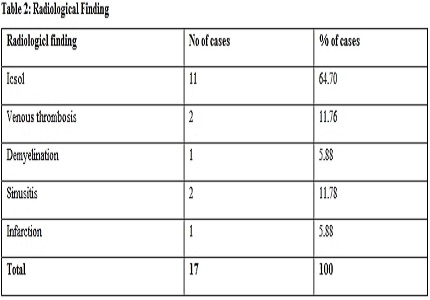
Result: In radiological study ICSOL is observed in 70.58% cases followed by 11.76% cases of sinusitis followed by 5.88% case each venous thrombosis,demyelination and infarction.
Conclusion: Early recognition of papilledema and elevated ICP is of paramount importance for ensuring restoration of vision. Newer advanced MR imaging techniques such as fMRI and DTI may prove useful in the future to assess the potential effects of papilledema on retinal and visual pathway integrity.
Downloads
References
2. Bruce BB, Biousse V, Newman NJ. Update on idiopathic intracranial hypertension. Am J Ophthalmol. 2011 Aug;152(2):163-9. doi: 10.1016/j.ajo.2011.03.020. Epub 2011 Jun 21. [PubMed]
3. Agid R, Farb RI, Willinsky RA, Mikulis DJ, Tomlinson G. Idiopathic intracranial hypertension: the validity of cross-sectional neuroimaging signs. Neuroradiology. 2006;48:521–527.
4. Wall M. Idiopathic intracranial hypertension. NeurolClin. 2010;28:593–617. [PubMed]
5. Silbergleit R, Junck L, Gebarski SS, Hatfield MK. Idiopathic intracranial hypertension (pseudotumor cerebri): MR imaging. Radiology. 1989 Jan;170(1 Pt 1):207-9.
6. George AE. Idiopathic intracranial hypertension: pathogenesis and the role of MR imaging. Radiology. 1989 Jan;170(1 Pt 1):21-2. [PubMed]
7. Brodsky MC, Vaphiades M. Magnetic resonance imaging in pseudotumor cerebri. Ophthalmology. 1998 Sep;105(9):1686-93. [PubMed]
8. Yuh WT, Zhu M, Taoka T, Quets JP, Maley JE, Muhonen MG, Schuster ME, Kardon RH. MR imaging of pituitary morphology in idiopathic intracranial hypertension. J MagnReson Imaging. 2000;12:808–813.
9. Degnan AJ1, Levy LM. Pseudotumor cerebri: brief review of clinical syndrome and imaging findings. AJNR Am J Neuroradiol. 2011 Dec;32(11):1986-93. doi: 10.3174/ajnr.A2404. Epub 2011 Jun 16. [PubMed]
10. Farb RI, Vanek I, Scott JN, Mikulis DJ, Willinsky RA, Tomlinson G, terBrugge KG. Idiopathic intracranial hypertension: the prevalence and morphology of sinovenous stenosis. Neurology. 2003 May 13;60(9):1418-24.
11. Higgins JN1, Cousins C, Owler BK, Sarkies N, Pickard JD. Idiopathic intracranial hypertension: 12 cases treated by venous sinus stenting. J Neurol Neurosurg Psychiatry. 2003 Dec;74(12):1662-6.
12. Jinkins JR, Athale S, Xiong L, Yuh WT, Rothman MI, Nguyen PT. MR of optic papilla protrusion in patients with high intracranial pressure. AJNR Am J Neuroradiol. 1996;17:665.
13. Gass A, Barker GJ, Riordan-Eva P, MacManus D, Sanders M, Tofts PS, McDonald WI, Moseley IF, Miller DH. MRI of the optic nerve in benign intracranial hypertension. Neuroradiology. 1996;38:769–773.
14. Watanabe A, Kinouchi H, Horikoshi T, Uchida M, Ishigame K. Effect of intracranial pressure on the diameter of the optic nerve sheath. J Neurosurg. 2008 Aug;109(2):255-8. doi: 10.3171/JNS/2008/109/8/0255.
15. Degnan AJ, Levy LM. Narrowing of Meckel's cave and cavernous sinus and enlargement of the optic nerve sheath in Pseudotumor Cerebri. J Comput Assist Tomogr. 2011 Mar-Apr;35(2):308-12. doi: 10.1097/RCT.0b013e31820d7a70.
16. Shofty B, Ben-Sira L, Constantini S, Freedman S, Kesler A. Optic nerve sheath diameter on MR Imaging: establishment of norms and comparison of pediatric patients with idiopathic intracranial hypertension with healthy controls. AJNR Am J Neuroradiol. 2012;33:366–369.
17. Rohr AC, Riedel C, Fruehauf MC, van Baalen A, Bartsch T, Hedderich J, Alfke K, Doerner L, Jansen O. MR imaging findings in patients with secondary intracranial hypertension. AJNR Am J Neuroradiol. 2011 Jun-Jul;32(6):1021-9. doi: 10.3174/ajnr.A2463. Epub 2011 Apr 21.
18. Biousse V, Ameri A, Bousser MG. Isolated intracranial hypertension as the only sign of cerebral venous thrombosis. Neurology. 1999 Oct 22;53(7):1537-42.
19. Killer HE, Jaggi GP, Miller NR. Papilledema revisited: is its pathophysiology really understood? Clin Experiment Ophthalmol. 2009 Jul;37(5):444-7. doi: 10.1111/j.1442-9071.2009.02059.x.
20. Seitz J, Held P, Strotzer M, et al. Magnetic resonance imaging in patients diagnosed with papilledema: a comparison of 6 different high-resolution T1- and T2(*)-weighted 3-dimensional and 2-dimensional sequences. J Neuroimaging 2002;12:164–71.
21. Bidot S, Saindane AM, Peragallo JH, Bruce BB, Newman NJ, Biousse V. Brain Imaging in Idiopathic Intracranial Hypertension. J Neuroophthalmol. 2015 Dec;35(4):400-11. doi: 10.1097/WNO.0000000000000303.
22. Digre KB, Nakamoto BK, Warner JE, Langeberg WJ, Baggaley SK, Katz BJ. A comparison of idiopathic intracranial hypertension with and without papilledema. Headache. 2009 Feb;49(2):185-93. doi: 10.1111/j.1526-4610.2008.01324.x.
23. Mashima Y, Oshitari K, Imamura Y, et al. High-resolution magnetic resonance imaging of the intraorbital optic nerve and subarachnoid space in patients with papilledema and optic atrophy. Arch Ophthalmol1996;114:1197–203.
24. Gibby WA, Cohen MS, Goldberg HI, et al. Pseudotumorcerebri: CT findings and correlation with vision loss. AJR Am J Roentgenol1993;160:143– 46. [PubMed]
25. Lagre`ze WA, Lazzaro A, Weigel M, et al. Morphometry of the retrobulbarhumanoptic nerve: comparison between conventional sonography and ultrafast magnetic resonance sequences. Invest Ophthalmol Vis Sci2007;48:1913–17. [PubMed]
26. Kimberly HH, Noble VE. Using MRI of the optic nerve sheath to detect elevated intracranial pressure. Crit Care. 2008;12 (5):181. doi: 10.1186/cc7008. Epub 2008 Sep 24. [PubMed]
27. Geeraerts T, Newcombe VF, Coles JP, Abate MG, Perkes IE, Hutchinson PJ, Outtrim JG, Chatfield DA, Menon DK. Use of T2-weighted magnetic resonance imaging of the optic nerve sheath to detect raised intracranial pressure. Crit Care. 2008;12(5):R114. doi: 10.1186/cc7006. Epub 2008 Sep 11.
28. Eliseeva NM, Serova NK, Arutiunov NV. [Magnetic resonance imaging of the orbital portion of the optic nerve at different stages of papilledema]. Vestn Oftalmol. 2005 Nov-Dec;121(6):5-9.
29. Mathew NT, Ravishankar K, Sanin LC. Coexistence of migraine and idiopathic intracranial hypertension without papilledema. Neurology 1996;46:1226–30. [PubMed]
30. Trobe JD. Papilledema: the vexing issues. J Neuroophthalmol. 2011 Jun;31(2):175-86. doi: 10.1097/WNO.0b013e31821a8b0b. [PubMed]
31. Winner P, Bello L. Idiopathic intracranial hypertension in a young child without visual symptoms or signs. Headache. 1996 Oct;36(9):574-6. [PubMed]
32. Hansen HC, Helmke K. Validation of the optic nerve sheath response to changing cerebrospinal fluid pressure: ultrasound findings during intrathecal infusion tests. J Neurosurg. 1997 Jul;87(1):34-40.
33. Passi N, Degnan AJ, Levy LM. MR imaging of papilledema and visual pathways: effects of increased intracranial pressure and pathophysiologic mechanisms. AJNR Am J Neuroradiol. 2013 May;34(5):919-24. doi: 10.3174/ajnr.A3022. Epub 2012 Mar 15.
34. Maysa A Ridha,a Amit M SaindaneMRI findings of elevated intracranial pressure in cerebral venous thrombosis versus idiopathic intracranial hypertension with transverse sinus stenosis Neuroophthalmology. 2013 Feb 1; 37(1): 1–6.doi: 10.3109/01658107.2012.738759.
35. AK Agarwal, PushpaYadav Papilledema (choked disc) Journal, Indian Academy of Clinical Medicine _ Vol. 1, No. 3 _ October-December 2000.
36. Julayanont P, Karukote A2, Ruthirago D1, Panikkath D3, Panikkath R3. Idiopathic intracranial hypertension: ongoing clinical challenges and future prospects. J Pain Res. 2016 Feb 19;9:87-99. doi: 10.2147/JPR.S60633. eCollection 2016.

 OAI - Open Archives Initiative