
A study to find the characteristic imaging findings of various ring-enhancing lesions using MR imaging
Abstract
Introduction: Multiple ring-enhancing lesions are one of the most commonly encountered neuroimaging abnormalities. A study was conducted to find the characteristic imaging findings of various ring-enhancing lesions on MRI.
Materials and methods: It was a prospective study conducted in the department of radiodiagnosis, GSL Medical College. All age groups of both gender with cerebral ring-enhancing lesions detected in contrast MR studies were taken up for spectroscopy. Patients with a history of claustrophobia and individuals with metallic implants insertion, cardiac pacemakers, and metallic foreign body in situ were excluded. The MRI scan was performed MRI PHILIPS ACHIEVA HDxt 1.5 T, the active shielded superconducting magnet of 1.5telsa magnetic field using SENSE coils for the acquisition of images.
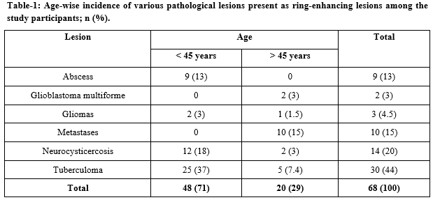
Results: Out of 30 study participants, under <45 years category tuberculoma was the common (37%) lesion whereas in >45 years category metastases was common (15%), 44% were female and 56% were males. Depicts T1 signal intensity showed hypointense signal intensity on T1W sequence in 65 (95.5%) cases. Only 4.6% of cases showed isointense signal intensity on T1W image. T2 signal intensity in 73.5% of cases showed hyperintense signal intensity.
Conclusions: MRI is an excellent, non-ionizing imaging modality with multiplanar imaging capabilities for excellent grey-white matter differentiation
Downloads
References
Omuro AM, Leite CC, Mokhtari K, Delattre JY. Pitfalls in the diagnosis of brain tumours. Lancet Neurol. 2006;5(11):937-948. doi: https://doi.org/10.1016/S1474-4422(06)70597-X.
Cunliffe CH, Fischer I, Monoky D, Law M, Revercomb C, Elrich S, et al. Intracranial lesions mimicking neoplasms. Arch Pathol Lab Med. 2009;133(1):101-123. doi: https://doi.org/10.1043/1543-2165-133.1.101.
Smirniotopoulos JG, Murphy FM, Rushing EJ, Rees JH, Schroeder JW. Patterns of contrast enhancement in the brain and meninges. Radiograph. 2007;27(2):525-551. doi: https://doi.org/10.1148/rg.272065155.
Bulakbasi N. Clinical applications of proton MR spectroscopy in the diagnosis of brain tumours. Spectro. 2004;18(2):143-153. doi: https://doi.org/10.1155/2004/381453.
Schwartz KM, Erickson BJ, Lucchinetti C. Pattern of T2 hypointensity associated with ring-enhancing brain lesions can help to differentiate pathology Neuroradiol. 2006;48(3):143-149. doi: https://doi.org/10.1007/s00234-005-0024-5.
Kim TK, Chang KH, Kim CJ, Goo JM, Kook MC, Han MH. Intracranial Tuberculoma: Comparison of MR with Pathologic Findings. AJNR Am J Neuroradiol. 1995;16(9):1903-1908.
Gupta RK, Husain M, Vatsal DK, Kumar R, Chawla S, Husain N. Comparative evaluation of magnetization transfer MR imaging and in vivo proton MR spectroscopy in brain tuberculomas. Magn Reson Imaging. 2002;20(5):375-381. doi: https://doi.org/10.1016/s0730-725x(02)00518-0.
Jayasundar R, Singh VP, Raghunathan P, Jain K, Banerji AK Inflammatory granulomas: evaluation with proton MRS NMR Biomed. 1999;12(3):139-144. doi: https://doi.org/10.1002/(sici)1099-1492(199905)12:3%3C139::aid-nbm550%3E3.0.co;2-h.
Vasudev MK, Jayakumar PN, SrikanthSG, Nagarajan K, Mohanty. A Quantitative magnetic resonance technique in the evaluation of intracranial tuberculomas Acta Radiol. 2007;48(2):200-206. doi: https://doi.org/10.1080/02841850601067678.
Amaral L, Maschietto M, Maschietto R, Cury R, Ferreira NF et al. Unusual manifestations of neurocysticercosis in MR imaging: analysis of 172 cases Arq Neuropsiquiatr. 2003;61(3A):533-541. doi: https://doi.org/10.1590/s0004-282x2003000400002.
Chang KH, Han MH. MRI of CNS parasitic diseases. J Magn Reson Imaging. 1998;8(2):297-307. doi: https://doi.org/10.1002/jmri.1880080209.
HR Martinez. R Rangel Guerra, G Elizondo, J Gonzalez, LE Todd et al. MR Imaging in neurocysticercosis: a study of 56 cases. Am J Neuroradiol. 1989;10(5):1011-1019.
Haris M, Gupta RK, Singh A, Husain N, Husain M et al. Differentiation of infective from neoplastic brain lesions by dynamic contrast-enhanced MRI Neuro Radiol. 2008;50(6):531-540. doi: https://doi.org/10.1007/s00234-008-0378-6.
Singhal, S.R., Nanda, S, Singhal SK. Neurocysticercosis as an important differential of seizures in pregnancy: two case reports. J Med Case Reports. 2011;5:206. doi: https://doi.org/10.1186/1752-1947-5-206.
Kumar A, Kaushik S, Tripathi RP, Kaur P, Khushu S. Role of in vivo proton MR spectroscopy in the evaluation of adult brain lesions: Our preliminary experience. Neurol India. 2003;51:474-478.
Halmes AB, Zimmerman RD, Morgello S, Weingarten K, Becker RD et al. MR Imaging of brain abscesses. Am J Roenlogenol AJR. 1989;152(5):1073-1085. doi: https://doi.org/10.2214/ajr.152.5.1073.
Zee CS, Segall HD, Boswell W, Ahmadi J, Nelson M et al. MR imaging of neurocysticercosis. J Comput Assist Tomogr. 1988;12:927-934. doi: https://doi.org/10.1097/00004728-198811000-00004.
Tsui EY, Chan JH, Cheung YK, Lai KF, Fong D, Ng SH. Evaluation of cerebral abscesses by diffusion-weighted MR imaging and MR spectroscopy; Comput Med Imaging Graph. 2002;26(5):347-351. doi: https://doi.org/10.1016/s0895-6111(02)00018-6.
Mishra AM, Gupta RK, Jaggi RS, Reddy JS, Jha DK et al. Role of diffusion-weighted imaging and in vivo proton magnetic resonance spectroscopy in the differential diagnosis of ring-enhancing intracranial cystic mass lesions. J Comput Assist Tomogr. 2004;28(4):540-547. doi: https://doi.org/10.1097/00004728-200407000-00017.
Tsougos I, Svolos P, Kousi E, Fountas K, Theodorou K, Fezoulidis I, et al. Differentiation of gioblastoma multiforme from metastatic brain tumor using proton magnetic resonance spectroscopy, diffusion and perfusion metrics at 3 T. Cancer Imaging. 2012;12(3):423-436. doi: https://doi.org/10.1102/1470-7330.2012.0038.
Kao HW, Chiang SW, Chung HW, Tsai FY, Chen CY. Advanced MR Imaging of Gliomas: An Update. Biomed Res Int. 2013;2013:970586. doi: https://doi.org/10.1155/2013/970586.
Copyright (c) 2020 Author (s). Published by Siddharth Health Research and Social Welfare Society

This work is licensed under a Creative Commons Attribution 4.0 International License.

 OAI - Open Archives Initiative