Role of Computed Tomography (CT) in traumatic head injury evaluation – a cross-sectional study
Abstract
Introduction: CT is the single primary modality in the evaluation of patients with acute head injuries. With these, a study was taken to find various clinico radiological patterns of head injuries and to correlate the CT features with clinical operative findings.
Materials and Methods: This was a cross-sectional study carried in patients of head injury. The patients with a head injury, craniofacial trauma who underwent CT scanning were included in the study. Patients on the ventilator and with Glasgow coma scale <6 were excluded. Patients were scanned using dual Slice CT, Siemens somatom Emotion duo. A P-value of less than 0.05 was considered statistically significant.
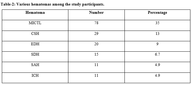
Results: Total 223 patients were included, 76.2% were males and 73.5% were abnormal scans. Among all intracranial traumatic lesions (ITL) the incidence of multiple ITLs were the most common (35%) and the death rate was 12.6%. Temporal bone fractures (15.2%) were the highest.
Conclusion: It was concluded that 21 – 40 years is the typical age group for head injuries, common among male and the incidence of mortality rate is more > 61 years. MICTLs are the most frequent type of hematomas.
Downloads
References
Osborn, Salzman, Barkovich, Katzman, Provenzale, Hansberger, diagnostic imaging brain - 2nd edition. Lippincott Williams & Wilkins 2009.
Hoyt DB, Holcomb J, Abraham E, Atkins J, Sopko G. Working Group on Trauma Research Program Summary Report: National Heart Lung Blood Institute (NHLBI), National Institute of General Medical Sciences (NIGMS), and National Institute of Neurological Disorders and Stroke (NINDS) of the National Institutes of Health (NIH), and the Department of Defense (DOD). J Trauma. 2004;57(2):410-415. doi: https://doi.org/10.1097/00005373-200408000-00038.
Andy Adam Adrian Dixon Jonathan Gillard Cornelia Schaefer-Prokop Ronald Grainger. Grainger and Allison's diagnostic radiology - 5th edition Elsevier 2014.pp 78.
John Haaga Daniel Boll. CT and MRI of the whole body - 5th edition Elsevier 2008. pp 295.
Miller EC, Holmes JF, Derlet RW. Utilizing clinical factors to reduce head CT scan ordering for minor head trauma patients. J Emerg Med. 1997;15(4):453-457. doi: https://doi.org/10.1016/s0736-4679(97)00071-1.
Kelly AB, Zimmerman RD, Snow RB, Gandy SE, Heier LA, Deck MD. Head trauma: comparison of MR and CT-experience in 100 patients. Am J Neuroradiol. 1988;9(4):699-708.
Glauser J. Head injury: which patients need imaging? Which test is best? Cleve Clin J Med 2004;71(4):353-357. doi: https://doi.org/10.3949/ccjm.71.4.353.
Jones TR, Kaplan RT, Lane B, Atlas SW, Rubin GD. Single versus multi detector row CT of the brain: quality assessment. Radiol. 2001;219(3):750-755. doi: https://doi.org/10.1148/radiology.219.3.r01jn47750.
Yealy DM, Hogan DE. Imaging after head trauma. Who needs what? Emerg Med Clin North Am. 1991;9(4):707-717.
Ambrose J, Honsfield G, Computed axial tomography. British J Radiol. 1973;46(542):148-149.
Asaleye CM, Famurewa OC, Komolafe EO, Komolafe MA, Amusa YB. The pattern of Computerized Topographic findings in moderate and severe head injuries in ILE- IFE, Nigeria. West Afr J Radiol. 2005;12:8-13.
Zimmerman RA, Bilaniuk LT, Hackney DB, Goldberg HI. Head injury: Early results of comparing CT and high field M.R. Amj Neuro Radiol. 1986;147(6):1215-1222. doi: https://doi.org/10.2214/ajr.147.6.1215.
Satish Prasad BS, Shama M Shetty. Evaluation of craniocerebral Trauma Using Computed Tomography. J Dental and Med Sci. 2014;13(9):57-62.
Van Dongen K.J, Reinder, Geert J. The prognostic value of CT in comatosed head injured patients. The J Neuro Surg. 1983;59(6):951-957. doi: https://doi.org/10.3171/jns.1983.59.6.0951.
Karibe H, Hayashi T, Hirano T, Kameyama M, Nakagawa A, Tominaga T. Surgical management of traumatic acute subdural hematoma in adults: a review. Neurol Med Chir (Tokyo). 2014;54(11):887-894. doi: https://dx.doi.org/10.2176%2Fnmc.ra.2014-0204.
Narayan RK, Greenberg RP, Miller JD, Enas GG, Choi SC, Kishore PR. Improved confidence of outcome prediction in severe head injury. A comparative analysis of the clinical examination, multi-modality evoked potentials, CT scanning, and intracranial pressure. J Neuro surg 1981;54(6):751-762. doi: https://doi.org/10.3171/jns.1981.54.6.0751.
Clifton GL, Grossman RG, Makela ME, Miner ME. Neurological course and correlated computed tomography findings after severe closed head injury. J Neuro Sur. 1980;52(5):611-624. doi: https://doi.org/10.3171/jns.1980.52.5.0611.
Kraus JF, Peek-Asa C, McArthur D: The independent effect of gender on outcomes following traumatic brain injury: a preliminary investigation. Neurosurg Focus. 2000;8(1):e5. doi: https://doi.org/10.3171/foc.2000.8.1.156.
Bharti P, Nagar A.M., Tyagi U. Pattern of trauma in western Uttar Pradesh. Neurol India. 1993;42:49-50.
Nayak A, Gupta MM, Shivam P. An analytic study of traumatic intra cerebral hematomas. Neurol India. 1993;41:217-222.
Seeling JM, Becker DP. Traumatic acute subdural haematoma. N Engl J Med. 1981;304(25):1511-1518. https://doi.org/10.1056/NEJM198106183042503
Takizawa T, Sato S. Traumatic subarachnoid haemorrhage. Neuro Med Chir 1984;24:390-395.
Koc RK, Akdemir H, Oktem IS, Meral M, Menku A. Acute subdural hematoma: outcome and outcome prediction. Neurosurg Rev 1997;20(4):239-244. doi: https://doi.org/10.1007/bf01105894.
Hirsh LF. Delayed traumatic intracerebral haematomas. Neurosurg. 1979;5(6):653-655. https://doi.org/10.1227/00006123-197912000-00001.


 OAI - Open Archives Initiative


