A study of the role of MRI and arthroscopy in the management of knee joint lesions
Abstract
Objectives: Aim of this study is to evaluate the types and incidence of injuries in internal derangement of the knee joint by MRI and to compare with arthroscopy findings in selected cases and to assess whether MRI can be used as a primary diagnostic tool for internal derangement of the knee joint.
Material and Method: This prospective study was done in the Department of Radiodiagnosis Chirayu Medical College and Hospital Bhopal, Madhya Pradesh, India. A total of 100 patients who were referred to the department with strong clinical suspicion of internal derangements of knee joint, underwent magnetic resonance imaging evaluation of knee followed by arthroscopy in selected cases, wherever indicated from August 2014 to July 2019.
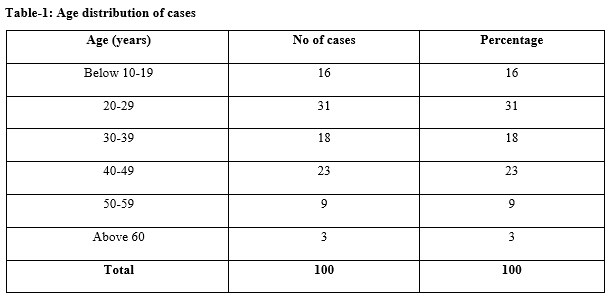
Results: Majority of patients in the current study group belonged to the age group 20-29 years (31%) with a mean age of 24.3 years. In this study, the majority of patients were males constituting 76 % of cases. The most common clinical presentation was that of pain in knee joint seen in 79% of cases. The second most common presentation was swelling seen in 54%. The most common positive clinical test was McMurray’s test for meniscal tear seen positive in 48% of cases. In the current study left knee involvement was more common than right knee, constituting 54%. Medial meniscal tears were more common than lateral meniscal tears 49 (73.2%).
Conclusion: MRI is a useful non-invasive modality having high sensitivity, specificity and accuracy in the diagnosis of meniscal and cruciate ligament injuries. MRI should be done in every patient of suspected internal derangement of the knee joint, to save a patient from unnecessary arthroscopy
Downloads
References
Biedert RM. Intrasubstance meniscal tears. Arch Orthop Trauma Surg. 1993;112(3):142-147. doi: https://doi.org/10.1007/bf00449992.
Hughes RJ, Houlihan-Burne DG. Clinical and MRI considerations in sports-related knee joint cartilage injury and cartilage repair. Semin Musculoskelet Radiol. 2011;15(1):69–88. doi: https://doi.org/10.1055/s-0031-1271960.
Crawford R, Walley G, Bridgman S, Maffulli N. Magnetic resonance imaging versus arthroscopy in the diagnosis of knee pathology, concentrating on meniscal lesions and ACL tears: a systematic review. British Med Bullet. 2007;84(1):5-23. doi: https://doi.org/10.1093/bmb/ldm022.
Khan Z, Faruqui Z, Oguynbiyi O, Rosset G, Iqbal J. Ultrasound assessment of internal derangement of the knee. Acta Orthop Belg. 2006;72(1):72-76.
Coumas JM, Palmer WE. Knee arthrography: evolution and current status. Radiol Clin North Am. 1998;36(4):703-728. doi: https://doi.org/10.1016/s0033-8389(05)70057-9.
Nikolaou VS, Chronopoulos E, Savvidou C, Plessas S, Giannoudis P, Efstathopoulos N, Papachristou G. MRI efficacy in diagnosing internal lesions of the knee: a retrospective analysis. J Traum Manag Outcomes. 2008;2(1):4. doi: https://doi.org/10.1186/1752-2897-2-4.
Ruwe PA, Wright J, Randall RL, Lynch JK, Jokl P, McCarthy S: Can MR imaging effectively replace diagnostic arthroscopy? Radiol. 1992;183(2):335-339. doi: https://doi.org/10.1148/radiology.183.2.1561332.
Stoller DW, Martin C, Crues 3rd JV, Kaplan L, Mink JH. Meniscal tears: pathologic correlation with MR imaging. Radiol. 1987;163(3):731-735. doi: https://doi.org/10.1148/radiology.163.3.3575724.
Zanetti M, Pfirrmann CW, Schmid MR, Romero J, Seifert B, Hodler J. Patients with suspected meniscal tears: prevalence of abnormalities seen on MRI of 100 symptomatic and 100 contralateral asymptomatic knees. Am J Roentgenol. 2003;181(3):635-641. doi: https://doi.org/10.2214/ajr.181.3.1810635.
Mesgarzadeh M, Moyer R, Leder DS, Revesz G, Russoniello A, Bonakdarpour A, et al. MR imaging of the knee: expanded classification and pitfalls to interpretation of meniscal tears. Radiograph. 1993;13(3):489-500. doi: https://doi.org/10.1148/radiographics.13.3.8316659.
Weiss CB, Lundberg M, Hamberg P, DeHaven KE, Gillquist J. Non operative treatment of meniscal tears. J Bone Joint Surg Am. 1989;71(6):811-822.
De Smet AA, Nathan DH, Graf BK, Haaland BA, Fine JP. Clinical and MRI findings associated with false-positive knee MR diagnoses of medial meniscal tears. Am J Roentgenol. 2008;191(1):93-99. doi: https://doi.org/10.2214/ajr.07.3034.
Kelly MA, Flock TJ, Kimmel JA, Kiernan Jr HA, Singson RS, Starron RB, Feldman F. MR imaging of the knee: clarification of its role. Arthroscopy: J Arthros Related Surg. 1991;7(1):78-85.
Cheung LP, Li KC, Hollett MD, Bergman AG, Herfkens RJ. Meniscal tears of the knee: accuracy of detection with fast spin-echo MR imaging and arthroscopic correlation in 293 patients. Radiol. 1997;203(2):508-512. doi: https://doi.org/10.1148/radiology.203.2.9114113.
JP Singh, Garg L, Garg L, Shrimali R, Setia V, Gupta V. MR Imaging of knee with arthroscopic correlation in twisting injuries. Indian J Radiol Imaging. 2004;14(1):33-40.
Naranje S, Mittal R, Sharma R. Arthroscopic and magnetic resonance imaging evaluation of meniscus lesions in the chronic anterior cruciate ligament-deficient knee.Arthoscopy. 2000;103(12):1079-1085. doi: https://doi.org/10.1016/j.arthro.2008.03.008.
Ha TP, Li KC, Beaulieu CF, Bergman G, Ch'en IY, Eller DJ, Cheung LP, Herfkens RJ. Anterior cruciate ligament injury: fast spin-echo MR imaging with arthroscopic correlation in 217 examinations. AJR. Am J Roentgenol. 1998;170(5):1215-1219. doi: https://doi.org/10.2214/ajr.170.5.9574587.
Schweitzer ME, Tran D, Deely DM, Hume EL. Medial collateral ligament injuries: evaluation of multiple signs, prevalence and location of associated bone bruises, and assessment with MR imaging. Radiol. 1995;194(3):825-829. doi: https://doi.org/10.1148/radiology.194.3.7862987.
Ochi M, Sumen Y, Kanda T, Ikuta Y, Itoh K. The diagnostic value and limitation of magnetic resonance imaging on chondral lesions in the knee joint. Arthroscopy: J Arthroscop Rel Surg. 1994;10(2):176-183. doi: https://doi.org/10.1016/s0749-8063(05)80090-8.
Heron CW, Calvert PT. Three-dimensional gradient-echo MR imaging of the knee: comparison with arthroscopy in 100 patients. Radiol. 1992;183(3):839-844. doi: https://doi.org/10.1148/radiology.183.3.1584944.


 OAI - Open Archives Initiative


