
Dosimetric comparison and clinical correlation between conventional four field radiotherapy versus three-dimensional conformal radiotherapy in cancer cervix
Abstract
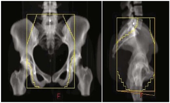
Introduction: With sectional imaging, wide variations are reported in pelvic anatomy of individual patients raising concerns over adequate coverage of target volume with conventional radiotherapy based on standard bony landmarks. Three-dimensional conformal radiotherapy (3DCRT) is reported to decrease normal tissue toxicity, along with decrease in chances of geographic miss. Present study is done for dosimetric comparison of Planning Target Volume (PTV) and Organs at Risk (OAR) in cancer cervix patients treated with conventional and conformal radiotherapy along with clinical correlation in terms of side effects and tumor response.
Materials and Methods: Fifty patients of cancer cervix underwent planning contrast enhanced CT scan. Target volumes & OAR were contoured. Patients were randomized into conventional & conformal arms. Conventional fields were planned using standard bony landmarks. CT based radiotherapy planning was done for 3DCRT arm. Field sizes &dose volume histogram (DVH) were recorded & compared for target coverage & OAR sparing in both arms. All patients received concurrent chemotherapy followed by brachytherapy.
Results: Field sizes used for the 3DCRT plans were significantly larger than those used for the conventional plans (p= 0.000). Optimal PTV coverage was significantly improved using 3DCRT as compared to conventional radiotherapy (p= 0.0001). Dose homogeneity in both arms were almost similar (p= 0.292), while conformity index was better in 3DCRT which was statistically significant between the groups (p= 0.000). Mean dose to the Planning Target Volume was increased significantly in the CT based plan when compared with the standard four field plan (p= 0.0001).Difference in doses to the organs at risk (urinary bladder, and small bowel)and their side effects were statistically significant across both groups. There was no difference in tumor response.
Conclusion: The present study showed significantly better target volume coverage & dose homogeneity with 3DCRT which may translate into better local control & survival but longer follow up is required to validate it.
Downloads
References
Zunino S, Rosato O, Lucino S, Jaurequi E, Rossi L, Valencia D. Anatomic study of the pelvis in carcinoma of the uterine cervix as related to the box technique. Int J Radiat Oncol Biol Phys. 1999;44(1):53-59. doi: https://doi.org/10.1016/S0360-3016(98)00538-0.
Kim RY, McGinnis S, Spencer SA, Meredith RF, Jennelle R, Salter MM. Conventional four-field pelvic radiotherapy technique without computer tomography-treatment planning in cancer of the cervix: Potential geographic miss and its impact on pelvic control. Int J Radiat Oncol Biol Phys. 1995;31(1):109-112. doi: https://doi.org/10.1016/0360-3016(94)00337-K.
Beadle BM, Jhingran A, Yom SY, Ramirez PT, Eifel PJ. Patterns of regional recurrence after definitive chemoradiotherapy for cervical cancer. Int J Radiat Oncol Biol Phys. 2010;76(5):1396-1403. doi: https://doi.org/10.1016/j.ijrobp.2009.04.009.
Gerstner N, Wachter S, Knocke TH, Fellner C, Wambersie A, P¨otter R. The benefit of Beam’s eye view-based 3D treatment planning for cervical cancer. Radiother Oncol 1999;51(1):71-78. doi: https://doi.org/10.1016/S0167-8140(99)00038-9.
Olofsen-van Acht M JJ, Quint S, Seven M, van Santvoort JP, van den Berg HA, Logmans A et al. Three-dimensional treatment planning for postoperative radiotherapy in patients with node-positive cervical cancer. Comparison between a conventional and a conformal technique. Strahlentherapie und Onkologie. 1999;175(9):462-469. doi: https://doi.org/10.1007/s000660050037.
Finlay MH, Ackerman I, Tirona RG, Hamilton P, Barbera L, Thomas G. Use of CT simulation for treatment of cervical cancer to assess the adequacy of lymph node coverage of conventional pelvic fields based on bony landmarks. Int J Radiat Oncol Biol Phys. 2006;64(1):205-209. doi: https://doi.org/10.1016/j.ijrobp.2005.06.025.
Gulia A, Patel F, Rai B, Bansal A, Sharma S C. Conventional four field radiotherapy versus computed tomography-based treatment planning in cancer cervix: A dosimetric study. South Asian J Cancer. 2013;2(3):132-135. doi: https://dx.doi.org/10.4103%2F2278-330X.114116.
Greer BE, Koh WJ, Figge DC, Russell AH, Cain JM, Tamimi HK. Gynaecologic radiotherapy fields defined by intra-operative measurements. Gynecol Oncol. 1990;38(3):421-424. doi: https://doi.org/10.1016/0090-8258(90)90084-X.
Pendlebury SC, Cahill S, Crandon AJ, Bull CA. Role of bipedal lymphangiogram in radiation treatment planning for cervix cancer. Int J Radiat Oncol Biol Phys. 1993;27(4):959-962. doi: https://doi.org/10.1016/0360-3016(93)90474-A.
Bonin SR, Lanciano RM, Corn BW, Hogan WM, Hartz WH, Hanks GE. Bony landmarks are not an adequate substitute for lymphangiography in defining pelvic lymph node location for the treatment of cervical cancer with radiotherapy. Int J Radiat Oncol Biol Phys. 1996; 34(1):167-172. doi: https://doi.org/10.1016/0360-3016(95)02055-1.
Mc Alpine A, Schlaereth JB, Lim P, Chen D, Eisenkop SM, Spiritos NM. Radiation fields in gynecologic oncology: Correlation of soft tissue (surgical) to radiologic landmarks. Gynecol Oncol. 2004;92(1):25-30. doi: https://doi.org/10.1016/j.ygyno.2003.09.008.
Boss EA, Barentsz JO, Massuger LF, Boonstra H. The role of MR imaging in invasive cervical carcinoma. Eur Radiol. 2000;10(2):256-270. doi: https://doi.org/10.1007/s003300050042.
Silva ML, Fogaroli R, Maia MA, Pellizzon AC, Novaes PE, Chojniak R, et al. A Comparative Analysis of the Conventional Pelvic Fields based on Bony Landmarks and Contrast-Enhanced CT Simulation for the Treatment of Cervical Cancer. Int J Rad Oncol* Biology* Physics. 2007;69(3):S407. doi: https://doi.org/10.1016/j.ijrobp.2007.07.1540.
WłodarczykH, Roszak A, Cikowska-WoźniakE, BratosK, Wojciechowska-ŁąckaA. Does conformal therapy improve dose distribution in comparison to old techniques in teleradiotherapy of cervical cancer patients?.Rep Pract Oncol Radiother. 2008;13(4):195-200. doi: https://doi.org/10.1016/S1507-1367(10)60009-0.

 OAI - Open Archives Initiative