
Role of MRI in evaluation of oral cavity cancers from central India
Abstract
Introduction: Squamous cell carcinoma of oral cavity ranks in the top three types of all cancer in the India. The diagnosisof it is basically clinical and bioptic, preoperative imaging is crucial for tumor staging that helps to decide appropriate therapeutic strategy and indicate prognosis.
Objectives: To evaluate the diagnostic accuracy of Magnetic resonance imaging (MRI) in staging of oral cavity cancers and to correlate the results with clinical and pathological data.
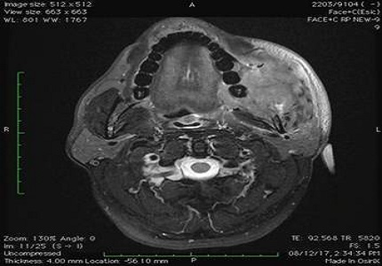
Methodology: This was a prospective study of 60 patients of oral cavity squamous cell carcinomas (SCC) where MRI was performed preoperatively to determine the tumor staging, tumor thickness, infiltration of surrounding structures and bone invasion. These findings were correlated with final pathological staging.
Results: Accurate correlation of T stages (TNM system) was obtained in 51 out of 60 cases on MRI. One lesion was classified as T2 on MRI but was found to be T4 on histopathology as MRI failed to detect subtle infiltration of the cortical bone. The accuracy of clinical data in T stage evaluation was 62 % (K- 0.470), however the MRI accuracy found to be 82 % (k 0.740). The sensitivity, specificity and accuracy of MRI in the detection of mandibular involvement were 94.1%, 60% and 81.5%, respectively.
Conclusion: MRI plays an important role in the evaluation of oral cavity carcinomas. MRIaccurately demonstrates the tumor size, depth of invasion, bone marrow involvement, perineural spread and lymphnode metastasis which are essential for treatment planning.
Downloads
References
Sankaranarayanan R, Masuyer E, Swaminathan R, et al. Head and neck cancer: a global perspective on epidemiology and prognosis. Anticancer Res. 1998 Nov-Dec;18(6B):4779-86.
Pintos J, Black MJ, Sadeghi N, Ghadirian P, Zeitouni AG, Raphael P, et al. Human papillomavirus infectionand oral cancer: a case-control study in Montreal Canada. Oral Oncology.2008;44(3):242–50.
Lenz M, Hermans R. Imaging of the oropharynx and oral cavity. Part II: Pathology. Eur Radiol. 1996;6(4):536-49.
Mari Hagiwara, Annette Nasbaum, Brian L Schmidt. Assessment of oral cavity carcinoma. Magnetic resonance imaging clinics august 2012: 20 (3):473- 494.
NCCN Guidelines Version 1.2012, Cancer of the Oral Cavity, National Comprehensive Cancer Care Clinical Practice Guidelines in Oncology (NCCN Guidelines). NCCN.org.
Tytor M, Olofsson J. Prognostic factors in oral cavity carcinomas. Acta Otolaryngol Suppl. 1992;492:75-8.
Hagiwara M, Nusbaum A, Schmidt BL. MR assessment of oral cavity carcinomas. DOI: https://doi.org/10.1016/j.mric.2012.05.003
Vidiri A, Ruscito P, Pichi B, Pellini R, Covello R, Sperduti I, Di Giovanni S, Spriano G, Crecco M. Oral and base of the tongue tumors. Correlation between clinical, MRI and pathological staging of primary tumor. Journal of Experimental and Clinical Cancer Research. 2007 Dec 1;26(4):575.
Moore MA, Yoo KY, Tuncer M. What is the future for the Asian Pacific Journal of Cancer Prevention (and Control) and the Asian Pacific Organization for Cancer Prevention (and Control)? Asian Pac J Cancer Prev. 2009 Jan-Mar;10(1):1-2.
Zeng H, Liang CH, Zhou ZG, Zheng JH, Zeng QX. Study of preoperative MRI staging of tongue carcinoma in relation to pathological findings. Di 1 junyi da xuexuebao= Academic journal of the first medical college of PLA. 2003 Aug;23(8):841-3.
Rodney R .Million, Nicholas J.Cassisi,ilius,J.BlipincottCo.,A Multidisciplinary Approach. Journal of the sciences and specialties of the head and neck .Volume 7 ,Issue3 January/February 1985 pages 260.
Okura M, Iida S, Aikawa T, Adachi T, Yoshimura N, Yamada T, Kogo M. Tumor thickness and paralingual distance of coronal MR imaging predicts cervical node metastases in oral tongue carcinoma. American Journal of Neuroradiology. 2008 Jan 1;29(1):45-50.
Chung TS, Yousem DM, Seigerman HM, Schlakman BN, Weinstein GS, Hayden RE. MR of mandibular invasion in patients with oral and oropharyngeal malignant neoplasms. American journal of neuroradiology. 1994 Nov 1;15(10):1949-55.
Kumaran PS, Thangaswamy SV, Navaneetham A. The need for early detection of neck nodal metastasis in squamous cell carcinoma of oral cavity. DOI: https://dx.doi.org/10.4103%2F0975-7406.100300.
de Bondt RB, Hoeberigs MC, Nelemans PJ, et al. Diagnostic accuracy and additional value of diffusion-weighted imaging for discrimination of malignant cervical lymph nodes in head and neck squamous cell carcinoma. doi: https://doi.org/10.1007/s00234-008-0487-2. Epub 2009 Jan 10.

 OAI - Open Archives Initiative