
A study to assess the distribution of cystic and solid hepatic lesionson contrast enhanced helical computed tomography
Abstract
Introduction: Focal liver lesions are defined as solid or liquid-containing masses foreign to the normal anatomy of the liver that may be told apart from the latter organ using imaging techniques.
Material and Methods: This prospective study was done in the Department of Radiodiagnosis and imaging at Bhopal Medical Centre, Bhopal, Madhya Pradesh, India. A total of 100 patients who were referred to our department with strong clinical suspicion of focal liver lesion and those diagnosed by ultrasonography underwent multiphasic contrast enhanced CT evaluation of abdomen using Single slice Spiral CT scanner from March 2010 to May 2012.
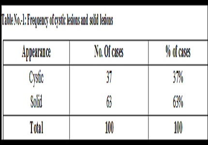
Results: The majority of lesions appeared as solid lesions on CT comprising 63% of lesions. The remaining were cystic lesions comprising 37% of cases. Among the cystic lesions, the most common CT diagnosis was Simple Cyst Seen in 49% of cases. The other diagnosis was liver abscess in 30% and hydatid cyst in 21%. Among the solid lesions, the most common CT diagnosis was that of metastases seen in 49% of cases. The next common lesions seen were HCC seen in 22.5% of cases and hemangioma seen in 19.5% of cases.
Discussion: Simple cysts are diagnosed by the presence of a well defined intra hepatic mass of fluid attenuation, no perceptible wall or internal septations and no enhancement. Among solid lesions, haemangiomas were diagnosed by the presence of a hypodense lesion with peripheral nodular enhancement of arterial attenuation with centripetal fill in.
Conclusion: Most of the lesions evaluated were solid compared to cystic. Among cystic lesions, simple cyst was the most common abnormality seen in around half the cases followed by liver abscess and hydatid cyst. Among the solid lesions, the most common abnormality detected was metastases seen in around half the cases followed by HCC and hemangioma.
Downloads
References
Pons F, Llovet JM. Approaching focal liver lesions. Rev EspEnferm Dig. 2004 Aug;96(8):567-73; 573-7.
Kumar PB, Hegde P, Kumar BNK et al. A Comparative Study of Ultrasound and CT findings in Focal Liver Lesions. Int J Biol Med Res. 2014; 5(3): 4362-4369.
Sica GT, Ji H, Ros PR. CT and MR imaging of hepatic metastases. AJR Am J Roentgenol. 2000 Mar;174 (3): 691-8. DOI: https://doi.org/10.2214/ajr.174.3.1740691.
Francis IR, Cohan RH, McNulty NJ et al. Multidetector CT of the liver and hepatic neoplasms: effect of multiphasic imaging on tumorconspicuity and vascular enhancement. AJR Am J Roentgenol. 2003 May;180(5):1217-24.
Zhu AX, Holalkere NS, Muzikansky A, Horgan K, Sahani DV. et al. Early antiangiogenic activity of bevacizumab evaluated by computed tomography perfusion scan in patients with advanced hepatocellular carcinoma. doi: https://doi.org/10.1634/theoncologist.2007-0174.
Ippolito D, Sironi S, Pozzi M, Antolini L, Ratti L, Alberzoni C, Leone EB, Meloni F, Valsecchi MG, Fazio F. et al. Hepatocellular carcinoma in cirrhotic liver disease: functional computed tomography with perfusion imaging in the assessment of tumor vascularization. doi: https://doi.org/10.1016/j.acra.2008.02.005.
Semelka RC, Martin DR, Balci C, et al. Focal liver lesions: comparison of dual-phase CT and multisequence multiplanar MR imaging including dynamic gadolinium enhancement. J MagnReson Imaging. 2001 Mar;13(3):397-401.
Murphy BJ, Casillas J, Ros PR, Morilloet al. The CT appearance of cystic masses of the liver. doi: https://doi.org/10.1148/radiographics.9.2.2538868.
J P Heiken, P J Weyman, J K Lee, et al. Detection of focal hepatic masses: prospective evaluation with CT, delayed CT, CT during arterial portography, and MR imaging. doi: https://doi.org/10.1148/radiology.171.1.2538862.
van Leeuwen MS, Noordzij J, Feldberg MA, et al. Focal liver lesions: characterization with triphasic spiral CT.DOI: https://doi.org/10.1148/radiology.201.2.8888219
Nino-Murcia M, Olcott EW, Jeffrey RB Jr, Lamm RL, et al.Focal liver lesions: pattern-based classification scheme for enhancement at arterial phase CT. DOI: https://doi.org/10.1148/radiology.215.3.r00jn03746.
Lee Joseph KT, Stuart S. Sagel, Robert J. Stanley et al. Computed body Tomography with MRI correlation 2004.
Fernandez MP, Redvanly RD.Primary hepatic malignant neoplasms.Radiol Clin North Am. 1998 Mar;36 (2): 333-48.
Loyer EM, Chin H, Du Brow RA, et al. Hepatocellular carcinoma and intrahepatic peripheral cholangiocarcinoma: enhancement patterns with quadruple phase helical CT--a comparative study. DOI: https://doi.org/10.1148/radiology.212.3.r99se32866.
Dachmann Abraham H, Lichtenstein JE, Friedman AC et al. Infantile hemangioendothelioma of the liver. A radiologic- pathologic- clinical correlation. AJR 1983; 140; 1091-1096.

 OAI - Open Archives Initiative