
Phenotypic characterization of Trichosporon species from clinical isolates
Abstract
Introduction: Trichosporon spp. arepresent as normal flora of skin and perigenital area in humans. They can cause invasive trichosporonosis in immuno-compromised individuals. Trichosporon infections are difficult to treat, since they do not respond to routinely used antifungal agents.
Aim: This study was undertaken to characterize clinical isolates of Trichosporonsp, using phenotypic methods.
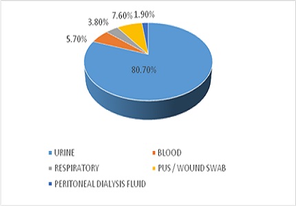
Material and Methods: Around 52 isolates of Trichosporon sp. obtained from various samples of patients in Sri Ramachandra Medical College & Research Institute, Chennai, were considered for the study. The isolates were studied for macroscopic morphology of the colonies on Sabouraud Dextrose Agar (SDA), microscopic morphology by Dalmau technique & Gram stain, ability to hydrolyze urea, sugar assimilation profile and growth at 37°C &0.1% cycloheximide.
Results and Conclusion: Two types of colony morphologies were observed, 47 isolates appeared flat with farinose covering and 5 were cerebriform with radial fissures. All isolates hydrolysed urea, and none of the isolates fermented but only assimilated sugars. The Trichosporon isolates were identified up to species level by phenotypic methods. Still the reliability of identification has to be confirmed with genotypic characterization techniques.
Downloads
References
2. Ruan SY, Chien JY, Hsueh PR.Invasive trichosporonosis caused by Trichosporonasahii and other unusual Trichosporon species at a medical center in Taiwan. Clin Infect Dis. 2009 Jul 1;49(1):e11-7. doi: 10.1086/599614. [PubMed]
3. Suzuki K, Nakase K, Kyo T, Kohara T, Sugawara Y, Shibazaki T, Oka K, Tsukada T, Katayama N.FatalTrichosporonfungemia in patients with hematologic malignancies. Eur J Haematol. 2010 May;84(5):441-7. doi: 10.1111/j.1600-0609.2010.01410.x.Epub 2010 Jan 13. [PubMed]
4. Colombo AL, Padovan AC, Chaves GM. Current knowledge of Trichosporon spp. and Trichosporonosis. ClinMicrobiol Rev. 2011 Oct;24(4):682-700. doi: 10.1128/CMR.00003-11. [PubMed]
5. Fell JW, Boekhout T, Fonseca A, Scorzetti G, Statzell-Tallman A.Biodiversity and systematics of basidiomycetousyeasts as determined by large-subunitrDNAD1/D2domainsequence analysis. Int J SystEvolMicrobiol. 2000 May;50 Pt 3:1351-71. [PubMed]
6. De Hoog G. S, J Guarro, J Gene, M. J. Figueres. Atlas of clinical fungi. Centraalbureauvoor Schimmelcultures Utrecht, The Netherlands, and UniversitatRoviraiVirgili, Reus, Italy.2000.
7. Kwon-Chung K. J, J. E. Bennett. Medical mycology. Lea &Febiger, Philadelphia, PA.1992.
8. Lacaz CS, Porto E, Martins JEC. MicologiaMédica. Fungos, actinomicetos e algas de interessemédico. 1984; 7th ed; Sarvier, São Paulo, 479 pp.
9. Guého E, de Hoog GS, Smith MT. Neotypification of the genus Trichosporon. Antonie Van Leeuwenhoek. 1992 May;61(4):285-8. [PubMed]
10. Magalhães AR, Mondino SS, Silva Md, Nishikawa MM.Morphological and biochemicalcharacterization of the aetiologicalagents of white piedra. Mem Inst Oswaldo Cruz. 2008 Dec;103(8):786-90. [PubMed]
11. McGinnis M. R. Mycology. In Clinical Microbiology Procedures Handbook.Edited by H. D. Isenberg. Washington, DC: American Society for Microbiology.1994; Section 6: pp. 6.1.1–6.1.6.1.12.
12. Fleming RV, Walsh TJ, Anaissie EJ. Emerging and less common fungal pathogens. Infect Dis Clin North Am. 2002 Dec;16(4):915-33, vi-vii.
13. Walsh TJ, Groll A, Hiemenz J, Fleming R, Roilides E, Anaissie E. Infections due to emerging and uncommon medically important fungal pathogens. ClinMicrobiol Infect. 2004 Mar;10 Suppl 1:48-66. [PubMed]
14. Barnett J. A, R. W. Payne, D. Yarrow. Yeasts: characteristics and Identification. Cambridge University Press, Cambridge, United Kingdom. 2000; 3rd ed.
15. Kurtzman C. P, J. W. Fell. The yeasts. A taxonomic study. Elsevier, Amsterdam, The Netherlands.1998.
16. Ichikawa T, Sugita T, Wang L, Yokoyama K, Nishimura K, Nishikawa A. Phenotypic switching and beta-N-acetylhexosaminidase activity of the pathogenic yeast Trichosporonasahii. Microbiol Immunol. 2004;48(4):237-42.
17. Ahmad S, Al-Mahmeed M, Khan ZU. Characterization of Trichosporon species isolated from clinical specimens in Kuwait. J Med Microbiol. 2005 Jul;54(Pt 7):639-46.
18. Lee J. W, Melcher G. A, Rinaldi M. G, Pizzo P. A, Walsh T. J. Patterns of morphologic variation among isolates of Trichosporonbeigelii. J ClinMicrobiol. 1990; 28: 2823–2827.
19. Pincus DH, Orenga S, Chatellier S. Yeast identification--past, present, and future methods. Med Mycol. 2007 Mar;45(2):97-121. [PubMed]
20. Sugita T, Nishikawa A, Shinoda T. Rapid detection of species of the opportunistic yeast Trichosporon by PCR. J ClinMicrobiol. 1998 May;36(5):1458-60.

 OAI - Open Archives Initiative