
Spectrum of MRI findings in traumaticknee
Abstract
Introduction: Injuries to the intraarticular structures like menisci and cruciate ligame nts are diagnosed with high sensitivity and specificity by MRI as compared with arthroscopy, which is still regarded as the gold standard. The acutely injured knee is readily imaged for the detection of meniscal and ligamentous injuries.
Aim: To describe the MRI features in various types of traumatic lesions andto analyze the types of knee joint abnormalities detected by MRI which will aid in making a proper diagnosis.
Materials and Methods: A total of 51 patients with history of trauma to the knee joint, who were referred to radiology department from orthopaedic department of Rajindra hospital Patiala, suspecting internal derangement of knee, served as subjects for this study. The inclusion and exclusion criteriafor this study was fixed as well as MRI protocol which included T1 and T2 saggital, PD weighted sequences in axial, Coronal and Saggital, Fat Suppressed T2 or STIR sequences if needed. The different types of lesions of all components of knee joint were categorized separately and data was assessed.
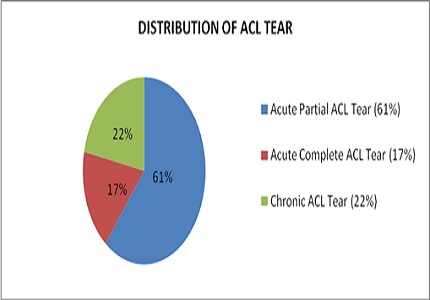
Results: Out of 51 patients who underwent MRI with history of trauma, 23 (45%) patients had abnormal ACL and 28 (54.9%) patients had normal ACL. PCL was found abnormal in only 3 (5.8%) patients. MCL was abnormal in 5 (9.8%), joint effusion was seen in maximum no of 44 (86.2%) patients. Muscular pathology was seen in 1patient and bone pathology in 23 (45%) patients. In medial meniscus tearsand lateral meniscus tears, posterior horn tears were the commonest. In ACL tears, acute partial tears were the commonest (61%).
Conclusion: In the setting of traumatic knee injury, MR imaging is the best noninvasive modality for the diagnosis of meniscal and ligamentous tears and to help guide the treatment of meniscal and ligament injuries.
Downloads
References
Prickett WD, Ward SI, Matava MJ. Magnetic resonance imaging of the knee. Sports Med. 2001; 31 (14): 997-1019.
Kean DM, Worthington BS, Preston BJ, Roebuck EJ, McKim-Thomas H, Hawkes RC, Holland GN, Moore WS. Nuclear magnetic resonance imaging of the knee: examples of normal anatomy and pathology. Br J Radiol. 1983 Jun; 56 (666): 355-64.
Soudry M, Lanir A, Angel D, Roffman M, Kaplan N, Mendes DG. Anatomy of the normal knee as seen by magnetic resonance imaging. Bone and Joint Journal. 1986; 68 (1):117-20.
Escobedo EM, Hunter JC, Zink-Brody GC, Wilson AJ, Harrison SD, Fisher DJ. Usefulness of turbospin-echo MR imaging in the evaluation of meniscialtears: comparison with a conventional spin-echosequence. AJR Am J Roentgenol. 1996 Nov; 167 (5):1223-7.
Nguyen JC, De Smet AA, Graf BK, Rosas HG. MRimaging-baseddiagnosis and classification of meniscaltears. Radiographics. 2014 Jul-Aug;34(4): 981-99. doi: https://doi.org/10.1148/rg.344125202.
Dandy DJ. The arthroscopic anatomy of symptomatic meniscal lesions. Bone and Joint Journal. 1990;72 (4): 628-33.
Kaplan AP, Helms CA, Dussault R. Anderson MV, Major NM: Musculoskeletal MRI. WB Saunders Company, Philadelphia 2009; 323-45.
Mansour MAM, Ahmed RM, Alaaibrahim, Elhussein N, Aljuaid SA. Magnetic resonance imaging diagnostic procedures for knee joint injuries. IOSR-JNHS. 2015;4 (2): 37-46.
Brandser EA, Riley MA, Berbaum KS, EL-Khoury GY, Bennett DL. MR imaging of anterior cruciate ligament injury: independent value of primary and secondary signs. AJR. American Journal of Roentgenology. 1996 Jul;167(1):121-6.
Vahey TN, Broome DR, Kayes KJ, Shelbourne KD. Acute and chronictears of the anterior cruciate ligament: differential features at MR imaging. Radiology. 1991; 181(1): 251-3.
Sonin AH, Fitzgerald SW, Friedman H, Hoff FL, Hendrix RW, Rogers LF. Posterior cruciate ligament injury: MR imaging diagnosis and patterns of injury. Radiology. 1994;190 (2):455-8.
Muller W. The Knee: Form, Function, and Ligament Reconstruction (1stedn.). Springer-Verlag 1983; 187-90.
Recht MP, Applegate G, Kaplan P, Dussault R, Schweitzer M, Dalinka MK, Resnick D. The MR appearance of cruciateganglion cysts: a report of 16 cases. Skeletal Radiology. 1994 Nov; 23(8):597-600.
Spindler KP, Schils JP, Bergfeld JA, Andrish JT, Weiker GG, Anderson TE, Piraino DW, Richmond BJ, Medendorp SV. Prospective study of osseous, articular, and meniscallesions in recent-anterior cruciate ligament tears by magnetic resonance imaging and arthro-scopy. The American Jourrnal of Sports Med. 1993; 21(4): 551-7.
McCauley TR, Moses M, Kier R, Lynch JK, Barton JW, Jokl P. MR diagnosis of tears of anterior cruciate ligament of the knee: importance of ancillary findings. AJR American Journal of Roentgenology. 1994; 162 (1): 115-9.
Mathis CE, Noonan K, Kayes K. "Bone bruises" of the knee: a review. Iowa Orthopaedic Journal. 1998; 18:112-7.
Singh JP, Garg L, Shrimali R, Setia V, Gupta V. MR Imaging of knee with arthroscopic correlation in twisting injuries. Indian journal of radiology and imaging. 2004;14 (1):33-40.
Yoon KH, Yoo JH, Kim KI. Bonecontusion and associatedmeniscal and medialcollateral ligament-injury in patients with anterior cruciate ligament rupture. J Bone Joint Surg Am. 2011;93 (16):1510-8.
Nasir AI. The role of Magnetic Resonance Imaging in the Knee Joint Injuries. International Research Journal of Medical Sciences 2013;1 (5):1-7.
Hetta W and Niazi G. MRI in assessment of sports related knee injuries. The Egyptian Society of Radiology and Nuclear Medicine 2014;45 (4):1153-1161.
Arumugam V, Ganesan GR, Natarajan P. MRI Evaluation of Acute Internal Derangement of Knee. Open Journal of Radiology 2015;5: 66-71.
Sohail K, Ayesha H, Shireen K, Zahir S, Ambreen S, Rehana B. Role of MRI in painful knee. Ann. Pak. Inst. Med Sci. 2015;11 (3):137-41.
Pame M, Gayan M, Hazarika K, Roy DKR. MRI evaluation of painful knee joint- the correlation of multiple coexisting pathologies, age and sex. J. Evid. Based Med. Healthc. 2017;4(18):1019-27.
Stoller DW, Martin C, Crues JV 3rd, Kaplan L, Mink JH. Meniscaltears: pathologic correlation with MR imaging. Radiology. 1987;163(3):731-5.
Crues JV, Richard R, Morgan FW. Meniscal pathology: The expanding role of magnetic resonance imaging. Clinical orthopaedics and related research 1990; 252 :80-86.

 OAI - Open Archives Initiative