Variation in patterns of superficial vein of cubital fossa
Abstract
Aim: To determine anatomical variations of superficial veins of cubital fossa.
Background: The cubital fossa Veins lie superficially in the subcutaneous tissue and not paired with any artery, they are easy to view and access. The superficial veins include the cephalic, basilic, median cubital, and antebrachial veins and their tributaries. The knowledge of superficial venous distribution of the cubital fossa is important, not only from an anatomical-clinical and surgical point of view, but also anthropologically and biologically.
Materials and Methods: The study done on120 males and 30 females were randomly selected from among the rural area of central India which also include the students and staff members of J.N.M. College Sawangi Wardha (M.S.) 150 cases of right upper limbs and 150 cases of left upper limbs. After taking the subject's consent, the superficial veins of the cubital fossa were made prominent by using tourniquet. The patterns of cubital veins were marked on skin and photograph are taken.
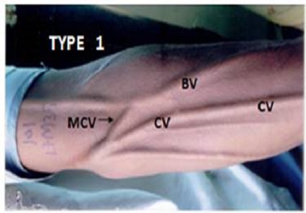
Results: the patterns of arrangement of superficial cubital vein observed are as follow. Type I –MCoV joining with the CV and BV in 51%., type II CV bifurcating intoMBV and MCV which join BV and ACV respectively, Type III-MVF bifurcating in MBV and MCV 9.66%., Type IV- 7.66%subjects of present study the CV terminates in to BV and ACV runs separately, In Type V- 3% of subjects the CV communicating directly with BV and ACV was absent and Type VI –A large single CV ascending upwards without any communication with other veins. 3 of 300 subjects i.e. 1%.
Conclusion: Numerous studies in different races and ethnic groups have shown similarities and differences in the deposition of the superficial veins of the cubital fossa. Various procedure should be perform with caution bearing in mind the anatomical variation present in these region.
Downloads
References
Standring S, editor. Gray's Anatomy. 41st ed. Edinburg: Elsevier Health Sciences; 2016.
Snell RS. Clinical anatomy by regions. , 9th ed. Baltimore: Lippincott Williams & Wilkins; 2012.
Williams PL, Bannister LH, Berry MM, Collins P, Dyson M, Dussek JE.et al. Gray’s Anatomy. 38th ed. Edinburgh UK: Churchill Livingstone; 1995.
Pyes KJ. Surgical Handicraft, 20th ed. Bristol: John Wright and Sons Ltd; 1977.
Moore KL, Dalley AF, Agur AM. Clinically oriented anatomy. Lippincott Williams & Wilkins; 2013.
Warwick R, Williams PL. Gray's anatomy, 35th ed. Edinburgh: Longman;1973.
Newman BH, Pichette S, Pichette D, Dzaka E. Adverse effects in blood donors after whole-blood donation: a study of 1000 blood donors interviewed 3 weeks after whole-blood donation. Transfusion. 2003;43:598–603.
Newman B, Waxman D. Blood donation-related neurologic needle injury: evaluation of 2 years' worth of data from a large blood center. Transfusion. 1996;36(3):213-215.
Warwick R,Williums R. Grays anatomy (36 edition) London, Churchill Livingstone.
Tewari SP, Singh SP, Singh S. The arrangement of superficial veins in cubital fossa in Indian subjects. J AnatSoc India. 1971;20(2):99-102.
Usha D, GopinathanandK, Dhall J.C.Superficial veins of the cubital fossa in Indian subjects.Anatomical Adjuncts. 1985;(1):39-45.
Okamoto P.The arrangement of the superficial veins of the cubittal fossa in Japanese men. Anat Rec. 1922;154:276‑8.
Singh SP, Ekandem GJ, Bose S. A study of the superficial veins of the cubital fossa in Nigerian subjects. Cells Tissues Organs. 1982;114(4):317-20.


 OAI - Open Archives Initiative


