
Comparison of CT only contour with MRI guided contouring in external beam radiotherapy for carcinoma rectum
Abstract
Aim: The purpose of the study is to compare CT only contour with MRI guided contour for delineation of the gross tumour volume (GTV) in carcinoma of rectum in external beam radiotherapy.
Materials and Methods: 18patients who underwent external beam radiotherapy treatment for carcinoma rectum were selected retrospectively. For all the patients, both CT and MRI were done as a part of planning process. Two sets of GTV were generated using only CT and with MRI assistance by a single oncologist. The generated contours were then compared and quantitatively analyzed using volume analysis and dice index.
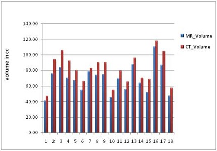
Results: The CT mean GTV was larger than the MRI mean GTV volume (68.54 ± 17.56 cc for CT versus 80.95 ± 19.19 cc for MRI). The dice index value between CT only GTV and MRI assisted GTV was 0.71 ± 0.13. The comparison of GTVs showed that the GTV_MRI was comparatively small and inside the GTV_CT except for six patients for whom GTV_MRI was marginally outside the GTV_CT.
Conclusion: The study showed that using MRI guidance for GTV delineation in carcinoma rectum is preferable and more accurate as compared to CT-only imaging because of superior soft tissue contrast.
Downloads
References
Evans PM.Anatomicalimaging for radiotherapy. Phys Med Biol.2008 Jun 21;53(12):R151-91. doi: https://doi.org/10.1088/0031-9155/53/12/R01. Epub 2008 May 21.
Njeh CF. Tumor delineation: The weakest link in the search for accuracy in radiotherapy. Journal of medical physics/Association of Medical Physicists of India. 2008 Oct; 33(4):136.
Khoo VS, Joon DL. New developments in MRI for target volume delineation in radiotherapy. Br J Radiol. 2006 Sep;79 Spec No 1:S2-15.
Devic S. MRI simulation for radiotherapy treatment planning. Med Phys. 2012 Nov;39(11):6701-11. doi: https://doi.org/10.1118/1.4758068.
Paulson ES, Erickson B, Schultz C, Allen Li X.ComprehensiveMRIsimulationmethodology using a dedicatedMRIscanner in radiation oncology for externalbeamradiationtreatmentplanning.Med Phys.2015Jan;42(1):28-39. doi: https://doi.org/10.1118/1.4896096.
Metcalfe P, Liney GP, Holloway L, Walker A, Barton M, Delaney GP, Vinod S, Tome W. The potential for an enhancedrole for MRI in radiation-therapytreatmentplanning.Technol Cancer Res Treat.2013Oct;12(5):429-46. doi: https://doi.org/10.7785%2Ftcrt.2012.500342. Epub 2013 Apr 24.
Beets-Tan RG, Beets GL.Rectal cancer: review with emphasis on MRimaging. Radiology.2004 Aug;232(2):335-46.
Varian Eclipse registration user guide.
Thada V, Jaglan V. Comparison of jaccard, dice, cosine similarity coefficient to find best fitness value for web retrieved documents using genetic algorithm. International Journal of Innovations in Engineering and Technology. 2013 Aug; 2(4):202-5.
Chang AE, Matory YL, Dwyer AJ, Hill SC, Girton ME, Steinberg SM, Knop RH, Frank JA, Hyams DA, Doppman JL. Magnetic resonance imaging versus computed tomography in the evaluation of soft tissue tumors of the extremities. Annals of surgery. 1987 Apr;205(4):340.
Aisen AM, Martel W, Braunstein EM, McMillin KI, Phillips WA, Kling TF. MRI and CT evaluation of primary bone and soft-tissue tumors. AJR Am J Roentgenol. 1986 Apr;146(4):749-56.
Goldblum JR, Weiss SW, Folpe AL. Enzinger and Weiss's Soft Tissue Tumors E-Book. Elsevier Health Sciences; 2013 Oct 11.
O'Neill BD, Salerno G, Thomas K, Tait DM, Brown G.MRvsCTimaging: lowrectal cancertumourdelineation for three-dimensional conformal radiotherapy. Br J Radiol.2009 Jun;82(978):509-13. doi: https://doi.org/10.1259/bjr/60198873. Epub 2009 Jan 19.
Kagawa K, Lee WR, Schultheiss TE, Hunt MA, Shaer AH, Hanks GE. Initial clinical assessment of CT-MRI image fusion software in localization of the prostate for 3D conformal radiation therapy. International Journal of Radiation Oncology, Biology Physics. 1997 May 1;38(2):319-25.
Kaus MR, Brock KK, Pekar V, Dawson LA, Nichol AM, Jaffray DA. Assessment of a model-based deformable image registration approach for radiation therapy planning. International Journal of Radiation Oncology Biology, Physics. 2007 Jun 1;68(2):572-80.
Van Herk M. Errors and margins in radiotherapy. Seminars in radiation oncology 2004 Jan 31 (Vol. 14, No. 1, pp. 52-64). WB Saunders.
Stroom JC, Heijmen BJ. Geometrical uncertainties, radiotherapy planning margins, and the ICRU-62 report. Radiotherapy and oncology. 2002 Jul 31;64(1):75-83.
Stroom JC, de Boer HC, Huizenga H, Visser AG.Inclusion of geometricaluncertainties in radiotherapytreatmentplanning by means of coverageprobability. Int J Radiat Oncol Biol Phys.1999Mar1;43(4):905-19.
Chao KS, Bhide S, Chen H, Asper J, Bush S, Franklin G, Kavadi V, Liengswangwong V, Gordon W, Raben A, Strasser J, Koprowski C, Frank S, Chronowski G, Ahamad A, Malyapa R, Zhang L, Dong L.Reduce in variation and improveefficiency of targetvolumedelineation by a computer-assistedsystem using a deformableimageregistrationapproach.Int J Radiat Oncol Biol Phys.2007Aug1;68(5):1512-21.
McJury M, O'Neill A, Lawson M, McGrath C, Grey A, Page W, O'Sullivan JM.Assessing the imagequality of pelvicMRimagesacquired with a flatcouch for radiotherapytreatmentplanning.Br J Radiol.2011 Aug;84(1004):750-5. doi: https://doi.org/10.1259/bjr/27295679.
Olch AJ, Gerig LLi H, Mihaylov I, Morgan A.Dosimetriceffectscaused bycouchtops and immobilizationdevices: report of AAPMTaskGroup176.Med Phys.2014Jun;41(6):061501. doi: https://doi.org/10.1118/1.4876299.
Steenbakkers RJ, Duppen JC, Betgen A, Lotz HT, Remeijer P, Fitton I, Nowak PJ, van Herk M, Rasch CR.Impact of kneesupport and shape of tabletop on rectum and prostateposition.Int J Radiat Oncol Biol Phys.2004Dec1;60(5):1364-72.
Viswanathan AN, Dimopoulos J, Kirisits C, Berger D, Pötter R. Computed tomography versus magnetic resonance imaging-based contouring in cervical cancer brachytherapy: results of a prospective trial and preliminary guidelines for standardized contours. International Journal of Radiation Oncology Biology, Physics. 2007 Jun 1;68(2):491-8.

 OAI - Open Archives Initiative