
Sonographic evaluation of abdominal organs in a sickle cell disease patient
Abstract
Background: SCD is one of the most common inherited hemoglobinopathies worldwide. It is a major health problem in Madhya Pradesh and surrounding states. The present study was undertaken to infer the value of ultrasonography in evaluating abdominal organs in a SCD patient.
Study design: This is a prospective hospital based study.
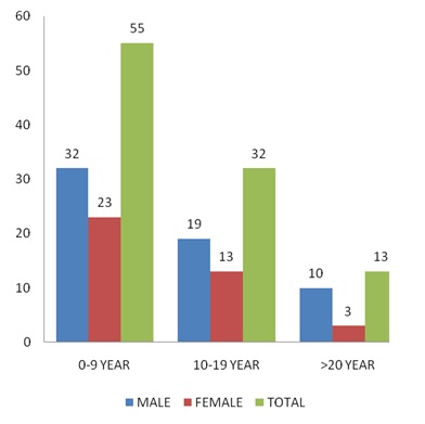
Method: 100 SCD patients were assessed for abdominal pathologies by USG, the results thus analyzed and prevalence was calculated.
Result: Hepatomegaly is the most common association observed in 69% of patients followed by splenomegaly in 31% patients & increased renal size in 29% patients. Cholelithiasis (17%), increased renal medullary echogenecity (14%), Auto-splenectomy (11%), Bright liver (9%), Splenic calcification (8%), Increased GB wall thickness (6%), Splenic infarct (5%) and Increased pancreatic echogenicity (3%) are the other associated findings.
Conclusion: Real time ultrasonography is a simple, cheap, rapid, easily accessible, non-invasive, non-ionizing screening procedure in all cases of SCD patient for assessment of pathological changes occurring in the various abdominal organs. The high rates of abdominal pathologies being diagnosed on ultrasonography underscores the need for it to be established as a standard screening procedure with established protocols.
Downloads
References
2. Bunn HF. Induction of fetal hemoglobin in sickle cell disease. Blood 1999; 93:1787–1789. [PubMed]
3. Rodgers GP. Overview of pathophysiology andrationale for treatment of sickle cell anemia. SeminHematol 1997; 34:2–7. [PubMed]
4.H. Lehman and M. Cutbush, “Sickle Cell Trait in Southern India,” British Medical Journal, Vol. 1, No.4755, 1952, pp. 404-405. [PubMed]
5. Dick R, Watkinson A. The liver and spleen. In: Sutton D, ed. Textbook of radiology and imaging. 7thed New York: Elsevier, 2002; 737-786.
6. Bhavna Dhingra, Suvasini Sharma, Devendra Mishra. 2009. Normal values of Liver and Spleen Size by Ultrasonography in Indian Children. PN:S097475590800697-1. [PubMed]
7. Miletic D, Fuckar Z, Sustic A, Mozetic V, Stimac D, Zauhar G. sonographic measurements of absolute and relative length in adults. J Clin Ultrasound 1998; 26:185-189.
8. Rumack CM, Wilson SR, Charboneau JW. Diagnostic ultrasound, 2nd ed. St. Louis, MO: Mosby, 1998:175-200. [PubMed]
9. Papadaki MG, Kattamis AC, Papadaki IG, Menegas DG, Georgakopoulou TP, Mavrommati-Metaxotou A, et al. Abdominal ultrasonographic findings in patients with Sickle cell anemia and thalassemia intermedia. PediatrRadiol 2003;33:515-21. [PubMed]
10. Balci A, Karazincir S, Sangün O, Gali E, Daplan T, Cingiz C. Prevalence of abdominal ultrasonographic abnormalities in patients with sickle cell disease. DiagnIntervRadiol 2008;14:133-7. [PubMed]
11. Mohanty J, Narayan J, Bhagat S, Panda BB, Satpathi G, Saha N. sonologicalevealuation of abdominal organs in sickle cell crisis in Western Orissa. Indian Journal of Radiology and Imaging 2004; 14:247-51.
12. Bakhieta IT. Sonographic findings in Sudanese children with sickle cell anaemia. J Diagn Med Sonography 2010;6:281-5.
13. Ma'aji SM, Jiya NM, Saidu SA, Danfulani M, Yunusa GH, Sani UM, Jibril B, Musa A, Gele HI, Baba MS, Bello S. Transabdominalultrasonographic findings in children with sickle cell anemia in Sokoto, North-Western Nigeria. Niger J Basic ClinSci [serial online] 2012 [cited 2013 Dec 17];9:14-7.
14. Hamim Abdul Rusheke. Abdominal ultrasonographic abnormalities in patients with sickle cell anaemia at muhimbili national hospital 2010; Muhimbili University of Health and Allied Science. ir.muhas.ac.tz:8080/jspui. http://hdl.handle.net/123456789/1046.
15. Lane PA. Sickle cell disease. PediatrClin North Am 1996; 43:639–664. [PubMed]
16. Ma'aji SM, Jiya NM, Saidu SA, Danfulani M, Yunusa GH, Sani UM, Jibril B, Musa A, Gele HI, Baba MS, Bello S. Transabdominalultrasonographic findings in children with sickle cell anemia in Sokoto, North-Western Nigeria. Niger J Basic ClinSci [serial online] 2012 [cited 2013 Dec 17];9:14-7.
17. Ahmed H.Salem et al: “Sonographic assessment of splenic size in Saudi patients with Sickle cell disease” Ann Saudi Med 1998;18(3):217-220. [PubMed]
18. Ibinaiye PO, Babadoko AA, Yusuf R, Hassan AA. Renal complications of sickle cell anemia in Zaria, Nigeria: An ultrasonographic assessment. West Afr J Radiol 2013;20:19-22.
19. D.A. Nzeh& M.A. Adedoyin et al. sonographic pattern of gall bladder disease in children with sickle cell disease. PediatrRadiol (1989) 19:290-292. [PubMed]
20. Namjoshi SP. Punctate echogenic foci in spleen and increase echogenicity in renal cortex in sickle cell Ameamia. J Clin Ultrasound 1999;27:52. [PubMed]
21. Walker TM, Searjeant GR. Increased renal reflectivityin sickle cell disease: prevalence and characteristics. ClinRadiol 1995; 195:566–569. [PubMed]
22. Namjoshi SP. Punctate echogenic foci in spleen and increase echogenicity in renal cortex in sickle cell Ameamia. J Clin Ultrasound 1999;27:52.

 OAI - Open Archives Initiative