
Comparative evaluation of ultrasonography and computed tomography findings in focal hepatic masses
Abstract
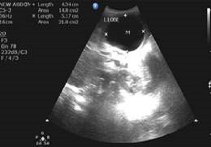
Introduction: Focal liver lesions are defined as solid or liquid-containing masses foreign to the normal anatomy of the liver that may be told apart from the latter organ using imaging techniques. They can have a number of etiologies including congenital, neoplastic and infectious and inflammatory conditions as well as trauma and miscellaneous entities. The data was analyzed to compare between ultrasonography (USG) and computed tomography (CT) findings in these cases.
Aims and Objectives: To evaluate the role of ultrasound and computed tomography in diagnosis of focal hepatic masses and compare the ultrasound and CT findings of focal hepatic masses and correlate with histopathological and surgical findings.
Material and Methods: USG and CT were performed on 40 focal hepatic mass patients. The diagnostic value of ultrasound was compared to those of CT. Final diagnosis was made after correlation with surgical findings, serological findings and histopathological examination. The data collected was analyzed statistically.
Results: Final diagnosis of focal hepatic masses was simple cysts (n=5), polycystic liver (n=1), metastasis (n=22), hydatid cysts(n=5), hemangioma (n=6), hepatocellular (n=11), focal nodular hyperplasia (n=1), abscess (n=16), cholangiocarcinoma (n=1). The sensitivity, specificity positive and negative likelihood ratio were 84.38%, 67.74%, 2.62 and 0.23 respectively, for USG and 100%, 97.14%, 35 and 0 respectively, for CT.
Conclusion: USG and CT are the modalities having comparable specificity and sensitivity, CT being slightly more accurate than USG in evaluation of focal hepatic lesions in atypical cases. USG is useful in follow-up cases.
Downloads
References
Tchelepi H, Ralls PW. Ultrasound of focal liver masses. Ultrasound Q. 2004 Dec;20(4):155-69.
Gupta AK, Chowdhury V, Khandelwal N. Benign Focal Lesions in Liver. In: Diagnostic Radiology gastrointestinal and hepatobiliary imaging. 3rd edn. Jaypee Brothers Medical Publishers (P) Ltd. 2011;13:238-45.
Sahani DV, Kalva SP. Imaging the liver. Oncologist. 2004;9(4):385-97.
Mortelé KJ, Ros PR. Cystic focal liver lesions in the adult: differential CT and MR imaging features. Radiographics. 2001 Jul-Aug;21(4):895-910.
Kar P and Jain R. Imaging of Space Occupying Lesions of Liver. Medicine update 2011:375-84.
Thimmaiah VT. Evaluation of focal liver lesions by ultrasound as a prime imaging modality. Sch. J. App. Med.Sci.,2013;1(6):1041-59.
Nino-Murcia M, Olcott EW, Jeffrey RB Jr, Lamm RL, Beaulieu CF, Jain KA. Focal liver lesions: pattern-based classification scheme for enhancement at arterial phase CT. Radiology. 2000 Jun;215(3):746-51.
Sukov RJ, Cohen LJ, Sample WF. Sonography of hepatic amebic abscesses.AJR Am J Roentgenol. 1980 May;134(5):911-5.
Gaines PA, Sampson MA. The prevalence and characterization of simple hepatic cysts by ultrasound examination. Br J Radiol. 1989 Apr;62(736):335-7.
Kalinova K. Imaging (ultrasonography, computer tomography) of patients with hydatid liver disease. Bulg. J. Vet. Med. 2007;10(1):45-51.
el-Tahir MI, Omojola MF, Malatani T, al-Saigh AH, Ogunbiyi OA. Hydatid disease of the liver: evaluation of ultrasound and computed tomography. Br J Radiol. 1992 May;65(773):390-2.
Alsaif HS, Venkatesh SK, Chan DS, Archuleta S. CT appearance of pyogenic liver abscesses caused by Klebsiella pneumoniae. Radiology. 2011 Jul;260(1):129-38. doi: https://doi.org/10.1148/radiol.11101876. Epub 2011 Apr 1.
Minami Y, Kudo M. Hepatic malignancies: Correlation between sonographic findings and pathological features. World J Radiol. 2010 Jul 28;2(7):249-56. doi: https://dx.doi.org/10.4329%2Fwjr.v2.i7.249.
Sica GT, Ji H, Ros PR. CT and MR imaging of hepatic metastases. AJR Am J Roentgenol. 2000 Mar;174(3):691-8.
Everson GT, Taylor MR, Doctor RB. Polycystic disease of the liver. Hepatology. 2004 Oct;40(4):774-82.
Kumar PB, Hegde P, Kumar BNK, Kumar MLH, Manjunatha YC. A comparative study of ultrasound and CT findings in focal liver lesions. Int Biol Med Res 2014;5(3):4362-69.
D'Onofrio M, Crosara S, De Robertis R, Canestrini S, Mucelli RP. Contrast-Enhanced Ultrasound of Focal Liver Lesions. AJR Am J Roentgenol. 2015 Jul;205(1):W56-66. doi: https://doi.org/10.2214/AJR.14.14203.

 OAI - Open Archives Initiative