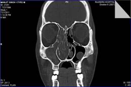
Comparison between preoperative computed tomography scan of paranasal sinuses and operative findings in functional endoscopic sinus surgery (FESS) in chronic sinusitis
Abstract
Background and objectives: The study was done to evaluate the role of computed tomography (CT) in clinically suspected cases of chronic sinusitis for detection, assessment of anatomical variants and pathological abnormalities of nasal cavity and paranasal sinuses. Also findings of CT were co-related with operative findings in functional endoscopic sinus surgery (FESS). The study was conducted on 60 cases attending CT section of Radiodiagnosis Department and ENT Department Rajindra Hospital, Patiala.
Materials and Methods: The study was conducted on 60 cases over a period 3 years from 2010 to 2013 irrespective of gender and age group attending the ENT and radiology department. Selections of patients were based on clinical features like nasal or post nasal discharge, nasal obstruction, headache anosmia, cough and hoarseness of voice. Relevant history, clinical examination and CT examination of every patient were done.
Results: In our study, the CT scan and operative findings correlated excellently in cases of paradoxical middle turbinate, bony destruction, concha bullosa, polypoidal changes in ethmoidal sinuses and osteomeatal complex normal widening. There was very good agreement between preoperative CT scan and operative findings for concha bullosa, paradoxical middle turbinate, bony destruction, osteomeatal complex normal widening, polypoidal change in sphenoid sinus. The most common pattern observed on CT scan was osteomeatal unit pattern in 35% followed by infundibular pattern (20%), sphenoethmoidal recess pattern (23.3%), unclassified pattern (15%) & sinonasal polyposis pattern (6.6%).
Conclusion: CT is the imaging modality of choice to reveal mucosal changes deeper in osteomeatal complex which are not visible endoscopically and to identify extent of paranasal sinus disease. However, endoscopy has complementary role with CT in evaluation of patients of chronic sinusitis.
Downloads
References
Lanza DC, Kennedy DW. Adult rhinusitis defined. Otolaryngol Head Neck Surg 1997;117 (3 Pt2):S1-7.doi: https://doi.org/10.1016/s0194-5998(97)70001-9.
Lund VJ, Kennedy DW, and the Staging and Therapy Group. Quantification for staging sinusitis. Ann Otol Rhinol Laryngol 1995;167(Suppl):17-21.
Shapiro ED, Milmoe GJ, Wald ER, Rodnan JB, Bowen AD. Bacteriology of the maxillary sinuses in patients with cystic fibrosis. J Infect Dis. 1982 Nov;146(5):589-93.doi: https://doi.org/10.1093/infdis/146.5.589.
Yousem DM. Imaging of sinonasal inflammatory disease. Radiology 1993;188:303-14.doi: https://doi.org/10.1148/radiology.188.2.8327669.
Davidson TM, Brahme FJ, Gallagher ME. Radiographic evaluation of nasal dysfunction: computed tomography versus plain films. Head Neck 1989;11:405-9.
Kronemer KA, McAlister WH. Sinusitis and its imaging in the pediatric population. Pediatr Radiol. 1997 Nov;27(11):837-46.
Duvoisin B, Landry M, Chapuis L, Krayenbuhl M, Schnyder P. Low-dose CT and inflammatory disease of the paranasal sinuses. Neuroradiology. 1991;33(5):403-6.doi: https://doi.org/10.1007/bf00598612.
Rao VM, el-Noueam KI. Sinonasal imaging: anatomy and pathology. Radiol Clin North Am. 1998 Sep;36(5):921-39.doi: https://doi.org/10.1016/s0033-8389(05)70069-5.
Tack D, Widelec J, De Maertelaer V, Bailly JM, Delcour C, Gevenois PA. Comparison between low-dose and standard-dose multidetector CT in patients with suspected chronic sinusitis. AJR Am J Roentgenol. 2003 Oct;181(4):939-44.doi: https://doi.org/10.2214/ajr.181.4.1810939.
Gotwald TF, Zinreich SJ, Corl F, Fishman EK. Three-dimensional volumetric display of the nasal ostiomeatal channels and paranasal sinuses. AJR 2001;176:241 –45.
Stammberger H. Endoscopic endonasal surgery- concepts in treatment of recurring rhinosinusitis. Part 1. Anatomic and pathophysiologic considerations. Otolaryngol Head Neck Surg 1986;94:143.doi: https://doi.org/10.1177/019459988609400202.
Shazo RD, Chapin K, Swain RE. Fungal sinusitis. N Engl J Med. 1997 Jul 24;337(4):254-9.doi: https://doi.org/10.1056/nejm199707243370407
Yousem DM. Imaging of sinonasal inflammatory disease. Radiology. 1993 Aug;188(2):303-14.
Elahi M, Frenkeil S, Remy H et al. Development of a standardized proforma for reporting computerized tomographic images of the paranasal sinuses. The Journal of Otolarygology 1996;25(2):113-20.
Bhattacharyya N. Test retest reliability of computed tomography in the assessment of chronic rhinosinusitis. Laryngoscope. 1999 Jul;109(7 Pt 1):1055-8.doi: https://doi.org/10.1097/00005537-199907000-00008.
Calhoun KH, Waggenspack GA, Simpson B et al. CT evaluation of the parnasal sinuses in symptomatic and asymptomatic populations. Otolaryngol Head Neck Surg. 1991 Apr;104(4):480-3.doi: https://doi.org/10.1177/019459989110400409.
Stoney p, Probst L, Shankar L et al. CT scanning for functional endoscopic sinus surgery: analysis of 200 cases with reporting scheme. J Otolaryngol. 1993 Apr;22(2):72-8.
Asruddin, Yadav SPS, Yadav RK et al.Low dose CT in chronic sinusitis. Indian J Otolaryngol Head Neck Surg. 1999 Dec;52(1):17-22. doi: 10.1007/BF02996425.
Yousem DM. Imaging of sinonasal inflammatory disease. Radiology. 1993 Aug;188(2):303-14.
Zinreich J. Imaging of inflammatory sinus disease. Otolaryngol Clin North Am. 1993 Aug;26(4):535-47.
Bolger WE, Butzin CA, Parsons DS. Paranasal sinus sinus bony anatomic variations and mucosal abnormalities:CT analysis for endoscopic sinus surgery. Laryngoscope. 1991 Jan;101(1 Pt 1):56-64.doi: https://doi.org/10.1288/00005537-199101000-00010.
Zinreich SJ, Mattox DE, Kennedy DW et al. Concha Bullosa: CT evaluation. J Comput Assist Tomogr. 1988 Sep-Oct;12(5):778-84.doi: https://doi.org/10.1097/00004728-198809010-00012.
Shroff MM, Kirtane MV, Shukla A et al. Anatomic variants and mucosal abnormalities in paranasal sinus CT –A study in patients with chronic sinusitis. Ind J Radiol Imag 1991;Nov:51-6.
Lusk RP, McAlister B,el-Fouley A. Anatomic variation in pediatric chronic sinusitis- A CT study. Otolaryngol Clin North Am. 1996 Feb;29(1):75-91.
Lazar RH, Younis RT, Parvey LS. Comparison of plain radiographs, coronal CT and intraoperative findings in children with chronic sinusitis. Otolaryngol Head Neck Surg. 1992 Jul;107(1):29-34.doi: https://doi.org/10.1177/019459989210700105.
Terrier F, Webei W, Ruefenacht D et al. Anatomy of ethmoid: CT, endoscopic and macroscopic . AJR Am J Roentgenol. 1985 Mar;144(3):493-500.doi: https://doi.org/10.2214/ajr.144.3.493.
Danielsen A, Olofsson J. Endoscopic sinus surgery. A long-term follow-up study. Acta Otolaryngol. 1996 Jul;116(4):611-9.doi: https://doi.org/10.3109/00016489609137898.
Damm M, Quante G et al. Impact of functional Endoscopy Sinus Surgery on Symptoms and Quality of Life in Chronic Rhinosinusitis. Laryngoscope. 2002 Feb;112(2):310-5.doi: https://doi.org/10.1097/00005537-200202000-00020.
Bhattacharyya N. Symptom Outcomes After Endoscopic Sinus Surgery for Chronic Rhinosinusitis. Arch Otolaryngol Head Neck Surg. 2004 Mar;130(3):329-33.doi: https://doi.org/10.1001/archotol.130.3.329.


 OAI - Open Archives Initiative


