
Role of susceptibility weighted imaging in characterization of intra-cranial lesions
Abstract
Background: Intrinsic differences between magnetic susceptibility of tissues can be used to generate a unique type of contrast in Magnetic Resonance Imaging (MRI), particularly to characterize tissues and improve the sensitivity for certain lesions, most notably hemorrhagic foci or calcification.
Methods: A blinded retrospective review of all brain imaging studies done at our centre between March 2014 and February 2016. One of them reviewed the MR images with exclusion of SWI, while the other radiologist reviewed the entire set of images including SWI. Their findings were then compared to study the efficacy of SWI in better detection and characterization of lesions.
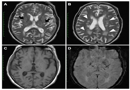
Results: The study included 3710 patients with different brain lesions. SWI revealed positive lesions in at least 619 of these patients. 21 patients with traumatic brain injury, 32 patients with cerebral microbleeds, 78 patients with brain tumors, 27 patients with venous malformations, 128 patients with intra-cranial calcifications, 277 patients with infarcts and 56 patients with extra-axial hemorrhage. Out of the 619 lesions, SWI played a significant role in lesion characterization in at least 198 patients which is about 5.33% of our total cohort of 3710 patients.
Conclusion: SWI images are fast sequences requiring no more than a total scan time of about 3 minutes. Inclusion of this sequence as a part of brain pathology canplay an important role in diagnosing different brain lesions as well as avoiding missing hemorrhagic lesions thus aviding inappropriate therapeutic regimens with catastrophic results.
Downloads
References
Haacke EM, Xu Y, Cheng YC, et al. Susceptibility weighted imaging (SWI).Magn Reson Med 2004;52(3):612–618. DOI: https://doi.org/10.1002/mrm.20198.
Akter M, Hirai T, Hiai Y, et al. Detection of hemorrhagic hypointense foci in the brain on susceptibility-weighted imaging: clinical and phantom studies. Acad Radiol 2007;14:1011–1019. DOI: https://doi.org/10.1016/j.acra.2007.05.013.
R. J. Robinson and S. Bhuta. Susceptibility-weighted imaging of the brain: current utility and potential applications. Journal of Neuroimaging, vol. 21, no. 4, pp. 189–204, 2011. doi: https://doi.org/10.1111/j.1552-6569.2010.00516.x. Epub 2011 Jan 31.
Haacke EM, Mittal S, Wu Z, Neelavalli J, Cheng YC. Susceptibility-weighted imaging: technical aspects and clinical applications, part 1. AJNR Am J Neuroradiol. 2009 Jan;30(1):19-30. doi: https://doi.org/10.3174/ajnr.A1400. Epub 2008 Nov 27.
Tong KA, Ashwal S, Obenaus A, Nickerson JP, Kido D, Haacke EM. Susceptibility-weighted MR imaging: a review of clinical applications in children. Am J Neuroradiol 2008;29(1):9–17. http://www.ajnr.org/content/29/1/9.long
Scheid R, Preul C, Gruber O, et al. Diffuse axonal injury associated with chronic traumatic brain injury: evidence from T2*-weighted gradient-echo imaging at 3T. AJNR Am J Neuroradiol 2003;24:1049–56. http://www.ajnr.org/content/24/6/1049.long
Tong KA, Ashwal S, Holshouser BA, et al. Diffuse axonal injury in children: clinical correlation with hemorrhagic lesions. Ann Neurol 2004;56:36–50. DOI: https://doi.org/10.1002/ana.20123.
Won Seo S, Hwa Lee B, Kim EJ, et al. Clinical significance of microbleeds in subcortical vascular dementia. Stroke. 2007;38(6):1949–1951. http://dx.doi.org/10.1161/STROKEAHA.106.477315.
Nighoghossian N, Hermier M, Adeleine P, Blanc-Lasserre K, Derex L, Honnorat J, et al. Old microbleeds are a potential risk factor for cerebral bleeding after ischemic stroke: a gradient-echo T2*-weighted brain MRI study. Stroke 2002;33(3):735. http://dx.doi.org/10.1161/hs0302.104615.
Sehgal V, Delproposto Z, Haddar D, et al. Susceptibility-weighted imaging to visualize blood products and improve tumor contrast in the study of brain masses. J Magn Reson Imaging 2006;24:41–51. https://www.ncbi.nlm.nih.gov/pubmed/16755540.
Lee BC, Vo KD, Kido DK, et al. MR high-resolution blood oxygenation level-dependent venography of occult (low-flow) vascular lesions. AJNR Am J Neuroradiol 1999;20:1239–42. PMID: 10472978.
New PFJ, Ojeman RG, Davis KR, et al. MR and CT of occult vascular malformations of the brain. . AJNR Am J Neuroradiol 1986;7:771-779. http://www.ajnr.org/content/7/5/771.abstract?ijkey=5dff92f11cfc2681980cbec7d7d83f3a2de04e71&keytype2=tf_ipsecsha#cited-by
Marchal G, Bosmans H, Fraeyenhoven LV, Wilms G, Hecke PV, Plets C.A.L.B. Intracranial vascular lesions. Optimization and clinical evaluation of three-dimensional time-of-flight MR angiography. Radiology 1990;175:443-448. DOI: http://dx.doi.org/10.1148/radiology.175.2.2326471.
Xu Y, Haacke EM. The role of voxel aspect ratio in determining apparent vascular phase behavior in susceptibility weighted imaging. Magn Reson Imaging 2006;24:155–60. DOI: doi: https://doi.org/10.1016/j.mri.2005.10.030 Epub 2005 Dec 27.
Neelavalli J, Chen YCN, Haacke EM. Improved Fourier based Method for Calculating Field Inhomogeneity from Known Susceptibility Distribution: Proceeding of the International Society of Magnetic Resonance in Medicine, May 19-25,2007.15:1016. http://cds.ismrm.org/ismrm-2007/files/1_program.pdf
Santhosh K, Kesavadas C, Thomas B, Gupta AK, Thamburaj K, Kapilamoorthy T. Susceptibility weighted imaging: a new tool in magnetic resonance imaging of stroke. Clin Radiol 2009;64(1):74–83. DOI: http://dx.doi.org/10.1016/j.crad.2008.04.022.
Mittal S, Wu Z, Neelavalli J, Haacke EM. Susceptibility-weighted imaging: technical aspects and clinical applications. Part 2. Am J Neuroradiol 2009;30(2):232. doi: https://doi.org/10.3174/ajnr.A1461. Epub 2009 Jan 8.
Yua X, Yuanc L, Jacksond A, et al. Prominence of Medullary Veins on Susceptibility-Weighted Images Provides Prognostic Information in Patients with Subacute Stroke. AJNR 2016 37: 423-429. doi: https://doi.org/10.3174/ajnr.A4541. Epub 2015 Oct 29.
Terasawa Y, Yamamoto N, Morigaki R, et al. Brush sign on 3-T T2*-weighted MRI as a potential predictor of hemorrhagic transformation after tissue plasminogen activator therapy. Stroke 2014;45:274–76. doi: https://doi.org/10.1161/STROKEAHA.113.002640. Epub 2013 Oct 30.

 OAI - Open Archives Initiative