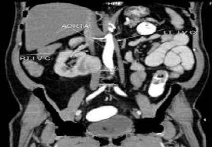
Double inferior vena cava with L-type crossed fused renal ectopia: A rare case report
Abstract
Duplication of the inferior vena cava (IVC), a rare anomaly reported to occur in 0.2-3% of the population and is known to be associated with various urogenital tract anomalies such as horseshoe kidneys, crossed fused ectopia and circum-aortic renal collar, retroaortic left renal vein and cloacal exstrophy. Inferior vena cava anomalies are rare, Some of such variations have significant clinical, surgical and radiological implications related to other cardiovascular anomalies and in some cases associated with venous thrombosis of lower limbs, particularly in young adults due to the inappropriate venous return increases the pressure, leading to blood stasis in lower extremities and development of varices. Left-sided IVC may cause misdiagnosis with para-aortic lymph node enalargement, and may cause difficulties in the transjugular approach for IVC filter implantation. So, these anomalies should be recognized carefully.
Downloads
References
2. Royal SA, Callen PW. CT evaluation of anomalies of the inferior vena cava and left renal vein. AJR Am J Roentgenol. 1979 May;132(5):759-63. [PubMed]
3. Smith TR, Frost A. Anomalous inferior vena cava associated with horseshoe kidneys. Clin Imaging. 1996 Oct-Dec;20(4):276-8. [PubMed]
4. Bass JE, Redwine MD, Kramer LA, Huynh PT, Harris JH Jr. Spectrum of congenital anomalies of the inferior vena cava: cross-sectional imaging findings. Radiographics. 2000 May-Jun;20(3):639-52.
5. Phillips E. Embryology, normal anatomy, and anomalies. In: Ferris EJ, Hipona FA, Kahn PC, Phillips E, Shapiro JH, editors. Venography of the inferior vena cava and its branches. Baltimore Md: Williams and Wilkins; 1969. pp. 1–32.
6. Pillari G, Wind ES, Wiener SL, Baron MG. Left inferior vena cava. AJR Am J Roentgenol. 1978 Feb;130(2):366-7. [PubMed]
7. Berkow AE, Henkin RE. Double inferior vena cava or iliac vein occlusion? A diagnostic problem in radionuclide venograms. AJR Am J Roentgenol. 1978 Mar;130(3):529-31. [PubMed]
8. Chuang VP, Mena CE, Hoskins PA. Congenital anomalies of the inferior vena cava. Review of embryogenesis and presentation of a simplified classification. Br J Radiol. 1974 Apr;47(556):206-13.
9. Bauer SB. Anomalies of the upper urinary tract. In :Walsh PC, Retik AB, Vaughan ED, Wein AJ, editors “ Campbell’s Urology”, 8th. Ed. Philadelphia, W.B.Saunders Co., 2002; p 1898-1902.
10. MCDONALD JH, MCCLELLAN DS. Crossed renal ectopia. Am J Surg. 1957 Jun;93(6):995-1002. [PubMed]
11. Mani N, Venkataramu NK, Singh P, Suri S. Duplication of IVC and associated renal anomalies. Indian Journal of Radiology and Imaging. 2000;10(3):157–58.
12. Brener BJ, Darling RC, Frederick PL, Linton RR. Major venous anomalies complicating abdominal aortic surgery. Arch Surg. 1974 Feb;108(2):159-65.


 OAI - Open Archives Initiative


