
High field 3T magnetic resonance imaging: MR-anatomy evaluation of traumatic knee injuries
Abstract
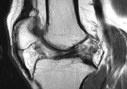
Background: MRI has high applicability to the knees, in comparison with other joints, and it provides excellent diagnostic capacity for evaluating lesions of different types, such as ligament, meniscal, tendon, bone and chondral injuries. MR imaging does not utilize ionizing radiation and therefore it is entirely safe; it is also noninvasive, painless, and allows acquisition of images in multiple planes without repositioning the patient. In addition, MR provides excellent spatial and contrast resolution of both intra- and extra-articular structures.
Objective: The purpose of the study was to review the normal MR anatomy of the internal structures of knee joint and to study MRI appearances of various meniscal and cruciate ligament injuries of knee.
Results: Knee injuries were more commonly encountered in males as compared to females. ACL was the most commonly injured ligament followed by tears of the menisci. Medial meniscal tears were twice as common as lateral meniscal tears. Vertical and complex tears are slightly more common than horizontal tears, which were predominantly seen in elderly population bearing degenerative changes. PCL tears and tears of the collateral ligaments were rare.
Conclusion: Magnetic resonance imaging is a good, accurate and non-invasive modality for the assessment of menisci and ligamenteous injuries. High field 3 T MRI is unique in its ability to evaluate the internal structures of knee with exquisite detail. MRI is highly accurate in the diagnosis of tears of the menisci and cruciate ligaments and thus, it is increasingly used as a first line investigation in patients with soft tissue trauma to knee.
Downloads
References
2. Mahmoud Karimi-Mobarake, Hamid Barani-Baravati. The Accuracy of Magnetic Imaging Compared with Arthroscopic Finding in Intra-articular Traumatic Knee Injury.Journal Applied Sciences 2005; 5(4): 686-688.
3. Winters K, Tregonning R. Reliability of magnetic resonance imaging of the traumatic knee as determined by arthroscopy. N Z Med J. 2005 Feb 11;118(1209):U1301. [PubMed]
4. Bryan S, Weatherburn G, Bungay H, Hatrick C, Salas C, Parry D, Field S, Heatley F. The cost-effectiveness of magnetic resonance imaging for investigation of the knee joint. Health Technol Assess. 2001;5(27):1-95. [PubMed]
5. Langer JE, Meyer SJ, Dalinka MK. Imaging of the knee. Radiol Clin North Am. 1990 Sep;28(5):975-90. [PubMed]
6. Raunest J, Hotzinger H, Burrig KF. Magnetic resonance imaging (MRI) and arthroscopy in the detection of meniscal degenerations correlation of arthroscopy and MRI with histology findings. Arthroscopy 1994; 10(6):634-640.
7. Lee JK, Yao L, Phelps CT, Wirth CR, Czajka J, Lozman J. Anterior cruciate ligament tears: MR imaging compared with arthroscopy and clinical tests. Radiology. 1988 Mar;166(3):861-4.
8. Mackenzie R, Dixon AK, Keene GS, Hollingworth W, Lomas DJ, Villar RN. Magnetic resonance imaging of the knee: assessment of effectiveness. Clin Radiol. 1996 Apr;51(4):245-50. [PubMed]
9. Munk B, Madsen F, Lundorf E, Staunstrup H, Schmidt SA, Bolvig L et al. Clinical magnetic resonance imaging and arthroscopic findings in knees: a comparative prospective study of meniscus anterior cruciate ligament and cartilage lesions. Arthroscopy, 1998; 14(2):171-175.
10. MR imaging of the posterior cruciate ligament: normal, abnormal, and associated injury patterns. Sonin AH, Fitzgerald SW, Hoff FL, Friedman H, Bresler ME. Radiographics. 1995 May;15(3):551-61. [PubMed]
11. Bencardino JT, Rosenberg ZS, Brown RR, Hassankhani A, Lustrin ES, Beltran J. Traumatic musculotendinous injuries of the knee: diagnosis with MR imaging. Radiographics. 2000 Oct;20 Spec No:S103-20.
12. JP Singh, L Garg, R Shrimali, V Setia, V Gupta. MR Imaging of knee with Arthroscopic Correlation in Twisting Injuries. Ind J Radio 2004 14; 1:33-40.
13. Ruth Crawford, Gayle Walley, Stephen Bridgman, Nicola Maffulli. Magnetic resonance imaging versus arthroscopy in the diagnosis of knee pathology, concentrating on meniscal lesions and ACL tears: a systematic review. British Medical Bulletin 2007; 84: 5-23.
14. Mesgarzadeh M, Moyer R, Leder DS, Revesz G, Russoniello A, Bonakdarpour A, Tehranzadeh J, Guttmann D. MR imaging of the knee: expanded classification and pitfalls to interpretation of meniscal tears. Radiographics. 1993 May;13(3):489-500. [PubMed]
15. Fox MG. MR imaging of the meniscus: review, current trends, and clinical implications. Magn Reson Imaging Clin N Am. 2007 Feb;15(1):103-23.

 OAI - Open Archives Initiative