
Utility of MRI in uterine and adnexal lesions
Abstract
Objective: To ascertain the role of MRI as a problem solving modality in characterising indeterminate uterine and adnexal masses from USG.
Methods: A prospective study was conducted with100 female patients with pelvic pathologies who underwent an ultrasound examination and proved to be diagnostic dilemmas.These patients were further investigated with an MRI of the pelvis without contrast. Routine sequences were performed in all patients including T1Wi ,T2Wi and GRE.
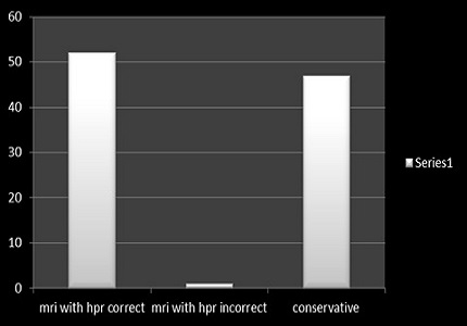
Results: There were 70 uterine lesions (fibroids -39, adenomyosis -14, Müllerian anomalies – 4,bulkiness of uterus -8,low lying placenta antenatal scan -2) and 30 adnexal lesions(malignant ovarian cysts -10,dermoid cyst – 6, chocolate cyst -8,tubovarian masses – 6,chronic torsion -1,chronic ectopic – 1, peritoneal inclusion cyst -1).The diagnosis made after the MRI scan was compared to the working diagnosis made from USG and these were separately compared with the HPR diagnoses in 53 patients who underwent surgery. The remaining 43 patients who were treated conservatively the MRI diagnoses were compared with the working USG diagnoses. The statistical data was analysed bycontingency coefficient, CC, method (variant of Chi square method). The CC for USG diagnoses and postoperative diagnoses was statistically significant. The CC for MRI diagnoses and postoperative diagnoses was 0.684 at a p value of 0.013.
Conclusions: The analysis of data from the study indicates a strong association between the MRI diagnoses and the postoperative diagnoses, more so than the USG diagnoses and the postoperative diagnoses. There is more variability between the USG diagnoses and the postoperative diagnoses by comparison.
Downloads
References
2. Patel V S, Somers S. MR imaging of the female pelvis: current perspectives and review of genital tract congenital anomalies, and benign and malignant diseases, Diagnostic Imaging 1997 Oct;38(5):417-99. [PubMed]
3. Jennifer Hubert, Diane Bergin MD. Imaging the female pelvis : when should MRI be considered? ,Applied radiology 2008: 37(1):9 – 24.
4. Haggerty AF, Hagemann AR, Chu C, Siegelman ES, Rubin SC .Correlation of pelvic magnetic resonance imaging diagnosis with pathology for indeterminate adnexal masses, International Journal of gynecological cancer 2014 Sep;24(7):1215-21.
5. Sohaib SA, Sahdev A, Van Trappen P, Jacobs IJ, Reznek RH. Characterization of adnexal mass lesions on MR imaging. AJR Am J Roentgenol. 2003 May;180(5):1297-304. [PubMed]
6. Amal Amin Abu El Maati ,Enas Abdel GhanyIbrahima, FatmaZeinhomMokhtarb .A two-stage imaging protocol for evaluating women presenting with acute pelvic pain ,The Egyptian Journal of Radiology and Nuclear Medicine 2013 Dec 44 (4): 923 – 936.
7. Al-Shukri M, Mathew M, Al-Ghafri W1, Al-Kalbani M, Al-Kharusi L, Gowri V1. A clinicopathological study of women with adnexal masses presenting with acute symptoms. Ann Med Health Sci Res. 2014 Mar;4(2):286-8. doi: 10.4103/2141-9248.129067.
8. Latthe P, Mignini L, Gray R, Hills R, Khan K. Factors predisposing women to chronic pelvic pain: systematic review. BMJ. 2006 Apr 1;332(7544):749-55. Epub 2006 Feb 16. [PubMed]
9. Khan AT, Shehmar M, Gupta JK. Uterine fibroids: current perspectives. Int J Womens Health. 2014 Jan 29;6:95-114. doi: 10.2147/IJWH.S51083. eCollection 2014. [PubMed]
10. Dueholm M, Lundorf E, Hansen ES, Ledertoug S, Olesen F. Accuracy of magnetic resonance imaging and transvaginal ultrasonography in the diagnosis, mapping, and measurement of uterine myomas. Am J Obstet Gynecol. 2002 Mar;186(3):409-15.
11. Wilde S, Scott-Barrett S. Radiological appearances of uterine fibroids. Indian J Radiol Imaging. 2009 Jul-Sep;19(3):222-31. doi: 10.4103/0971-3026.54887. [PubMed]
12. Eric D. Levens, MD, Robert Wesley, PhD, AhalyaPremkumar, MD, Wendy Blocker, MSN, and Lynnette K. Nieman, MD. Magnetic resonance imaging and transvaginal ultrasound for determining fibroid burden: implications for clinical research ,American Journal of Obstetric Gynaecology 2009 May 200 (5) 537.
13. Bazot M, Cortez A, Darai E, Rouger J, Chopier J, Antoine JM, Uzan S. Ultrasonography compared with magnetic resonance imaging for the diagnosis of adenomyosis: correlation with histopathology. Hum Reprod. 2001 Nov;16(11):2427-33.
14. Spencer JA, Ghattamaneni S. MR imaging of the sonographically indeterminate adnexal mass. Radiology. 2010 Sep;256(3):677-94. doi: 10.1148/radiol.10090397. [PubMed]
15. Noha Mohamed AbdelMaboud Ibrahim .The role of MRI in the diagnosis of endometriosis, The Egyptian Journal of Radiology and Nuclear Medicine 2012 December 43 (4) :631-36.
16. aiyeoba O, Soper DE. A practical approach to the diagnosis of pelvic inflammatory disease. Infect Dis Obstet Gynecol. 2011;2011:753037. doi: 10.1155/2011/753037. Epub 2011 Jul 26. [PubMed]
17. Rha SE, Byun JY, Jung SE, Jung JI, Choi BG, Kim BS, Kim H, Lee JM. CT and MR imaging features of adnexal torsion. Radiographics. 2002 Mar-Apr;22(2):283-94. [PubMed]
18. Adusumilli S, Hussain HK, Caoili EM, Weadock WJ, Murray JP, Johnson TD, Chen Q, Desjardins B. MRI of sonographically indeterminate adnexal masses. AJR Am J Roentgenol. 2006 Sep;187(3):732-40. [PubMed]
19. Mueller GC, Hussain HK, Smith YR, Quint EH, Carlos RC, Johnson TD, DeLancey JO. Müllerian duct anomalies: comparison of MRI diagnosis and clinical diagnosis. AJR Am J Roentgenol. 2007 Dec;189(6):1294-302. [PubMed]
20. Pellerito JS, McCarthy SM, Doyle MB, Glickman MG, DeCherney AH. Diagnosis of uterine anomalies: relative accuracy of MR imaging, endovaginal sonography, and hysterosalpingography. Radiology. 1992 Jun;183(3):795-800.

 OAI - Open Archives Initiative