
Detection of malignant tissue in mammography image using morphology based segmentation technique
Abstract
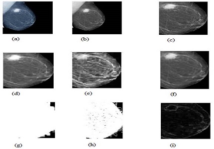
Breast cancer is the leading cause of the death among the women. Mammography is the best diagnostic technique for the breast cancer. But not all breast cancer can be seen by mammogram. Although breast cancer can be mortal, people have the highest chances to survive if cancer could be detected at the early stages. But there are certain limitations of the segmentation technique it is difficult to find the effected region perfectly. The proposed work deals with an approach for extracting the malignant masses in the mammography image for the detection of earlier breast cancer. The steps involve in this work are removal of noise from the background information, thresholding and retrieving the largest region of interest, performing morphological operations and extracting the ROI and identifying the malignant masses from the image. This method is compared with Enhancement, edge detection, Region Growing, Watershed Transformation techniques and found more accurate, sensitive, and precise in comparison to the others.
Downloads
References
2. American Cancer Society. Breast Cancer Facts & Figs. American Cancer Society, Inc, 2013. Available online at: http://www.cancer.org/acs/groups/content/@epidemiologysurveilance/documents/document/acspc-036845.pdf. cited on 23th March 2016.
3. Bronzino, J. The biomedical engineering handbook (2nd ed., vol.1). US. 2000.CRC press.
4. H.P. Chan, B. Sahiner, K.L. Lam. Computerized analysis of mammographic microcalcifications in morphological and texture feature spaces. Med. Phys. vol -25, no. 10, 2007-2019.
5. N. Petrick, H. P. Chan, B. and D. Wei. An adaptive density weighted contrast enhancement filter for mammographic breast mass detection. IEEE Trans. Med. Image, vol. 15, pp. 59-67, 2000.
6. A. Sahakyan, H. Sarukhanyan. Segmentation of the breast region in digital mammograms and detection of masses”, International journal of advanced computer science and applications, pp.102, vol- 3, no. 2, 2012.
7. Warfield, S. K. Zou and K. H.Wells. Simultaneous truth and performance level estimation (STAPLE): an algorithm for the validation of image segmentation. IEEE Transactions on Medical Imaging.vol.23, no.7, July2004.
8. Yasmin, M. Sharif, and S. Mohsin. Survey Paper on Diagnosis of Breast Cancer Using Image Processing Technique. Research Journal Of Recent Sciences, vol. 2, no. 10, pp. 88-98, October 2013.
9. I.K. Maitra, S. Nag and S.K. Bandhopadhyay. Identification of abnormal masses in digital mammography images. International Journal Of Computer Graphics, vol. 2, no. 1, 2011.

 OAI - Open Archives Initiative