Morphological Variations of Human Spleen and its Clinical Significance
Abstract
Introduction: The spleen is an important lymphatic organ in the human body. Its immunological and haematological functions are being well realized now-a-days. Aim of the present study to compare morphological variations of spleen with other workers and try to correlate these variations with some important clinical conditions.
Materials and Methods: The present study included 62 human cadaveric spleens. The morphological features of the spleen like its length, breadth, width and weight were measured. The shape, poles, borders, surfaces and the impressions on the spleen were observed.
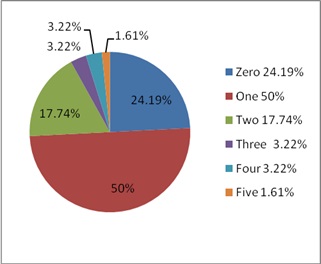
Results: The lengths of the spleens varied between 6 cm and 14 cm, with an average length of 9.59 cm. Their breadth was observed to vary between 3.5 cm and 8.5 cm, with an average breadth of 6.58 cm. their widths of the spleens varied from 2 cm to 7 cm, with an average width of 4.54 cm. Various shapes of the spleens were observed in the present study. out of 62 spleens tetrahedral shaped (33.87% ) was most common followed by wedge (32.25%) and triangular(19.35%) shape. In (50%) the cases notches were found on the superior border. The number of notches varied from zero to five, but in most of the cases (67.74%), there were 1 or 2 notches.
Conclusion: The findings of the present study will be of fundamental importance to the physicians, surgeons and radiologists and gives clue for various clinical diseases.
Downloads
References
2. Hollinshead WH. Anatomy for Surgeons. 3rd ed. vol-2. New York: Harper and Row, 1982; 436-45.
3. Michels NA. The variational anatomy of the spleen and the splenic artery. American Journal of Anatomy 1942; 70: 21-72.
4. Lamb PM, Lund A, Kanagasabay RR, Martin A, Webb JAW, Reznek RH. Spleen size: how well do linear ultrasound measurements correlate with three-dimensional CT volume assessments? Br J Radiol 2002; 75(895): 573-7. [PubMed]
5. Kumar V, Abbas AK, Fausto N. Aster JC. eds. Robbins and Cotran pathologic basis of disease. 8th ed. New Delhi: Saunders Elsevier; 2010. p. 632.
6. Beauchamp RD, Holzman MD. Fafiam TC, Spleen. In: Townsend CM, Beauchamp RD. Evers BM, Mattox KL. eds. Sabiston Text book of surgery: The biological basis of modern surgical practice. Vol. 2 17th ed. Bangalore: Saunders Elsevier ; 1994. p. 1679-1708.
7. Maier RV. Spleen. In: Mulholland MW, Lillemoe KD, Doherty GM, Maier RV, Upchurch GR Jr. eds. Greenfield’s surgery: scientific principles and practice. 4th ed. Philadelphia: Lippincott Williams & Wilkins; 2005.
8. Keith L.Moore,T.v.n Persaud.The digestive system. The developing Human:Clinically oriented Embryology.Eighth Edition.New yourk.New delhi,Elsevier publication,2008;224.
9. G.J.Romanes, The abdominal cavity, Cunningham manual of practical anatomy, fifteenth edition,New yourk,Oxford university press,2008;126-127.
10. Ungor B, Malas MA, Sulak O, Albay S. Development of spleen during the fetal period. Surg Radiol Anat. 2007;29(7):543-550. [PubMed]
11. Charware PN, Belsare SM, Kulkarni YR, Pandit SV, Ughade JM. The Morphological Variations of the Human Spleen. Journal of Clinical and Diagnostic Research. 2012 April, Vol-6(2): 159-162.
12. Sant S. Embryology for medical students. New Delhi: Jaypee brothers medical publishers (p) ltd., 2002; 203-04.
13. Rao S, Setty S, Katikireddi RS. Morphometric Study of Human Spleen. Int J Biol Med Res. 2013.
14. Skandalakis PN, Colborn GL, Skandalakis LJ, Richardson DD, Mitchell WE, Skandalakis JE. The surgical anatomy of the Spleen. Surg Clin North Am. 1993;73(4):747-68. [PubMed]
15. Soyluo lu AY, Tanyeli E,Marur T,Ertem AD, Ozku K, Akkyn SM. Splenic artery and relation between the tail of the pancrease and spleen in a surgical anatomy view. Karadeniz Typ Dergisi 1996;9:103-107.


 OAI - Open Archives Initiative


