
Study of variations in posterior communicating artery in human brain
Abstract
Introduction: Brain is normally supplied by two internal carotid arteries and two vertebral arteries which unite to form the circle of Willis. The danger of intracranial vascular lesion has increased, so a thorough knowledge of arterial circulation of brain is essential. There are variations in the length, diameter and branches of vessels forming the circle of Willis. Posterior communicating arteries are important arteries connecting the carotid and vertebral systems.
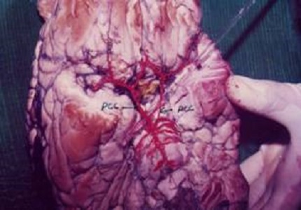
Materials and Methods: One hundred and four brain specimens were studied. The major blood vessels forming the circle of willis were traced by dissection. Their length, origin, branching pattern and anastomosis were studied, painted and photographed.
Results: It was observed that the variations in arteries forming circle of Willis was 54%. Maximum variations were observed in the communicating arteries (29%), of these the variations were more (15%) in the posterior communicating arteries.
Conclusion: A gross anatomical study of arteries forming the circle of willis was conducted. The origin course and branching pattern of major arteries forming the circle was studied and tabulated. Maximum numbers of variations were observed in the posterior communicating artery.
Downloads
References
2. Crowell RM, Morawetz RB. The anterior communicating artery has significant branches. Stroke. 1977 Mar-Apr;8(2):272-3. [PubMed]
3. LEWIS OJ. The form and development of the blood vessels of the mammalian cerebral cortex. J Anat. 1957 Jan;91(1):40-6. [PubMed]
4. Padget 1948, The development of the cranial arteries in the human embryo. Contr. Embryol, 32, 205-61. [PubMed]
5. Abbie AA. The Blood Supply of the Lateral Geniculate Body, with a Note on the Morphology of the Choroidal Arteries. J Anat. 1933 Jul;67(Pt 4):491-521. [PubMed]
6. Abbie, 1934, The morphology of forebrain arteries with a special reference to the evolution of basal ganglia. J. Anat. 68, 433-70. [PubMed]
7. Fawcett, Blac ford, 1906, The circle of willis and examination of 700 specimens, J. Anat. 40, 63-9.
8. Kaplan H.A., Ford D.H., 1966, Brain vascular system, Elsevier publishing Co., Amsterdam pp. 230.
9. Mc. Cullough, A.W., 1962, Some anomalies of the cerebral arterial circle (of willis) and related vessels. Anat. Rec. 142:537.
10. Lang E.K., Hann E.C. 1965 , Angiographic and isotope pool circulation study of the cerebral hemisphere after internal carotid artery occlusion .Qty. in Gerentology & Geriatrics 1966 pg. 510.
11. Anubha Saha, Bovindala Bhagyalakshmi et al (2013)- Gomal Journal of Medical Sciences January – June 2013, Vol.11, No.1.

 OAI - Open Archives Initiative