
A Cross-sectional Histo-morphological Study of Vesiculobullous Lesions of Skin
Abstract
Histopathology study of skin biopsies is one of the useful techniques in the investigation of vesiculobullous lesions. Out of the 70 cases of vesicobullous studied, pemphigus vulgaris was the most common type with 44 cases (61.4%) which is followed by 25 cases (35.7%) of pemphigus foliaceus and least percent was found with 1 cases (2.9%) for pemphigus vegetans. Case study showed that disease was predominately observed in male in the ratio of 1.4:1 (25 males and 18 females). It was observed in Pemphigus vulgaris cases that initial lesions involves mucous membranes in 37.2% cases and 13.9% cases involves lesions including both skin and mucosae in 13.9% cases. Mucosal involvement at one time or the other was seen in 31 patients (72.1 %). In despite of this, in pemphigus foliaceus, only 4% cases showed initial mucosal involvement and involvement of both skin and mucous membrane was observed in 12% cases. This establishes that along with clinical correlation, histo-morphological study is also a important tool in the diagnosis of Vesiculobullous skin disorders.
Downloads
References
2. Tani M, Shimizu R, Ban M, Murata Y, Tamaki A. Systemic lupus erythematosus with vesiculobullous lesions. Immunoelectron microscopic studies. Arch Dermatol. 1984 Nov;120(11):1497-501.
3. Kabir AN. Direct immunofluorescence test of skin biopsy samples results of 204 cases. Dinajpur Medical College Journal. 2009; 2(1): 8-21.
4. Minz RW, Chhabra S, Singh S, Radotra BD, Kumar B. Direct immunofluorescence of skin biopsy: perspective of an immunopathologist. Indian J Dermatol Venereol Leprol. 2010 Mar-Apr;76(2):150-7. doi: 10.4103/0378-6323.60561.
5. Zuber TJ. Punch biopsy of the skin. Am Fam Physician. 2002 Mar 15;65(6):1155-8, 1161-2, 1164. [PubMed]
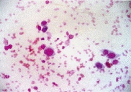
6. Gupta LK, Singhi MK. Tzanck smear: a useful diagnostic tool. Indian J Dermatol Venereol Leprol. 2005 Jul-Aug;71(4):295-9. [PubMed]
7. Korman N. Pemphigus. J Am Acad Dermatol. 1988 Jun;18(6):1219-38. [PubMed]
8. Shafi M, Khatri ML, Mashina ML et al. Pemphigus: A clinical study of 109 cases from Tripoli; Libya. Indian journal of Dermatology Vernerology and Leprology. 1994; 60:140-143.
9. Fabbri P, Lotti T, Panconesi E . Pathogenesis of pemphigus the role of epidermal plasminogen activators in acantholysis. International Journal of Dermatology. 1985; 422-425.
10. Bean SF, Lynch FW. Senear-Usher syndrome (pemphigus erythematosus). Immunofluorescent studies in a patient. Arch Dermatol. 1970 Jun;101(6):642-5.
11. Lynch PJ, Gallego RE, Saied NK. Pemphigus -- a review. Ariz Med. 1976 Dec;33(12):1030-7.

 OAI - Open Archives Initiative