
Cytomorphological study of palpable Breast Lumps by FNAC
Abstract
Aim and Objective: To assess the distribution of various cytomorphological patters of clinically palpable breast lumps by Fine Needle Aspiration Cytology technique.
Materials and Methods: A retrospective study over a period of 3 years 270 breast aspirates who attended the surgery out patient department in government medical college Anantapur from January 2012 to December 2014 were studied with clinical correlation and cytological analysis with FNAC. Smears were stained with H &E stain, and correlation with imaging studies, including mammography was done.
Results and Analysis: Total of 270 cases were studied, out of these 191 (70.74%) were found benign and 50 (18.51%) were malignant. 20 (7.40%) cases given un- satisfactory results. Out of 270 cases of analysis, Fibroadenoma was the most common benign lesion found in 133 (49.25%) patients, followed by fibrocystic disease 28(10.37%) and mastitis/Breast abscess 8(2.96%) were common breast lesions on cytology. Malignant breast lesions constitute 50(18.51%) cases, among which Duct cell carcinoma 47(17.40%) cases were commonest type.
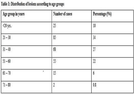
Conclusion: Benign breast lesions are common than malignant lesions, fibroadenoma and fibrocystic disease are more common in benign disease. Maximum number of lesions (34%) was seen in age group of 20 to 30 years, whereas IDC accounts for the highest number of malignant lesions. Fine-needle aspiration cytology is a rapid and effective method for the primary categorization of palpable breast lumps into benign, malignant, atypical, suspicious, and unsatisfactory categories.
Downloads
References
2. Chopra R. The Indian scene.J ClinOncol.2001; 19:106–11. [PubMed]
3. Meena SP, Hemrajani DK, Joshi N.A comparative and evaluative study of cytological and histological grading system profile in malignant neoplasm of breast--an important prognostic factor.Indian J PatholMicrobiol. 2006 Apr;49(2):199-202.
4. Mohammed AZ, Edino ST, Ochicha O, Alhassan SU. Value of fine needle aspiration biopsy in preoperative diagnosis of palpable breast lumps in resource-poor countries: A Nigerianexperience. Ann Afr Med. 2005; 4:19–22.
5. Yeoh GP, Chan KW. Fine needle aspiration of breast masses: an analysis of 1533 cases in private practice.Hong Kong Med J. 1998 Sep;4(3):283-288.
6. Park IA, Ham EK. Fine needle aspiration cytology of palpable breast lesions. Histologic subtype in false negative cases.ActaCytol. 1997 Jul-Aug;41(4):1131-8. [PubMed]
7. Rocha PD, Nadkarni NS, Menezes S. Fine needle aspiration biopsy of breast lesions and histopathologic correlation.An analysis of 837 cases in four years.ActaCytol. 1997 May-Jun;41(3):705-12. [PubMed]
8. Domínguez F, Riera JR, Tojo S, Junco P. Fine needle aspiration of breast masses.An analysis of 1,398 patients in a community hospital.ActaCytol. 1997 Mar-Apr;41(2):341-7. [PubMed]
9. Tiwari M. Role of fine needle aspiration cytology in diagnosis of breast lumps.Kathmandu Univ Med J (KUMJ). 2007 Apr-Jun;5(2):215-7. [PubMed]
10. Qasim M, Ali J, Akbar SA, Mustafa S. Lump breast: Role of FNAC in diagnosis. Prof Med J.2009; 16:235–8. [PubMed]
11. Zuk JA, Maudsley G, Zakhour HD. Rapid reporting on fine needle aspiration of breast lumps in outpatients. J ClinPathol. 1989 Sep;42(9):906-11. [PubMed]
12. Tabbara SO, Frost AR, Stoler MH, Sneige N, Sidawy MK.Changing trends in breast fine-needle aspiration: results of the Papanicolaou Society of Cytopathology Survey.DiagnCytopathol. 2000 Feb;22(2):126-30. [PubMed]
13. Khemka A, Chakrabarti N, Shah S, Patel V. Palpable breast lumps: Fine-needle aspirationcytology versus histopathology: A correlation of diagnostic accuracy. Internet J Surg. 2009; 18:1.
14. MacIntosh RF, Merrimen JL, Barnes PJ.Application of the probabilistic approach to reporting breast fine needle aspiration in males.ActaCytol. 2008 Sep-Oct;52(5):530-4.
15. Reddy DG, Reddy CR. Carcinoma of the breast, its incidence and histological variants among South Indians.Indian J Med Sci. 1958 Apr;12(4):228-34. [PubMed]
16. Clegg-Lamptey J, Hodasi W. A study of breast cancer in korlebu teaching hospital: Assessingthe impact of health education. Ghana Med J. 2007; 41:72–7. [PMC free article] [PubMed]
17. Kim A, Lee J, Choi JS, Won NH, Koo BH.Fine needle aspiration cytology of the breast. Experience at an outpatient breast clinic.ActaCytol. 2000 May-Jun;44(3):361-7.
18. Choi YD, Choi YH, Lee JH, Nam JH, Juhng SW, Choi C.Analysis of fine needle aspiration cytology of the breast: a review of 1,297 cases and correlation with histologicdiagnoses.ActaCytol. 2004 Nov-Dec;48(6):801-6. [PubMed]
19. Khan N, Rana F, Afroz N, Khan MA.Cytohistomorphological grading of breast carcinoma with special reference to apoptotic rates and lymph node metastasis.Indian J PatholMicrobiol. 2007 Jul;50(3):613-8. [PubMed]
20. Dey P, Luthra UK, Prasad A, Sheikh ZA, George SS.Cytologic grading and DNA image cytometry of breast carcinoma on fine needle aspiration cytology smears.Anal Quant CytolHistol. 1999 Feb;21(1):17-20.
21. Sankaye S, Kachewar S. Pathological panorama of Breast Cysts. Med J.2014; 39(3);458-463.
22. Kocjan G, Bourgain C, Fassina A, Hagmar B, Herbert A, Kapila K, Kardum-Skelin I, Kloboves-Prevodnik V, Krishnamurthy S, Koutselini H, Majak B, OlszewskiW,Onal B, Pohar-Marinsek Z, Shabalova I, Smith J, Tani E, Vielh P, Wiener H, Schenck U, Schmitt F.The role of breast FNAC in diagnosis and clinical management: a survey of current practice.Cytopathology. 2008 Oct;19(5):271-8. doi: 10.1111/j.1365-2303.2008.00610.x.

 OAI - Open Archives Initiative