
Cytology of lesions arising in and around operative scars: short series of four cases
Abstract
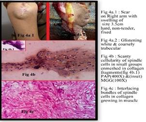
Fine needle aspiration cytology (FNAC) is an established and valuable method for morphological diagnosis of soft tissue masses and confirmation of their local recurrence or metastasis. This study aims to highlight role of FNAC in confirmation of soft tissue recurrences that occurred in and around operative scars. Wet fixed & air dried smears were made from the material aspirated from four representative cases and stained routinely. Preoperative cytodiagnosis could be given in all cases. In two cases arising in previous scars of benign nerve sheath tumors, FNAC confirmed recurrence in one case and could suggest increased grade of lesion in the other. In the third case, in which initial nature of the lesion was not known, FNAC diagnosed scar endometriosis on two occasions. In fourth case local recurrence of extra abdominal fibromatosis was confirmed. Histopathological correlation was available in all cases. FNAC confirmed recurrence and could suggest diagnosis on scanty material.
Downloads
References
2. Domanski HA. Fine-needle aspiration cytology of soft tissue lesions: diagnostic challenges.DiagnCytopathol.2007 Dec;35(12):768-73. [PubMed]
3. Dey P, Mallik MK, Gupta SK, Vasishta RK. Role of fine needle aspiration cytology in the diagnosis of soft tissue tumours and tumour-like lesions. Cytopathology.2004 Feb;15(1):32-7. [PubMed]
4. Pant A, Julfiqar, Huda N, Aslam M. Benign solitary Schwannoma of right ulnar nerve – A case report. ActaMedica International. 2015; 2(1):164-167.
5. Qian X. Soft tissue. In: Cytology: Diagnostic Principles and Clinical Correlates. Cibas E, Ducatman B, eds.3rd Edition. Philadelphia Pa:Saunders; 2009. Pg451-453.
6. Geller DS, Gebhardt M. Malignant Peripheral Nerve Sheath Tumors (MPNST)[Internet].2006 [Cited 2006]. Availiable from sarcomahelp.org/mpnst.html.
7. CzerniakB,Tuziak T. Soft Tissue Lesions. In: Koss' Diagnostic Cytology and Its Histopathologic Bases. Koss, Leopold G,Melamed, Myron R eds. 5th Edition. Philadelphia Pa:Lippincott Williams & Wilkins;2006. Pg1325-1328.
8. Dimitrova V, Yordanova I, Pavlova V, Valchev V, GospodinovD,Parashkevova B, Balabanov Ch. A case of neurofibromatosis type 1. J of IMAB 2008; 14(1):63-67. doi: 10.5272/jimab.14-1.
9. Kar M, Deo SV, Shukla NK, Malik A, DattaGupta S, Mohanti BK et al. Malignant peripheral nerve sheath tumours (MPNST)- Clinicopathological study and treatment outcome of twenty four cases. World J Surg Oncol2006 Aug 22;4:55.
10. Female reproductive system: Uterus-corpus. In: Rosai and Ackerman's Surgical Pathology.Rosai J ed. 10th ed. St. Louis: Mosby; 2012. Pg1486-7.
11. Medeiros Fd1, CavalcanteDI, Medeiros MA, Eleutério J Jr. Fine-needle aspiration cytology of scar endometriosis: study of seven cases and literature review. DiagnCytopathol 2011 Jan;39(1):18-21. doi: 10.1002/dc.21319. [PubMed]
12. Poflee S, Bode A, Mahana S. Cytodiagnosis of scar endometriosis. CytoJournal 2014;11:1. doi: 10.4103/1742-6413.126222. [PubMed]
13. Zampieri N, Cecchetto M, Zorzi MG, Pietrobelli A, Ottolenghi A, Camoglio F. An unusual case of extra-abdominal desmoidtumour. Eur J Cancer Care (Engl).2010 May;19(3):410-2. doi: 10.1111/j.1365-2354.2008.01002.x. Epub 2009 Aug 26. [PubMed]
14.Stoeckle E, Coindre JM, Longy M, Binh MB, Kantor G, Kind M et al. A critical analysis of treatment strategies in desmoidtumours: a review of a series of 106 cases.Eur J SurgOncol.2009 Feb;35(2):129-34.doi: 10.1016/j.ejso.2008.06.1495. Epub 2008 Aug 29. [PubMed]
15.Fibromatoses, In:Enzinger& Weiss’s Soft tissue tumors. Weiss SW, Goldblum JR eds. 5th Edition. Philadelphia PA:Mosby 2008. Pg237-246. [PubMed]
16. Satsuma S, Yamamoto T, Kobayashi D, Yoshiya S, Marui T, Akisue T et al. Extraabdominaldesmoid tumor in a surgical scar of a patient with Sprengel's deformity. J Pediatr Surg. 2003 Oct;38(10):1540-2.

 OAI - Open Archives Initiative