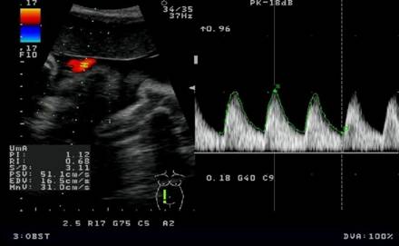
Best color Doppler indices in prediction of fetal hypoxia in IUGR fetuses
Abstract
Objective: To determine the best predictor of fetal hypoxia amongst the color Doppler indices resistivity index(RI), pulsality index(PI) and systolic/ diastolic (S/D) ration of umbilical artery (UA), middle cerebral artery(MCA), descending abdominal aorta (DAA), MCA/UA PI ratio and abnormal flow pattern, absent end diastolic flow/reverse end diastolic flow (AEDF/REDF) in umbilical artery and descending abdominal aorta, in prediction of adverse perinatal outcome in normal and intrauterine growth retardation (IUGR) fetuses with or without pregnancy induced hypertension (PIH).
Material and Method: 100 women with normal Singleton pregnancies and 100 women with IUGR with or without PIH or both were prospectively examined with Doppler Ultrasonography of the umbilical artery, middle cerebral artery & descending abdominal aorta and perinatal outcomes was evaluated in relation to their indices and compared with each other.
Observation: In study group sixty four fetuses (64%) had one major or minor adverse perinatal outcome in comparison to control group which had only 6% adverse perinatal outcome. In study group premature delivery was 46%, lower segment cesarean section (LSCS) was 38%, and perinatal death was 22%. Fetuses with absent end diastolic flow/reverse end diastolic flow (AEDF/REDF) in umbilical artery and descending abdominal aorta had 100% perinatal mortality.
Conclusion: Umbilical artery SD Ratio (cut off 3), middle cerebral artery / umbilical artery PI < 1.08, AEDF / REDF abnormal flow pattern in umbilical artery and descending abdominal aorta were found to be the best Doppler indices for prediction of adverse perinatal outcome in women with PIH and IUGR.
Downloads
References
2. Stuart B, Drumm J, Fitzgerald DE and Duignan NM. Fetal blood velocity waveforms in normal pregnancy. British journal of obstetrics and Gynaecology, Sep 1980, Vol. 87, pp.780-5. [PubMed]
3. Lakhkar BN, Rajagopal KV, Gourisankar PT. Doppler Prediction of Adverse Perinatal Outcome in PIH and IUGR. IJRI 2006 16:1:109-116. [PubMed]
4. Gramellini D, Folli MC, Raboni S, Vadora E, Merialdi A. Cerebral-umbilical Doppler ratio as a predictor of adverse perinatal outcome. Obstet Gynecol. 1992 Mar;79(3):416-20. [PubMed]
5. Chauhan R, Samiksha Trivedi- Role of Doppler study in high risk pregnancy- Journal of obstetric and gynecology of india Vol 52, No.30:may/june 2002. [PubMed]
6. Mari G, Deter RL. Middle cerebral artery flow velocity waveforms in normal and small-for-gestational-age fetuses. Am J Obstet Gynecol. 1992 Apr;166(4):1262-70. [PubMed]
7. Ott WJ. Comparison of the non-stress test with the evaluation of centralization of blood flow for the prediction of neonatal compromise. Ultrasound Obstet Gynecol. 1999 Jul;14(1):38-41.
8. Fong KW, Ohlsson A, Hannah ME, Grisaru S, Kingdom J, Cohen H, Ryan M, Windrim R, Foster G, Amankwah K. Prediction of perinatal outcome in fetuses suspected to have intrauterine growth restriction: Doppler US study of fetal cerebral, renal, and umbilical arteries. Radiology. 1999 Dec;213(3):681-9. [PubMed]
9. Acharya G, Wilsgaard T, Berntsen GKR, Maltau M, Kiserud T. Reference ranges for serial measurements of umbilical artery doppler indices in second half of pregnancy. Am J Obstet Gynecol. 2005 192:154-8.
10. Piazze J, Padula F, Cerekja A, Cosmi EV, Anceschi MM. Prognostic value of umbilical-middle cerebral artery pulsatility index ratio in fetuses with growth restriction. Int J Gynaecol Obstet. 2005 Dec;91(3):233-7. Epub 2005 Oct 7.
11. Rizvi S.M.R.,Iqbal Nasir,Yasmeen Naila. Small for gestational age fetus Role of colour Doppler in Ultrasund in the management. Professional Med J Dec 2006; 13(4);705-709. [PubMed]
12. Ertan AK, He JP, Tanriverdi HA, Hendrik J, Limbach HG, Schmidt W. Comparison of perinatal outcome in fetuses with reverse or absent enddiastolic flow in the umbilical arteryand/or fetal descending aorta. J Perinat Med. 2003;31(4):307-12. [PubMed]
13. Gerber S, Hohlfeld P, Viquerat F, Tolsa JF, Vial Y. Intrauterine growth restriction and absent or reverse end-diastolic blood flow in umbilical artery (Doppler class II or III): A retrospective study of short- and long-term fetal morbidity and mortality. Eur J Obstet Gynecol Reprod Biol. 2006 May 1;126(1):20-6. Epub 2005 Aug 31.
14. Strigini FA, De Luca G, Lencioni G, Scida P, Giusti G, Genazzani AR. Middle cerebral artery velocimetry: different clinical relevance depending on umbilical velocimetry. Obstet Gynecol. 1997 Dec;90(6):953-7.
15. Bahado Singh RO, Kovanci E, Jeffres A et al.The Doppler cerebroplacental ratio and perinatal outcome in intrauterine growth restriction. American Journal of Obstetric & Gynecol 1999; 180: 750-6.
16. Odibo AO, Riddick C, Pare E, Stamilio DM, Macones GA. Cerebroplacental Doppler ratio and adverse perinatal outcomes in intrauterine growth restriction: evaluating the impact of using gestational age-specific reference values. J Ultrasound Med. 2005 Sep;24(9):1223-8.
17. Narula Harneet, Kapila AK, Mohi Manjeet Kour. Cerebral and umbilical arterial blood flow velocity in normal and growth retarted pregnancy. Obstet Gynecol India Vol. 59,No.1: January/Fabruary 2009 pg 47-52.
18. Bhatt CJ Arora J,Shah M.S. Role of colour Doppler in pregnancy induced hypertention(a study of 100 cases). Indian journal of radiology imaging 2003 Vol.13, issue-4 pg 417-420.
19. Acharya D, Nagraj K,Nair NS, Bhatt HV. Maternal Determinants of intrauterine growth retardation: a case control study in Udupi district Karnataka. Indian journal of community medicine 2004;29(4):10-12.


 OAI - Open Archives Initiative


