
Role of liquid-based cytology and cell block study of pleural fluid in the evaluation of cases of malignant Pleural effusion with special reference to immunohistochemistry
Abstract
Introduction- Lung cancer is the most common primary tumor associated with malignant pleural effusion (MPE). In this study, we aim to use cell remnants for cell block preparation after performing liquid-based cytology (LBC) of effusion fluid. Immunohistochemistry was helpful to evaluate those cases having diagnostic dilemmas in LBC and cell block.
Method: It was a cross-sectional, prospective, single institution-based study, conducted in the department of Pathology in collaboration with the Department of Respiratory Medicine IPGMER & SSKM Hospital, from January 2018 to June 2019 in the institution.
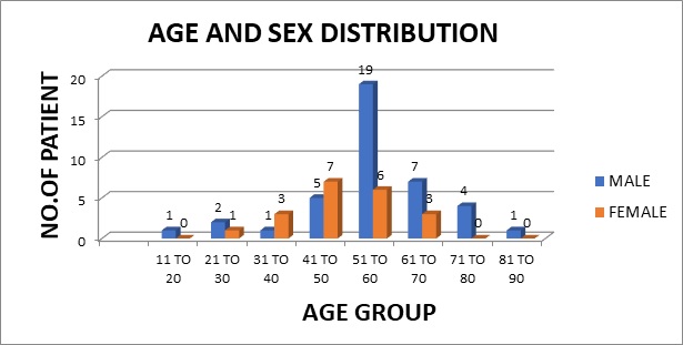
Result: Most of the study population were in between the age group of 51 to 60 years with male predominance and with fever and cough being the predominant symptoms. Liquid-based cytology was positive for malignancy in 58% of cases and suspicious of malignancy in 22% of cases of malignant pleural effusion and it had 95.35% sensitivity, 58.82% specificity in diagnosing malignant pleural effusion.LBC was done followed by cell block preparations are studied further by Immunohistochemistry.
Discussion: Morphological features were better identified by the cell block method when compared to LBC. Multiple sections can be obtained for special stain or IHC study which bridges the gap between cytology and histology.
Downloads
References
Sahn SA. Malignancy metastatic to the pleura. Clin Chest Med. 1998 Jun;19(2):351-61. doi: 10.1016/s0272-5231(05)70082-4.
Matthay RA, Coppage L, Shaw C, Filderman AE. Malignancies metastatic to the pleura. Invest Radiol. 1990 May;25(5):601-19. doi: 10.1097/00004424-199005000-00025.
Leuallen Ec, Carr Dt. Pleural effusion; a statistical study of 436 patients. N Engl J Med. 1955 Jan 20;252(3):79-83. doi: 10.1056/NEJM195501202520301.
Light RW, Macgregor MI, Luchsinger PC, Ball WC Jr. Pleural effusions: the diagnostic separation of transudates and exudates. Ann Intern Med. 1972 Oct;77(4):507-13. doi: 10.7326/0003-4819-77-4-507.
Heffner JE, Brown LK, Barbieri CA. Diagnostic value of tests that discriminate between exudative and transudative pleural effusions. Primary Study Investigators. Chest. 1997 Apr;111(4):970-80. doi: 10.1378/chest.111.4.970.
Bishop JA, Teruya-Feldstein J, Westra WH, Pelosi G, Travis WD, Rekhtman N. p40 (ΔNp63) is superior to p63 for the diagnosis of pulmonary squamous cell carcinoma. Mod Pathol. 2012 Mar;25(3):405-15. doi: 10.1038/modpathol.2011.173.
-9. Leuallen Ec, Carr Dt. Pleural effusion; a statistical study of 436 patients. N Engl J Med. 1955 Jan 20;252(3):79-83. doi: 10.1056/NEJM195501202520301.
Light, Richard W., and YC Gary Lee, eds. Textbook of Pleural Diseases Second Edition. CRC Press, 2008.
Noppen M, De Waele M, Li R, Gucht KV, D'Haese J, Gerlo E, et al. Volume and cellular content of normal pleural fluid in humans examined by pleural lavage. Am J Respir Crit Care Med. 2000 Sep;162(3 Pt 1):1023-6. doi: 10.1164/ajrccm.162.3.9910050.
Chen YS, Lee JM, Chao YK. Introduction: 2017 Taiwan Association of Thoracic & Cardiovascular Surgery. J Thorac Dis. 2017 Oct;9(Suppl 14):S1397-S1398. doi: 10.21037/jtd.2017.10.133.
Thapar M, Mishra RK, Sharma A, Goyal V, Goyal V. Critical analysis of cell block versus smear examination in effusions. J Cytol. 2009 Apr;26(2):60-4. doi: 10.4103/0970-9371.55223.
Shivakumarswamy U, Arakeri SU, Karigowdar MH, Yelikar B. Diagnostic utility of the cell block method versus the conventional smear study in pleural fluid cytology. J Cytol. 2012 Jan;29(1):11-5. doi: 10.4103/0970-9371.93210.
Karahalli E, Yilmaz A, Türker H, Ozvaran K. Usefulness of various diagnostic techniques during fiberoptic bronchoscopy for endoscopically visible lung cancer: should cytologic examinations be performed routinely? Respiration. 2001;68(6):611-4. doi: 10.1159/000050581.
Ma Y, Fan M, Dai L, Kang X, Liu Y, Sun Y, et al. The expression of TTF-1 and Napsin A in early-stage lung adenocarcinoma correlates with the results of surgical treatment. Tumor Biol. 2015 Sep;36(10):8085-92. doi: 10.1007/s13277-015-3478-z.
ApplImmunohistochemMolMorphol.2011Jul;19(4):313-7.doi:10.1097/PAI.0b013e318205b05,PubMed.gov.
Travis WD, et al. Arch Pathol Lab Med. 2012 Sep 12. [Epub ahead of print].
Zhang C, Schmidt LA, Hatanaka K, Thomas D, Lagstein A, Myers JL. Evaluation of napsin A, TTF-1, p63, p40, and CK5/6 immunohistochemical stains in pulmonary neuroendocrine tumors. Am J Clin Pathol. 2014 Sep;142(3):320-4. doi: 10.1309/AJCPGA0IUA8BHQEZ.
HumPathol. 2014 May; 45(5): 926–934.Published online 2014 Jan 25. doi: 10.1016/j.humpath.2014.01.005.
Heidari B, Bijani K, Eissazadeh M, Heidari P. Exudative pleural effusion: effectiveness of pleural fluid analysis and pleural biopsy. East Mediterr Health J. 2007 Jul-Aug;13(4):765-73.
Al-Alusi F. Pleural effusion in Iraq: a prospective study of 100 cases. Thorax. 1986 Jun;41(6):492-3. doi: 10.1136/thx.41.6.492.
Hierholzer J, Luo L, Bittner RC, Stroszczynski C, Schröder RJ, Schoenfeld N, et al. MRI and CT in the differential diagnosis of pleural disease. Chest. 2000 Sep;118(3):604-9. doi: 10.1378/chest.118.3.604.
Valdés L, Alvarez D, Valle JM, Pose A, San José E. The etiology of pleural effusions in an area with high incidence of tuberculosis. Chest. 1996 Jan;109(1):158-62. doi: 10.1378/chest.109.1.158.
Luo L, Hierholzer J, Bittner RC, Chen J, Huang L. Magnetic resonance imaging in distinguishing malignant from benign pleural disease. Chin Med J (Engl). 2001 Jun;114(6):645-9.
Ghosh I, Dey SK, Das A, Bhattacharjee D, Gangopadhyay S. Cell block cytology in pleural effusion. J Indian Med Assoc. 2012 Jun;110(6):390-2, 396.
Thapar M, Mishra RK, Sharma A, Goyal V, Goyal V. Critical analysis of cell block versus smear examination in effusions. J Cytol. 2009 Apr;26(2):60-4. doi: 10.4103/0970-9371.55223.
Woo CG, Son SM, Han HS, Lee KH, Choe KH, An JY, et al. Diagnostic benefits of the combined use of liquid-based cytology, cell block, and carcinoembryonic antigen immunocytochemistry in malignant pleural effusion. J Thorac Dis. 2018 Aug;10(8):4931-4939. doi: 10.21037/jtd.2018.07.139.
Daudu, O. U., et al. "IOSR Journal of Dental and Medical Sciences (IOSR-JDMS) e-ISSN: 2279-0853, p-ISSN: 2279-0861. Volume 16, Issue 5 Ver. VI (May. 2017), PP 34-35 www. iosrjournals. org."
Copyright (c) 2021 Author (s). Published by Siddharth Health Research and Social Welfare Society

This work is licensed under a Creative Commons Attribution 4.0 International License.

 OAI - Open Archives Initiative