
Evaluation of spectrum of MRI findings in children with Hypoxic Ischemic Encephalopathy and its comparison with transcranial sonography
Abstract
Background: Hypoxic ischemic encephalopathy is a serious concern among asphyxiated newborns due to the associated long term sequelae like cognitive impairment and cerebral palsy. Though the incidence of hypoxic injury remains higher in preterm babies due to incomplete brain maturation, it can occur in term babies as well despite institutional deliveries due to an array of unavoidable fetal, maternal and placental causes.
Aims: This study was conducted as an attempt to evaluate the risk factors, to study the correlation between the term of pregnancy with TCUS and MRI imaging findings in HIE and characterise patterns of CNS involvement.
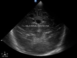
Materials and methods: It was a cross-sectional study carried on 50 neonates with clinically diagnosed HIE presenting to the Department of Radiodiagnosis, Rajindra Hospital Patiala who were subjected to transcranial sonography and MRI.
Results and Conclusion: This study demonstrated term infants have significant involvement of basal ganglia thalamus type (central) pattern of involvement and preterm infants have periventricular leukomalacia type (white matter injury) of a pattern of involvement. The overall sensitivity and specificity of TCUS in detecting imaging findings in children with clinically diagnosed HIE compared to MRI was found to be 70.45% and 50% respectively, yielding the overall diagnostic accuracy of TCUS as 68% compared to MRI. TCUS can depict central and white matter abnormalities better than peripheral lesions. However MRI provides additional diagnostic information in many cases and can detect precisely the extent of brain injury.
Downloads
References
Thoker AH, Sheikh M, Thoker PA, Thoker M. Neurodevelopmental outcome in perinatal asphyxia. American Journal of Experimental and Clinical Research. 2017 Apr 8;4(2):206-9.
Pathiraja RP, Guneskera D. Perinatal asphyxia and Hypoxic Ischemic Encephalopathy-The Current Situation. Sri Lanka Journal of Obstetrics and Gynaecology April2017; 39(1),pp.8-11
Vélez SM, García JM, Marín LC, Arango JM, White CA, Pineda DS. Hypoxic ischemic encephalopathy in the neonatal period, evaluation with magnetic resonance. Rev. Colomb. Radiol. 2018; 29(4): 5025-31
Pathiraja RP, Guneskera D. Perinatal asphyxia and Hypoxic Ischemic Encephalopathy-The Current Situation. Sri Lanka Journal of Obstetrics and Gynaecology April, 2017; 39(1):8-11
Rutherford MA. The asphyxiated term infant. MRI of the neonatal brain. London: WB Saunders. 2002 Jan:99-128.
Bundi LB, Mwango G, Oliver VO, Mulama B. Clinical neonatal hypoxic ischemic injury: Cranial ultrasound spectrum of findings in neonates admitted to a Newborn Unit in Nairobi, Kenya. West African Journal of Radiology. 2020 Jul 1;27(2):108.
Liauw L, van der Grond J, van den Berg-Huysmans AA, Palm-Meinders IH, van Buchem MA, van Wezel-Meijler G. Hypoxic-ischemic encephalopathy: diagnostic value of conventional MR imaging pulse sequences in term-born neonates. Radiology. 2008 Apr;247(1):204-12. doi: 10.1148/radiol.2471070812. Epub 2008 Feb 27. PMID: 18305189.
Genedi EA, Osman NM, El-deeb MT. Magnetic resonance imaging versus transcranial ultrasound in early identification of cerebral injuries in neonatal encephalopathy. The Egyptian Journal of Radiology and Nuclear Medicine. 2016 Mar 1;47(1):297-304.
Aun AE, Hassan HA, Ali WI, Ataky MM. Transcranial Ultrasound in Comparison to MRI in Evaluation of Hypoxic Ischemic Injury in Neonates. The Egyptian Journal of Hospital Medicine. 2019 Jan 1;74(4):842-52.
Jose O, Sheena V. MRI changes of brain in newborns with hypoxic ischemic encephalopathy clinical stage II or stage III-a descriptive study. International Journal of Medical Paediatrics and Oncology. 2017;3(1):29-33.
Bhagat H, Kawade R, Sachdev YP. Study of role of MRI brain in evaluation of hypoxic ischemic encephalopathy. Indian Journal of Basic and Applied Medical Research; Radiology and Imaging -December 2017: 7(1), 28-33.
Steggerda SJ, Leijser LM, Wiggers-de Bruïne FT, van der Grond J, Walther FJ, van Wezel-Meijler G. Cerebellar injury in preterm infants: incidence and findings on US and MR images. Radiology. 2009 Jul;252(1):190-9. doi: 10.1148/radiol.2521081525. Epub 2009 May 6. PMID: 19420320.
Giri S, Sau R, Das S, et al. Correlation of transcraial ultrasound and magnetic resonance imaging in evaluation of imaging patterns of clinically diagnosed hypoxic ischemic encephalopathy in neonates. J. Evid. Based Med. Healthc. 20 20 ;7(19)938-42 .
Copyright (c) 2021 Author (s). Published by Siddharth Health Research and Social Welfare Society

This work is licensed under a Creative Commons Attribution 4.0 International License.

 OAI - Open Archives Initiative