
Application of Thyroid Peroxidase (TPO) and Hector Battifora Mesothelial-1 (HBME-1) immunohistochemical markers in the diagnosis of papillary thyroid carcinoma of the thyroid
Abstract
Background: Thyroid cancer is the most common endocrine malignancy accounting for >90% of malignancies of endocrine glands. The inter and intraobserver variation in the histomorphological diagnosis of Papillary Thyroid Carcinomas may sometimes pose a diagnostic difficulty. Application of IHC biomarkers may play an active or complementary role in their accurate classification.
Aim: The present study was conducted to evaluate if HBME-1 and TPO immunohistochemical analysis can reliably differentiate papillary carcinomas from other thyroid lesions.
Material and Methods: 50 cases of benign and malignant thyroid lesions were taken. Immunohistochemical staining for HBME-1 and TPO was performed. HBME-1 and TPO score was interpreted as absent and positive. Medical records were retrieved and their clinical data, surgical treatment, and pathological findings were noted.
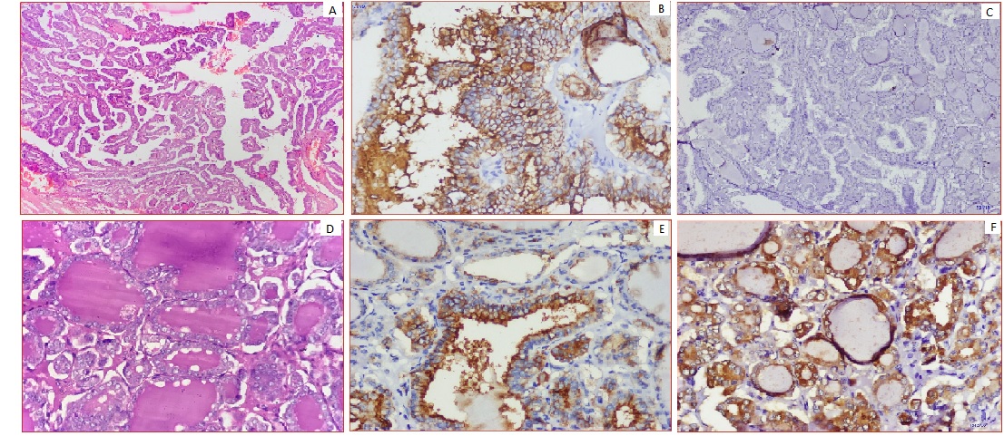
Results: Out of 50 cases, 19 (73.1%) cases were diagnosed PTC, 4 (15.4%) cases were FTC, 3(11.5%) cases were of MTC and 24 cases of benign thyroid lesions. TPO expression was found positive in 91.7% of cases of Benign thyroid lesions. In malignant thyroid lesions, negative expression of TPO was seen in 63.16%, 0% and 33.33% of PTC, FCT, and MCT respectively. HBME-1 showed negative expression in 83.3% of cases of benign thyroid lesions. Whereas, in malignant thyroid lesion HBME-1 expression was positive in 78.95%, 50% and 0% cases of PTC, FCT, and MCT respectively.
Conclusion: Testing for expression of HBME-1 has been shown to improve the diagnostic accuracy for thyroid malignant nodules. The combination of HBME-1, and TPO may contribute to an accurate diagnosis of papillary thyroid carcinoma.
Downloads
References
Hodgson NC, Button J, Solorzano CC. Thyroid cancer: is the incidence still increasing? Ann Surg Oncol. 2004;11(12):1093-1097. doi: https://doi.org/10.1245/ASO.2004.03.066.
Pellegriti G, Frasca F, Regalbuto C, Squatrito S, Vigneri R. Worldwide increasing incidence of thyroid cancer: update on epidemiology and risk factors. J Cancer Epidemiol. 2013;2013. doi: https://doi.org/10.1155/2013/965212.
Dinets A, Hulchiy M, Sofiadis A, Ghaderi M, Höög A, Larsson C, et al. Clinical, genetic, and immunohistochemical characterization of 70 Ukrainian adult cases with post-Chornobyl papillary thyroid carcinoma. Eur J Endocrinol. 2012;166(6):1049-1060. doi: https://doi.org/10.1530/EJE-12-0144.
Nguyen QT, Lee EJ, Huang MG, Park YI, Khullar A, Plodkowski RA. Diagnosis and treatment of patients with thyroid cancer. Am Health Drug Benefits. 2015;8(1):30.
Stathathos N, Ringel. Molecular markers of thyroid nodules. In Adv Mol Cell Endocrinol. 2006:4;19-34. doi: https://doi.org/10.1016/S1569-2566(04)04002-5.
De Micco C, Vasko V, Garcia S, Zoro P, Denizot A, Henry JF. Fine-needle aspiration of thyroid follicular neoplasm: diagnostic use of thyroid peroxidase immunocytochemistry with monoclonal antibody 47. Surg. 1994;116(6):1031-1035.
Wu G, Wang J, Zhou Z, Li T, Tang F. Combined staining for immunohistochemical markers in the diagnosis of papillary thyroid carcinoma: improvement in the sensitivity or specificity? J Int Med Res. 2013;41(4):975-983. doi: https://doi.org/10.1177/0300060513490617.
Fedchenko N, Reifenrath J. Different approaches for interpretation and reporting of immunohistochemistry analysis results in the bone tissue - a review. Diagn Pathol. 2014;9:221. doi: https://doi.org/10.1186/s13000-014-0221-9.
Kilfoy BA, Zheng T, Holford TR, Han X, Ward MH, Sjodin A, et al. International patterns and trends in thyroid cancer incidence, 1973-2002. Cancer Causes Control. 2009; 20(5):525-531. doi: https://doi.org/10.1007/s10552-008-9260-4.
Papotti M, Rodriguez J, De Pompa R, Bartolazzi A, Rosai J. Galectin-3 and HBME-1 expression in well-differentiated thyroid tumors with follicular architecture of uncertain malignant potential. Mod Pathol. 2005;18(4):541-546. doi: https://doi.org/10.1038/modpathol.3800321.
Al Zaher N, Al Salam S, El Teraifi H. Thyroid carcinoma in the United Arab Emirates: perspectives and experience of a tertiary care hospital. Hematol Oncol Stem Cell Ther. 2008;1(1):14-21. doi: https://doi.org/10.1016/s1658-3876(08)50055-0.
Sumana BS, Shashidhar S, Shivarudrappa AS. Galectin-3 Immunohistochemical Expression in Thyroid Neoplasms. J Clin Diagn Res. 2015;9(11):EC07-EC11. doi: https://doi.org/10.7860/JCDR/2015/16277.6760.
Dwivedi SS, Khandeparkar SG, Joshi AR, Kulkarni MM, Bhayekar P, Jadhav A et al. Study of immunohistochemical markers (CK-19, CD-56, Ki-67, p53) in differentiating benign and malignant solitary thyroid nodules with special reference to papillary thyroid carcinomas J Clin Diagn Res. 2016;10(12):EC14-EC19. doi: https://doi.org/10.7860/JCDR/2016/22428.9114.
Sack MJ, Astengo-Osuna C, Lin BT, Battifora H, LiVolsi VA. HBME-1 immunostaining in thyroid fine-needle aspirations: a useful marker in the diagnosis of carcinoma. Mod path: 1997;10(7):668-674.
Miettinen M, Kärkkäinen P. Differential reactivity of HBME-1 and CD15 antibodies in benign and malignant thyroid tumours. Virchows Archiv. 1996;429(4-5):213-219. doi: https://doi.org/10.1007/BF00198336.
Cheung, C, Ezzat, S., Freeman JL, Rosen BI, Asa SL. Immunohistochemical Diagnosis of Papillary Thyroid Carcinoma. Mod Pathol, 2001;14(4):338-342. doi: https://doi.org/10.1038/modpathol.3880312.
De Micco C, Savchenko V, Giorgi R, Sebag F, Henry JF. Utility of malignancy markers in fine-needle aspiration cytology of thyroid nodules: comparison of Hector Battifora mesothelial antigen-1, thyroid peroxidase and dipeptidyl aminopeptidase IV. Br J Cancer. 2008;98(4):818-823. doi: https://doi.org/10.1038/sj.bjc.6604194.
Nga ME, Lim GS, Soh CH, Kumarasinghe MP. HBME-1 and CK19 are highly discriminatory in the cytological diagnosis of papillary thyroid carcinoma. Diagn Cytopathol. 2008;36(8):550-556. doi: https://doi.org/10.1002/dc.20841.
Mase T, Funahashi H, Koshikawa T, Imai T, Nara Y, Tanaka Y, et al. HBME-1 immunostaining in thyroid tumors especially in follicular neoplasm. Endocr J. 2003;50(2):173-177. doi: https://doi.org/10.1507/endocrj.50.173.
Liu Z, Xun X, Wang Y, Mei L, He L, Zeng W, et al. MRI and ultrasonography detection of cervical lymph node metastases in differentiated thyroid carcinoma before reoperation. Am J Transl Res. 2014;6(2):147-154.
De Matos PS, Ferreira AP, de Oliveira Facuri F, Assumpcao LV, Metze K, et al Usefulness of HBME‐1, cytokeratin 19 and galectin-3 immunostaining in the diagnosis of thyroid malignancy. Histopathology. 2005;47(4):391-401. doi: https://doi.org/10.1111/j.1365-2559.2005.02221.x.
Palo S, Biligi DS. Differential diagnostic significance of HBME-1, CK19 and S100 in various thyroid lesions.Malays J Pathol. 2017;39(1):55-67.
Zhu X, Sun T, Lu H, Zhou X, Lu Y, Cai X et al. Diagnostic significance of CK19, RET, galectin-3 and HBME-1 expression for papillary thyroid carcinoma. J Clin Pathol. 2010;63(9):786-789. doi: http://dx.doi.org/10.1136/jcp.2010.076901.
Griffith OL, Melck A, Jones SJ, Wiseman SM. Meta-analysis and meta-review of thyroid cancer gene expression profiling studies identifies important diagnostic biomarkers. J Clin Oncol. 2006;24(31):5043-5051. doi: https://doi.org/10.1200/JCO.2006.06.7330.
Weber KB, Shroyer KR, Heinz DE, Nawaz S, Said MS, Haugen BR. The use of a combination of galectin-3 and thyroid peroxidase for the diagnosis and prognosis of thyroid cancer. Am J Clin Pathol. 2004;122(4):524-31. doi: https://doi.org/10.1309/UUQTE505PTN5QJ7M.
Arcolia V, Journe F, Renaud F, Leteurtre E, Gabius HJ, Remmelink M, et al. Combination of galectin-3, CK19 and HBME-1 immunostaining improves the diagnosis of thyroid cancer. Onco lett 2017;14(4):4183-4189. doi: https://doi.org/10.3892/ol.2017.6719.
Henry JF, Denizot A, Porcelli A, Villafane M, ZoroP, Garcia S et al. Thyroperoxidase immunodetection for the diagnosis of malignancy on fine-needle aspiration of thyroid nodules. World J. Surg.1994;18(4):529-534. doi: https://doi.org/10.1007/BF00353756.
Schmitt AC, Cohen C, Siddiqui MT. Paired box gene 8, HBME-1, and cytokeratin 19 expression in preoperative fine-needle aspiration of papillary thyroid carcinoma: diagnostic utility. Cancer Cytopathol. 2010;118(4):196-202. doi: https://doi.org/10.1002/cncy.20082.
Raggio E, Camandona M, Solerio D, Martino P, Franchello A, Orlandi F, et al. The diagnostic accuracy of the immunocytochemical markers in the pre-operative evaluation of follicular thyroid lesions. J Endocrinol Invest. 2010;33:378–381. doi: https://doi.org/10.1007/BF03346607.

 OAI - Open Archives Initiative