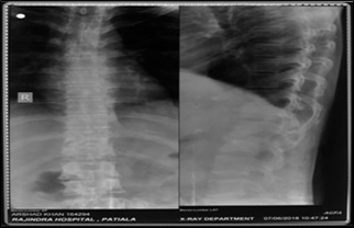
Comparison of MRI with x-ray in the evaluation of tuberculosis of spine
Abstract
Introduction: MRI is the most valuable method for detecting early disease and is preferred technique to define the activity and extent of infection followed by x–ray.
Aim: To evaluate MRI as a valuable noninvasive diagnostic tool in spinal tuberculosis and to correlate with plain radiograph for the early detection of spinal tuberculosis.
Material and method: This cross-sectional study was carried out on 40 patients who were suspected as cases of spinal tuberculosis. Plain X-ray were done before the MRI examination.
Results: The comparison of X-ray and MRI for evaluating spinal TB on the basis of end plate irregularity, thecal sac compression, cord compression and cord changes was statistically highly significant. It was statistically significant on the basis of Disk Space Narrowing/Disk Involvement, paravertebral Widening/Psoas abscess and Posterior Element Involvement. X-ray when compared to MRI was found to have a sensitivity of 48.72% and a specificity of 100% in detection of end plate irregularities, sensitivity of 89.47% and specificity of 100% in detection of vertebral height reduction, sensitivity of 78.79% and specificity of 100% in detection of disk Space narrowing / disk Involvement and sensitivity of 28.57% and specificity of 92.31% in detection of paravertebral widening/psoas abscess.
Conclusion: MRI is a better and more Informative imaging modality in evaluation of patients of Pott’s spine providing the diagnosis earlier than conventional methods.
Downloads
References
Chauhan A, Gupta BB. Spinal tuberculosis. Indian AcadClin Med. 2007;8:110-4.
McLain RF, Isada C. Spinal tuberculosis deserves a place on the radar screen. Cleve Clin J Med. 2004;71 (7): 543-549. doi: https://doi.org/10.3949/ccjm.71.7.537
Boachie-Adjei O, Squillante RG. Tuberculosis of the spine. Orthop Clin North Am. 1996;27(1):95-103.
Schirmer P, Renault CA, Holodniy M. Is spinal tuberculosis contagious? Int J Infect Dis. 2010;14(8): e 659-66. doi: https://doi.org/10.1016/j.ijid.2009.11.009. Epub 2010 Feb 23
Garg RK, Somvanshi DS. Spinal tuberculosis: a review. J Spinal Cord Med. 2011;34(5):440-454. doi: https://doi.org/10.1179/2045772311Y.0000000023.
Moorthy S, Prabhu NK. Spectrum of MR imaging findings in spinal tuberculosis. AJR Am J Roentgenol. 2002;179(4):979-983. doi: https://doi.org/10.2214/ajr.179.4.1790979
Moon MS. Tuberculosis of the spine. Controversies and a new challenge. Spine (Phila Pa 1976). 1997;22 (15): 1791-1797.doi: https://doi.org/10.1097/00007632-199708010-00022.
Ansari S, Amanullah MF, Ahmad K, Rauniyar RK. Pott's Spine: Diagnostic Imaging Modalities and Technology Advancements. N Am J Med Sci. 2013; 5 (7): 404-411. doi: https://dx.doi.org/10.4103%2F1947-2714.115775.
Sharma G, Ghode R. Tubercular Spondylitis: Prospective Comparative Imaging Analysis on Conventional Radiograph and MRI. Int J Anat, Radiol Surg.2016;5(3):41-46. doi: 10.7860/IJARS/2016/18136:2169.
Jain AK. Tuberculosis of the spine: a fresh look at an old disease. J Bone Joint Surg Br. 2010;92(7):905913. doi: https://doi.org/10.1302/0301-620X.92B7.24668.
Lifeso RM, Weaver P, Harder EH. Tuberculous spondylitis in adults. J Bone Joint Surg Am. 1985; 67 (9): 1405-1413.
Wang H, Li C, Wang J, Zhang Z, Zhou Y. Characteristics of patients with spinal tuberculosis: seven-year experience of a teaching hospital in Southwest China. Int Orthop. 2012;36(7):1429-1434. doi: https://doi.org/10.1007/s00264-012-1511-z. Epub 2012 Feb 23.
Sinan T, Al-Khawari H, Ismail M, Ben-Nakhi A, Sheikh M. Spinal tuberculosis: CT and MRI feature. Ann Saudi Med. 2004; 24(6):437-441. doi: https://doi.org/10.5144/0256-4947.2004.437
Pallewatte A S, Wickramasinghe NA. Magnetic resonance imaging findings of patients with suspected tuberculosis from a tertiary care centre in Sri Lanka Ceylon Med J. 2016;61(4):185-188. doi: http://doi.org/10.4038/cmj.v61i4.8387.
Owolabi LF, Nagoda MM, Samaila AA, Aliyu I. Spinal tuberculosis in adults: a study of 87 cases in Northwestern Nigeria. Neurol Asia 2010;15(3): 239-244
Burrill J, Williams CJ, Bain G, Conder G, Hine AL, Misra RR. Tuberculosis: a radiologic review. Radiograph.2007;27(5):1255-1273.doi: https://doi.org/10.1148/rg.275065176
Sharif HS. Role of MR imaging in the management of spinal infections. AJR Am J Roentgenol. 1992;158 (6) : 1333-1345. doi: https://doi.org/10.2214/ajr.158.6.1590137
Desai SS. Early diagnosis of spinal tuberculosis by MRI. J Bone Joint Surg Br. 1994;76(6):863-869.
Dagirmanjian A, Schils J, McHenry M, Modic MT. MR imaging of vertebral osteomyelitis revisited. AJR Am J Roentgenol. 1996;167(6):1539-1543. doi: https://doi.org/10.2214/ajr.167.6.8956593
Bajwa GR. Evaluation of the role of MRI in spinal Tuberculosis: A study of 60 cases. Pak J Med Sci. 2009;25(6):944-947.
Osborn AG. Nonneoplastic disorders of the spine and spinal cord. In: Diagnostic Neuroradiology. 1st edn. Elsevier; 2009;20:820-875.
Yasaratne BM, Wijesinghe SN, Madegedara RM. Spinal tuberculosis: A study of the disease pattern, diagnosis and outcome of medical management in Sri Lanka. Indian J Tuberc. 2013;60:208-216.
Kukreja R, Mital M, Gupta PK. Evaluation of Spinal Tuberculosis by Plain X-Rays and Magnetic Resonance Imaging in a Tertiary Care Hospital in Northern India - A Prospective Study. Int J Contemp Med Res. 2018;5(2):B4-B9.


 OAI - Open Archives Initiative


