
Clinical correlation of retinal nerve fiber layer thickness in multiple sclerosis patients- A North Indian study
Abstract
Background: Axonal loss is thought to occur early in the course multiple sclerosis (MS) and is supposed to be associated with, and predictive of, neurologic deficits progressing to permanent disability.Axonal loss in the retinal nerve fiber layer (RNFL) is measured by optical coherence tomography (OCT).
Material and Methods: A longitudinal observational study, conducted on 30 MS patients. All subjects underwent neurological examination, including expanded disability status scale (EDSS) scoring and OCT on two visits, minimum 2 months apart.
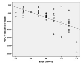
Results: Total of 60 eyes of 30 patients were subdivided into 21 eyes having optic neuritis (ON) [‘MS – ON’] and 39 eyes without ON. The RNFL thickness (RNFLt) was found to be significantly reduced in all parameters except in temporal quadrant, as the duration of disease increases. Average RNFLt were found to have negative correlation (r = -0.450) with disease duration. Negative correlation (r=-0.657) was also found between EDSS score change and average RNFLt change. The eyes having ON showed statistically significant RNFL thinning as compared to the non – ON fellow eyes. The baseline EDSS score was found to be negatively correlated (moderate degree, r = -0.348) with baseline average RNFL thickness, with p-value of 0.006.
Conclusions: The RNFLt is not only significantly thinner in those with history of ON, but it is also affected remarkably even in absence of prior ON, suggesting subclinical ongoing axonal loss in patients with MS. The EDSS score is inversely correlated with RNFL thickness.
Downloads
References
De Stefano N, Matthews PM, Antel JP, et al. Chemical pathology of acute demyelinating lesions and its correlation with disability. Ann Neurol. 1995 Dec;38(6):901-9. DOI: https://doi.org/10.1002/ana.410380610.
Sergott RC. Optical coherence tomography: measuring in-vivo axonal survival and neuroprotection in multiple sclerosis and optic neuritis. Curr Opin Ophthalmol. 2005 Dec;16(6):346-50.
The clinical profile of optic neuritis. Experience of the Optic Neuritis Treatment Trial. Optic Neuritis Study Group. Arch Ophthalmol. 1991 Dec;109(12):1673-8.
Miller D, Barkhof F, Montalban X, et al. Clinically isolated syndromes suggestive of multiple sclerosis, part I: natural history, pathogenesis, diagnosis, and prognosis. Lancet Neurol. 2005 May;4(5):281-8. DOI: https://doi.org/10.1016/S1474-4422(05)70071-5.
Frohman EM, Fujimoto JG, Frohman TC, et al. Optical coherence tomography: a window into the mechanisms of multiple sclerosis. Nat Clin Pract Neurol. 2008 Dec;4(12):664-75. doi: https://doi.org/10.1038/ncpneuro0950.
Trip SA, Schlottmann PG, Jones SJ, et al. Retinal nerve fiber layer axonal loss and visual dysfunction in optic neuritis. Ann Neurol. 2005 Sep;58(3):383-91. DOI: https://doi.org/10.1002/ana.20575.
Trip SA, Wheeler-Kingshott C, Jones SJ, et al. Optic nerve diffusion tensor imaging in optic neuritis. Neuroimage. 2006 Apr 1;30(2):498-505. Epub 2005 Oct 20.
Costello F, Coupland S, Hodge W, et al. Quantifying axonal loss after optic neuritis with optical coherence tomography. Ann Neurol. 2006 Jun;59(6):963-9. DOI: https://doi.org/10.1002/ana.20851.
Parisi V, Manni G, Spadaro M, et al. Correlation between morphological and functional retinal impairment in multiple sclerosis patients. Invest Ophthalmol Vis Sci. 1999 Oct;40(11):2520-7.
Spain RI, Maltenfort M, Sergott RC, et al. Thickness of retinal nerve fiber layer correlates with disease duration in parallel with corticospinal tract dysfunction in untreated multiple sclerosis. J Rehabil Res Dev. 2009;46(5):633-42.
Pueyo V, Martin J, Fernandez J, et al. Axonal loss in the retinal nerve fiber layer in patients with multiple sclerosis. MultScler. 2008 Jun;14(5):609-14. doi: https://doi.org/10.1177%2F1352458507087326. Epub 2008 Apr 18.
Sepulcre J, Murie-Fernandez M, Salinas-Alaman A, et al. Diagnostic accuracy of retinal abnormalities in predicting disease activity in MS. Neurology. 2007 May 1;68(18):1488-94.
Gencer M, Delipoyraz I, Buttanri B, Turkoglu R, Cetinkaya Y, Tireli H. Association of Retinal Nerve Fiber Layer Thickness with Disease Disability, Disease Duration, SF-36, and PASAT Scores in Multiple Sclerosis. J Neurol Sci [Turk] 2013; 30(4): 702-10.
Pulicken M, Gordon-Lipkin E, Balcer LJ, Frohman E, Cutter G, Calabresi PA. Optical coherence tomography and disease subtype in multiple sclerosis. Neurology 2007Nov; 69(22):2085–92. https://doi.org/10.1212/01.wnl.0000294876.49861.dc
Costello F, Hodge W, Pan YI, et al. Tracking retinal nerve fiber layer loss after optic neuritis: a prospective study using optical coherence tomography. MultScler. 2008 Aug;14(7):893-905. doi: https://doi.org/10.1177%2F1352458508091367. Epub 2008 Jun 23.
Fisher JB, Jacobs DA, Markowitz CE, et al. Relation of visual function to retinal nerve fiber layer thickness in multiple sclerosis. Ophthalmology. 2006 Feb;113(2):324-32. Epub 2006 Jan 10.
Petzold A, de Boer JF, Schippling S, Vermersch P, Kardon R, Green A, et al. Optical coherence tomography in multiple sclerosis:a systematic review and meta-analysis. Lancet Neurol 2010; 9(9):921-932. https://doi.org/10.1016/S1474-4422(10)70168-X
Singh S, Sharma R, Gurunadh VS, Shankar S. OCT based evaluation of retinal changes in multiple sclerosis. Int J Res Med Sci 2017;5:4117-21. http://dx.doi.org/10.18203/2320-6012.ijrms20173994
Pulicken M, Gordon-Lipkin E, Balcer LJ, et al. Optical coherence tomography and disease subtype in multiple sclerosis. Neurology. 2007 Nov 27;69(22):2085-92.
Henderson AP, Trip SA, Schlottmann PG, et al. An investigation of the retinal nerve fibre layer in progressive multiple sclerosis using optical coherence tomography. Brain. 2008 Jan;131(Pt 1):277-87. Epub 2007 Dec 4.

 OAI - Open Archives Initiative