Ultrasonographic analysis of the anatomical relationship between femoral vessels in the upper part of thigh in critically ill patients– a cross sectional study
Abstract
Objective: Femoral vessels are one of the frequently used sites of cannulation in intensive care units. In resource limited settings cannulations are done blindly without ultrasonographic guidance based on a traditional belief that in the upper thigh vein keeps a medial relationship to artery. In this trial we tried to analyse the anatomical relationship of femoral vein to femoral artery using ultrasound in critically ill patients.
Methods: This cross sectional study analysed the anatomical relationship of femoral vein to femoral artery at 2cm, 4 cm and 6 cm from the mid inguinal point in both thighs of the patients using ultrasonography. The study was done among patients admitted in a multidisciplinary intensive care unit.
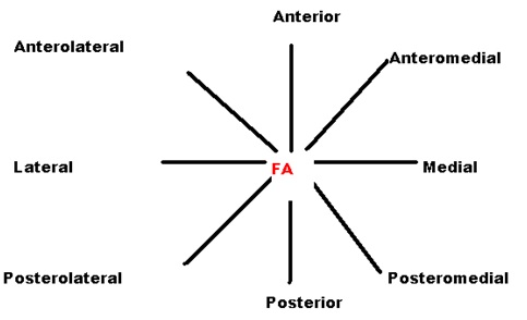
Results: Three hundred limbs of one hundred and fifty patients were analysed by ultrasonography. A total of 900 measurements were taken at three different levels of both legs. At 2 cm below the mid inguinal point, in 256 limbs (85.3%) femoral vein was medial to femoral artery (95% Confidence Interval82.82% to 89.14%), at 4 cm below the mid inguinal point, in 210 limbs (70%) femoral vein was postero medial to femoral artery (95% CI64.47% to 75.13%),and at6 cm below the mid inguinal point in 200 limbs(66.7%)femoral vein was posterior to femoral artery(95% CI 61.02% to 71.98%).
Conclusion: Femoral vein showed variable relationship to femoral artery in the upper part of the thigh. As the distance increased from mid inguinal point, variation from normal relationship was also found to be increasing.
Downloads
References
Troianos CA, Hartman GS, Glas KE, et al. Guidelines for performing ultrasound guided vascular cannulation: recommendations of the American Society of Echocardiography and the Society of Cardiovascular Anesthesiologists. J Am SocEchocardiogr. 2011 Dec;24(12):1291-318. doi: https://doi.org/10.1016/j.echo.2011.09.021.
Chaurasya B D. Human anatomy; regional and applied dissection and clinical. New Delhi: CBS publishers & Distributors; 2010.
Komolafe O, Olatise O. Utility of blind percutaneous jugular venous cannulation in resource-limited settings. J Vasc Access. 2017 Jan 18;18(1):26-29. doi: https://doi.org/10.5301%2Fjva.5000627. Epub 2016 Nov 14.
Buchanan MS, Backlund B, Liao MM, et al. Use of ultrasound guidance for central venous catheter placement: survey from the American Board of Emergency Medicine Longitudinal Study of Emergency Physicians. AcadEmerg Med. 2014 Apr;21(4):416-21. doi: https://doi.org/10.1111/acem.12350.
Maizel J, Bastide MA, Richecoeur J, et al. Practice of ultrasound-guided central venous catheter technique by the French intensivists: a survey from the BoReal study group. Ann Intensive Care. 2016 Dec;6(1):76. doi: https://doi.org/10.1186/s13613-016-0177-x. Epub 2016 Aug 8.
Grant J, Basmajian J. Grant's Method of Anatomy. Baltimore: Williams & Wilkins; 1980.
Baum PA, Matsumoto AH, Teitelbaum GP, Zuurbier RA, Barth KH. Anatomic relationship between the common femoral artery and vein: CT evaluation and clinical significance. Radiology. 1989;173(3):775-77.
P Souza Neto E, Grousson S, Duflo F, et al. Ultrasonographic anatomic variations of the major veins in paediatric patients. Br J Anaesth. 2014 May;112(5):879-84. doi: https://doi.org/10.1093/bja/aet482. Epub 2014 Feb 10.
Hughes P, Scott C, Bodenham A. Ultrasonography of the femoral vessels in the groin: implications for vascular access. Anaesthesia. 2000 Dec;55(12):1198-202.
Getzen LC, Pollak EW. Short-term femoral vein catheterization.A safe alternative venous access?Am J Surg. 1979 Dec; 138(6):875-8.
Williams JF, Seneff MG , Friedman BC, McGrath, Brian j, Gregg, Richard, Sunner, Jennie RN; Zimmerman, Jack E. Use of femoral venous catheters in critically ill adults: prospective study. Critical Care Medicine 1991April; 19(4): 550–53.
Gilston A. Letter: Cannulation of the femoral vessels. Br J Anaesth. 1976 May; 48(5):500-1.
Fuller TJ, Mahoney JJ, Juncos LI, et al. Arteriovenous fistula after femoral vein catheterization. JAMA. 1976 Dec 27; 236(26):2943-4.
Grassi CJ, Bettmann MA, Rogoff P, Reagan K , Harrington DP. Femoral arteriovenous fistula after placement of a Kimrayâ€Greenfield filter. American Journal of Roentgenology. 1988 Nov; 151(4): 681–2.
Hessel SJ, Adams DF, Abrams HL.Complications of angiography.Radiology. 1981 Feb;138(2):273-81.
Grier D, Hartnell G. Percutaneous femoral artery puncture: practice and anatomy. British Journal of Radiology. 1990; 63(752): 602–4.
Rapoport S, Sniderman KW, Morse SS, et al. Pseudoaneurysm: a complication of faulty technique in femoral arterial puncture. Radiology. 1985 Feb;154(2):529-30. DOI: https://doi.org/10.1148/radiology.154.2.3966139.
Altin RS, Flicker S, Naidech HJ. Pseudoaneurysm and arteriovenous fistula after femoral artery catheterization: association with low femoral punctures. AJR Am J Roentgenol. 1989 Mar;152(3):629-31. DOI: https://doi.org/10.2214/ajr.152.3.629.


 OAI - Open Archives Initiative


