E-ISSN:2320-8686
P-ISSN:2321-127X
RNI:MPENG/2017/74037

Research Article

Endoscopic Diagnosis

International Journal of Medical Research and Review

2025 Volume 13 Number 3 Jul-Sep
Publisherwww.medresearch.in

Evaluating the Accuracy of Endoscopic diagnosis: A comparative analysis with Histopathology in Upper and Lower GI Tract Disorders

Rathore S1*, Singh PK2
DOI:https://doi.org/10.17511/ijmrr.2025.i03.01

1* Smriti Rathore, MD Pathology, Assistant Professor, Department of Pathology, Shri Balaji Institute of Medical Sciences, Raipur, Chhattisgarh, India.

2 Prashant Kumar Singh, DNB Gastroenterology, MD Medicine, Assistant Professor, Department of Medicine, Shri Balaji Institute of Medical Sciences, Raipur, Chhattisgarh, India.

Background: Endoscopy and histopathology are complementary tools in diagnosing gastrointestinal (GI) disorders. While endoscopy provides macroscopic visualization, histopathology offers definitive microscopic diagnosis. This study aims to evaluate the correlation between endoscopic findings and histopathological results in upper and lower GI disorders.

Objectives: 1. To assess the diagnostic correlation between endoscopic impressions and histopathological diagnosis. 2. To determine the accuracy of endoscopy in detecting various upper and lower GI tract pathologies.

Methods: A retrospective study was conducted on 200 patients undergoing upper GI endoscopy (UGIE) and Colonoscopy for GI symptoms. Endoscopic findings were documented and correlated with histopathology reports of biopsy specimens.

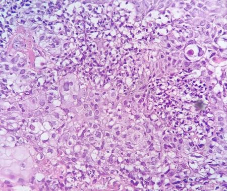
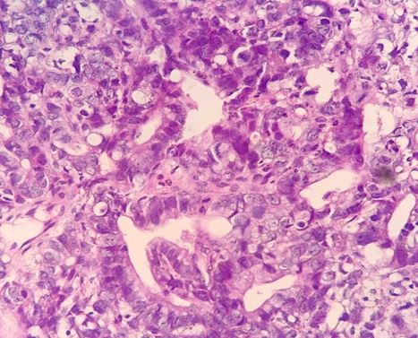
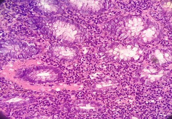
Results: Endoscopic and histopathological findings were consistent in 92% of upper GI cases and 90.6% of lower GI cases. Among 125 upper GI cases, 115 were concordant, and among 75 lower GI cases, 68 were concordant. The highest correlation was observed in cases of gastric ulcers, esophagitis, colorectal polyps, inflammatory bowel disease (IBD) and malignancy.

Conclusion: Endoscopy is a reliable initial diagnostic tool. However, histopathological confirmation is essential, especially in cases with subtle or ambiguous endoscopic findings.

Keywords: Endoscopy, Histopathology, GI Disorders, Upper GI, Colonoscopy

Corresponding Author How to Cite this Article To Browse
Smriti Rathore, MD Pathology, Assistant Professor, Department of Pathology, Shri Balaji Institute of Medical Sciences, Raipur, Chhattisgarh, India.
Email:
Rathore S, Singh PK, Evaluating the Accuracy of Endoscopic diagnosis: A comparative analysis with Histopathology in Upper and Lower GI Tract Disorders. Int J Med Res Rev. 2025;13(3):1-6.
Available From
https://ijmrr.medresearch.in/index.php/ijmrr/article/view/1553

Manuscript Received Review Round 1 Review Round 2 Review Round 3 Accepted
2025-06-05 2025-06-13 2025-06-21 2025-06-30 2025-07-07
Conflict of Interest Funding Ethical Approval Plagiarism X-checker Note
None Nil Yes 11.98

© 2025 by Rathore S, Singh PK and Published by Siddharth Health Research and Social Welfare Society. This is an Open Access article licensed under a Creative Commons Attribution 4.0 International License https://creativecommons.org/licenses/by/4.0/ unported [CC BY 4.0].

Download PDFBack To ArticleIntroductionMaterials and MethodsResultDiscussionConclusionReferences

Introduction

The Human Gastrointestinal (GI) tract is a common site for various lesions, including congenital, inflammatory, and neoplastic conditions. These lesions contribute significantly to morbidity and mortality.[1]

To diagnose GI diseases, investigative methods such as endoscopy, colonoscopy, cytopathology, gastric secretory function and absorption of vitamin B12, etc., are comfortably used. However, all of the above have some shortcomings and limitations.[2]

The introduction of endoscopes in the 1960s has greatly improved the diagnostic facility for fiberoptic endoscopy because they are readily accessible and can easily be sampled for specific histopathological or microbiologic investigation with available biopsy forceps.[3] It is a diagnostic procedure used to visualize the oropharynx, esophagus, stomach, and proximal duodenum.[4] Gastric symptoms like dyspepsia, vomiting, abdominal pain, ulcers, etc, are very common causes of discomfort amongst patients & are the common reasons for referral for endoscopic examination. GIT endoscopy, along with biopsy, is not only used to diagnose malignant and inflammatory lesions but is also used for monitoring the course, extent of the disease, response to the therapy and early detection of complications.[5] Colonoscopy is a diagnostic as well as a therapeutic procedure performed to evaluate the large intestine (i.e., colon, rectum, and anus) as well as the distal portion of the small intestine (terminal ileum).[6]

The aim and objectives of this study are to assess the diagnostic correlation between endoscopic impressions and histopathological diagnosis, and to determine the accuracy of endoscopy in detecting various upper and lower GI tract pathologies.

Materials and Methods

This was a retrospective study conducted from March 2024 to April 2024. The study was conducted among 200 cases who underwent upper gastrointestinal endoscopy(125) or colonoscopy(75) at Sri Balaji Institute of Medical Sciences(SBIMS), Raipur, Chhattisgarh. All endoscopic and colonoscopic biopsies of the upper and lower GI tract were included in the study, and biopsies were taken from abnormal or suspicious lesions. Biopsy was done for therapeutic purposes,

cases where biopsy cannot be done, or where consent was not given and autolysed specimens were excluded. Inadequate biopsies in terms of no glands, only fibrocollagenous tissue, inadequate or inconclusive histopathology reports, along with incomplete procedures or lost follow-up, were also excluded from the study.

Endoscopies were done with a flexible fibre-optic endoscope. The lesions were noted on gross visualization using an endoscope and colonoscope. Patients suspected for any lesions were taken up for biopsy. The biopsies were fixed in 10% formalin and routinely processed and embedded in paraffin with mucosal surfaces uppermost. 5-micron-thick sections were cut perpendicular to this surface. Routine staining with hematoxylin and eosin was performed. Microscopic findings were studied by two pathologists independently.

Statistical Analysis: This was a descriptive study to correlate endoscopic appearances/ diagnosis with the final histologic diagnosis. Hence, the data collected was analysed as a percentage of concordance between the two. The discordant cases were analysed to obtain reasons for discordance. SPSS was used to perform statistical analyses.

Ethical considerations: Informed consent was waived due to the retrospective nature of the study, and patient data were anonymised to protect confidentiality.

Limitations: Limitations included the retrospective study design, potential biases in patient selection, and reliance on existing medical records, which may have affected data completeness and accuracy.

Result

The current study was conducted for 1 year at SBIMS, Raipur. A total of 125 endoscopic and 75 colonoscopic biopsies were taken from different sites of the upper and lower GI tract.

Age group, gender and site distribution- In the present study, the peak incidence of the GI lesions was in the age group between 40-60 years. The mean age was 52.5 years. In this study, out of 125 endoscopy patients, 77 were males and 48 were females. The male-to-female ratio was 1.6:1. Out of 75 colonoscopy patients, 48 were males and 27 were females. Male to female ratio was 1.7:1. (Tables 1& 2).


Table 1: Age and Sex Distribution of Endoscopy Patients

Age Group  Sex of the patientTotal
MaleFemale
0-20202(1.6%)
21-40251338(30.4%)
41-60362258(46.4%)
61-80151227(21.6%)
78(62.4%)47(37.6%)

Table 2: Age and Sex Distribution of Colonoscopy Patients

Age Group  Sex of the patientTotal
MaleFemale
0-20000
21-4011819(25.3%)
41-60291443(57.3%)
61-809413(17.3%)
49(65.3)26(34.6%)

Maximum number of upper GI patients presented with abdominal pain(65%), regurgitation(25%), vomiting(10%), dyspepsia(10%). Most of the colonoscopy patients presented with altered bowel habits(68%), abdominal pain(45%), blood in stools(15%), weight loss(10%).

Table 3: Correlation in Upper GI Endoscopy

Endoscopy diagnosisCases(n)Histopathology diagnosisconcordance
Esophagitis2019(95%)
Gastric ulcer2524(96%
Chronic Gastritis3531(88.5%
Non specific Duodenitis1513(80%)
Malignancy2221(95.4%)
normal87(87.5%)

Out of 125 upper GI cases, 115 endoscopy reports correlated with histopathology reports. Concordance Rate was 92%. (Table 3) In 10 cases, endoscopy reports didn’t correlate with histopathology reports. (Table 4)

Table 4: Discordance in endoscopy and histopathology reports

No of casesEndoscopy diagnosisHistopathology diagnosis
1EsophagitisEsophageal Candidiasis
1Gastric ulcerdysplasia
1Chronic GastritisAtrophic Gastritis
3Chronic GastritisIntestinal Metaplasia
2Non specific duodenitisBrunner Gland Hyperplasia
1Suspected MalignancyReactive changes
1NormalReactive changes

ijmrr-1553-01.JPG
Figure 1: Microscopic image of Squamous cell Carcinoma

ijmrr-1553-02.JPG
Figure 2: Moderately differentiated Adenocarcinoma

Table 5: Lower GI Endoscopy Findings and Concordance

Colonoscopic diagnosisCases(n)Histopathological diagnosis concordance
Non-specific colitis2825(89.2%)
Colorectal polyp2018(90%)
Malignancy1917(89.4%)
Ulcerative colitis55(100%)
Tuberculosis22(100%)
Chron’s disease11(100%)

Out of 75 upper GI cases, 68 endoscopy reports correlated with histopathology reports. Concordance Rate was 90.6%.

(Table 5) In 7 cases, endoscopy reports didn’t correlate with histopathology reports. (Table 6)


ijmrr-1553-03.JPG
Figure 3: Microscopic image of Infective Colitis

Table 6: Discordance in Colonoscopy and Histopathology Reports

No of casesColonoscopy diagnosisHistopathology diagnosis
3Non-specific colitisAmoebic Colitis
2Colorectal polypDysplasia
2MalignancyIschemic colitis

Discussion

The GIT presents a spectrum of disorders ranging from benign inflammatory conditions to malignant neoplasms, with dyspepsia and GERD being common indications for upper GIT endoscopy. Dyspepsia, characterised by upper abdominal pain or discomfort, can signify various underlying issues such as peptic ulcer disease, gastritis, or gastric cancer.

In contrast, GERD involves reflux of stomach acid into the oesophagus, leading to symptoms such as heartburn and regurgitation, potentially progressing to Barrett’s oesophagus and increasing the risk of oesophageal adenocarcinoma. Endoscopic techniques play a crucial role in diagnosing and managing these conditions, aiding the early detection of malignancies and guiding appropriate patient management.[7]

The modern endoscope has evolved from a rigid hollow metal tube to a light, flexible fibreoptic system using self-illumination. It not only allows visual inspection of the GI tract, but also permits access to the suspected tissue area with the aid of a biopsy needle. Endoscopic biopsy is an easy, minimally invasive & cost-effective procedure when it comes to arriving at a specific diagnosis of a patient with non-specific symptoms. Expertise on the part of endoscopists in choosing the appropriate site is therefore needed,

along with proper processing of biopsy tissue and meticulous reporting by the histopathologist for interpretation of endoscopic biopsies. Ultrasound can be performed with the help of an endoscope in selected cases whose biopsies are negative for malignancy but have suspicious findings on endoscopy and have atypical presentation like profound weight loss, advanced age, short duration of symptoms, etc[8].

In the present study, the peak incidence of the GI lesions was in the age group between 40-60 years. The mean age was of 52.5 years which almost simulates the study conducted by Patel et al[8] and Rauta et et al[9]. The youngest patient was 18 years old and the oldest patient was 84 years old.

In the present study, out of the 125 gastroduodenal biopsies, 30 cases were from the oesophagus,79 cases were from the gastric region, and the remaining 16 cases were from the duodenum. The present study is consistent with the studies made by Sarma et al[9], Mishra et al[10], which shows that gastric biopsies are more common compared to oesophagus and duodenal biopsies.

Oesophagal lesions: Among the oesophagal biopsies received in the present study (30cases), 23 were non-neoplastic and 7 were malignancies. This data is similar to other studies by Mishra et al [10] and Abilash SC et al[11].

Gastric Lesions: Of the total 79 biopsies from the stomach, 64 were non-neoplastic and 15 were neoplastic. The most common lesion in gastric biopsy in this study was chronic gastritis, which is similar to the study done by Mishra et al[10] and Islam et al[12].

Duodenal lesions: In the present study, 16 duodenal biopsies were received, out of which 15 showed chronic non-specific duodenitis and 1 case showed villous atrophy, which is similar to a study done by Mishra et al[10] and Krishnappa R et al[13].

Colonic Lesions: Among the lower gastrointestinal biopsies, colonic biopsies were the most common. 75 colorectal biopsies were received. The most common lesions in the colonic biopsies in the present study were chronic non-specific colitis (37.3%) and colorectal polyp(26.6%), followed by malignancy(25.3%), ulcerative colitis(6%), tuberculosis(2%) and Crohn’s disease(1%). Our findings are correlating with the study of Sharma et al(14).


In the present study, endoscopy and histopathology reports were concordant in 92% and 90.6% in upper and lower GI lesions, respectively, which is very high and similar to the studies done by Mishra et al[10], Islam et al[12] and Krishnappa et al[13].

10(8%) upper GI cases and 7(9.6%) lower GI cases were discordant. These discrepancies highlight the importance of considering the various factors, such as histologic heterogeneity, limitations in human interpretation. The data presented in the tables and supported by previous studies demonstrate a high concordance between endoscopic and histological diagnoses in the oesophagus, stomach, duodenum and colon, indicating the reliability of endoscopic procedures in diagnosing gastrointestinal lesions. Variability in concordance rates highlights the need for histopathological confirmation to ensure diagnostic accuracy and appropriate treatment.

Conclusion

The upper GI endoscopy and colonoscopy are the diagnostic tools of choice to detect upper and lower GI diseases. Gross visualisation through endoscopy and colonoscopy cannot always detect the mucosal lesions, whether it is benign or malignant; histopathologic examination can provide an accurate diagnosis. This study provides critical insights into the diagnostic concordance between endoscopic and histopathological findings in patients with upper and lower GIT lesions. Although endoscopy and colonoscopy are the primary diagnostic tools for GIT pathologies, our findings highlight the importance of histopathological confirmation to ensure diagnostic accuracy.

Permission from the Institutional Research Board: Yes

Funding: Nil

Conflict of interest: None Initiated

References

1. Chag S, Patel N, Rathod K. Endoscopic correlation with histological findings in upper gastrointestinal biopsies. International Journal of Clinical and Diagnostic Pathology. 2025;8(1):108-13. [Crossref][PubMed][Google Scholar]

2. Shankar U, Kumaria R, Sarmaikb A, Maribashetti K. A clinicopathological correlation of gastrointestinal lesions on endoscopic biopsies with positron emission tomography (pet) findings. International Journal of Medical Reviews and Case Reports (2022) 6(7):1-8. . [Crossref][PubMed][Google Scholar]

3. Rashmi K, Horakerappa MS, Karar A, Mangala G. A study on histopathological spectrum of upper gastrointestinal tract endoscopic biopsies. Int J Med Res Health Sci. 2013; 2:418-24. [Crossref][PubMed][Google Scholar]

4. Ahlawat R, Hoilat GJ, Ross AB. Esophagogastroduodenoscopy Updated 2023 Aug 8]. In: StatPearls [Internet]. Treasure Island (FL): StatPearls Publishing; 2025 Jan-. Available from: [Article][Crossref][PubMed][Google Scholar]

5. Mohan B, Manjunath HK, Geethamani V, et al. Histomorphological Analysis of Upper GI Endoscopic Biopsies: A Retrospective Study of 106 Cases. National Journal of Laboratory Medicine. 2019 Oct, Vol-8(4): 4-7. [Crossref][PubMed][Google Scholar]

6. Stauffer CM, Pfeifer C. Colonoscopy. [Updated 2023 Jul 24]. In: StatPearls [Internet]. Treasure Island (FL): StatPearls Publishing; 2025 Jan-. Available from: [Article][Crossref][PubMed][Google Scholar]

7. Rauta S, Baisakh P, Sahoo A K, et al. Correlation of Endoscopic and Histopathological Diagnoses in Upper Gastrointestinal Tract Lesions: A Cross-Sectional Study. Cureus 16(9): e69553. . [Crossref][PubMed][Google Scholar]

8. Patel J, Patel A, Throt R, et al. Study of Endoscopic and Histopathologic correlation in Upper Gastrointestinal lesions in a Tertiary Care Hospital. IJCRT | Volume 10, Issue 1 January 2022 | ISSN: 2320-882. . [Crossref][PubMed][Google Scholar]

9. Sarma U, Kalita u. Clinicopathological Study of Gastroduodenal Biopsies and Correlate with Endoscopic. Pac J Cancer Biol, 9 (3), 265-9. . [Crossref][PubMed][Google Scholar]

10. Mishra R, Vahikar S, Shrivastava K, et al. Histopathological spectrum of gastrointestinal lesions in patients undergoing gastrointestinal endoscopic biopsies- A ospective study. Trop J Pathol Microbiol. 2022;8(1):22-28. [Crossref][PubMed][Google Scholar]


11. Abilash, S. C. , et al. Histopathologic spectrum of upper gastrointestinal tract mucosal biopsies: A retrospective study. " Sch. J. App. Med. Sci 4.5E (2016): 1807-13 [Crossref][PubMed][Google Scholar]

12. Islam, Sk Md Jaynul, et al. Endoscopic and histologic diagnosis of upper gastrointestinal lesions, experience in a port city of Bangladesh. Chattagram Maa-O-Shishu Hospital Medical College Journal 13. 3 (2014): 11-14. [Crossref][PubMed][Google Scholar]

13. Krishnappa, Rashmi, et al. A study on histopathological spectrum of upper gastrointestinal tract endoscopic biopsies. International Journal of Medical Research & Health Sciences 2. 3 (2013): 418-24. [Crossref][PubMed][Google Scholar]

14. Sharma V, Bansal R, Sharma S, Khare A. Histopathological spectrum with clinical correlation of lower gastrointestinal tract endoscopic biopsies. International Journal of Clinical and Diagnostic Pathology 2022; 5(1): 89-93. . [Crossref][PubMed][Google Scholar]

Disclaimer / Publisher's Note The statements, opinions and data contained in all publications are solely those of the individual author(s) and contributor(s) and not of Journals and/or the editor(s). Journals and/or the editor(s) disclaim responsibility for any injury to people or property resulting from any ideas, methods, instructions or products referred to in the content.