E-ISSN:2320-8686
P-ISSN:2321-127X
RNI:MPENG/2017/74037

Case Report

Pancreatoblastoma

International Journal of Medical Research and Review

2025 Volume 13 Number 2 Apr-Jun
Publisherwww.medresearch.in

A Case Report on Treatment of Pancreatoblastoma in a 55-year-old female with acute pancreatitis

Priyanka1*, Hemanth Vudayaraju2, V. Sai Sindhuja3
DOI:https://doi.org/10.17511/ijmrr.2025.i02.09

1* Priyanka, MS (General Surgery), DNB (General Surgery), DrNB (Surgical Oncology), FALS (Oncology), Yashoda Hospitals, Secunderabad, Telangana, India.

2 Hemanth Vudayaraju, MS (General Surgery), MCh (Surgical Oncology), DNB (Surgical Oncology), Director-Surgical Oncology and Minimal Access Onco Surgery and Robotic Surgeon, Yashoda Hospitals, Secunderabad, Telangana, India.

3 V. Sai Sindhuja, MS (General Surgery), Resident DrNB (Surgical Oncology), Yashoda Hospitals, Secunderabad, Telangana, India.

Background: Pancreatoblastoma is a rare, malignant tumor that develops in the pancreas and is distinguished by lobular structures which contain acinar cells and squamoid corpuscles. The most common site was the head of the pancreas, and the most common symptom is abdominal pain and vomiting.

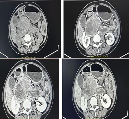
Case Presentation: A 55-year-old female presented with complaints of abdominal pain and vomitings with vitals in normal range. However, her serum amylase and lipase levels were found to be elevated. A contrast-enhanced computed tomography (CECT) revealed a large exophytic mass lesion in her pancreas, measuring 6.5 x 9.3 x 8.5 cm and causing distortion of the SMV medially and stretching of the anterior superior pancreaticoduodenal vein. The main pancreatic duct is severely compressed. Our patient underwent a Whipple procedure and exhibited an unremarkable recovery.

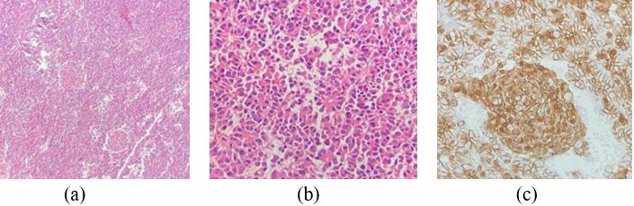
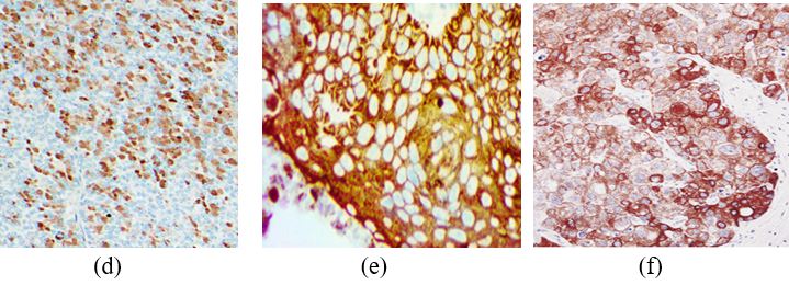
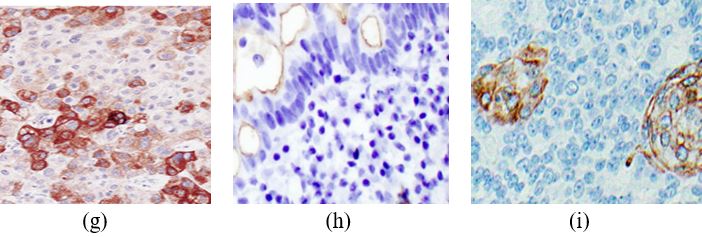
Conclusion: Pancreatoblastoma is a curable tumor that requires early diagnosis through a multi-disciplinary approach. The tumor is removed completely by surgery and pathology; Immunohistochemistry confirms the diagnosis. Following surgery, the patient is closely monitored to look for any residual disease, metastases, or recurrences.

Keywords: Pancreatoblastoma, amylase, lipase, Whipple procedure, immuno-histochemistry

Corresponding Author How to Cite this Article To Browse
Priyanka, MS (General Surgery), DNB (General Surgery), DrNB (Surgical Oncology), FALS (Oncology), , Yashoda Hospitals, Secunderabad, Telangana, India.
Email:
Priyanka, Hemanth Vudayaraju, V. Sai Sindhuja, A Case Report on Treatment of Pancreatoblastoma in a 55-year-old female with acute pancreatitis. Int J Med Res Rev. 2025;13(2):41-45.
Available From
https://ijmrr.medresearch.in/index.php/ijmrr/article/view/1552

Manuscript Received Review Round 1 Review Round 2 Review Round 3 Accepted
2025-05-19 2025-05-27 2025-06-04 2025-06-12 2025-06-20
Conflict of Interest Funding Ethical Approval Plagiarism X-checker Note
None Nil Yes 11.32

© 2025 by Priyanka, Hemanth Vudayaraju, V. Sai Sindhuja and Published by Siddharth Health Research and Social Welfare Society. This is an Open Access article licensed under a Creative Commons Attribution 4.0 International License https://creativecommons.org/licenses/by/4.0/ unported [CC BY 4.0].

Download PDFBack To ArticleIntroductionCase PresentationDiscussionConclusionReferences

Introduction

Pancreatoblastoma (PB) is a rare form of pancreatic tumor, accounting for < 1 % with an annual incidence rate of approximately 0.004 per 100,000 individuals.[1] It is often misdiagnosed as a neuroblastoma.[2] In the year 2014, the Papanicolaou Society of Cytopathology (PSC) officially recognised the name "Pancreatoblastoma" for this particular type of tumor. PB is found to occur more frequently among Asian individuals compared to Caucasian people.[3] It is more aggressive in adults compared to pediatric cases and bears a poor prognosis.[4] Because of the lack of specific symptoms, it often leads to a delay in diagnosis.[5] PB is inconsistently documented in the medical literature, with case reports being the most common and a comprehensive systematic analysis is required.[6] In this case report, we described the clinical characteristics, pathologic features and treatment of pancreatoblastoma in a 55-year-old female patient.

Case Presentation

A 55-year-old female presented with complaints of vomiting and abdominal pain radiating posteriorly. She had experienced a similar episode 4 months ago, which was relieved with medication. The patient was admitted to our department for further management. There was no family history of comorbidities or any malignancies.

On physical examination, the baseline vitals were stable. Baseline investigations were done; all were within normal limits, but her serum amylase (383 U/L) and lipase levels (1634 U/L) were found elevated.

Contrast-enhanced computed tomography (CECT) abdomen revealed a large exophytic well well-marginated mass lesion arising from the inferior aspect of the head of the pancreas in the periampullary region and pancreaticoduodenal groove. The lesion measures 6.5 x 9.3 x 8.5 cm (Figure 1). Mild post-contrast enhancement, which is slightly hypoenhancing to the pancreas, is seen with multiple eccentric non-enhancement areas. The fat plane with the corresponding, surrounding structure was well maintained. The distal common bile duct and the main pancreatic duct are severely compressed by the mass lesion, with upstream dilation of the main pancreatic duct.

The lesion distorts the mesenteric vein medially (abutting for < 180 degrees) and stretches the anterior superior pancreaticoduodenal vein, the first jejunal branch of the superior mesenteric vein.

Reports show the possibility of solid pseudo papillary epithelial neoplasm, duodenal gastrointestinal stromal tumour (GIST). The rest of the pancreas shows normal enhancement with main pancreatic duct dilation at the neck, approximately 6 mm.

Mild peripancreatic fluid and fat stranding is found extended around the D3 segment of the duodenum and peri-gastric regions, and multiple small volume para-aortic lymph nodes are noted in the upper abdomen.

There is mild omental fat stranding in the left hypochondrium, along with a tiny enhancing nodule that resembles the spleen. With acute symptoms, the patient was observed for a week, with no symptomatic improvement. After discussing the outcomes patient was planned for a surgery.

The patient underwent Whipple's procedure, a lesion measuring 10 x 8 cm was observed originating from the head and uncinate process of the pancreas, and there was 1 litre of hemorrhagic ascitic fluid present intraoperatively.

Tumour found adhered to the right Gerota fascia, which we have completely resected. The fat planes with the superior mesenteric artery (SMA) and superior mesenteric vein (SMV)were preserved. Histological evaluation of the tumor had sheets and nests comprised of tiny, homogeneous primitive cells, frequently with squamous nests and central keratinisation.

Immunohistochemical examination revealed that tumour cells strongly expressed cytokeratin AE1/AE3, CK19, CK7, CEA, SYN, CD56 and trypsin but were negative for chromogranin (Figure 2). The squamous differentiation areas were also highlighted by CK5 and EMA.

Finally, a definitive diagnosis of pancreatoblastoma was established following the identification of specific histological and immunohistochemical markers. The postoperative period was uneventful, and the patient was discharged on POD-5. Upon follow-up, her health status remained satisfactory, with no signs of residual tumour or recurrence.


ijmrr-1552-01.JPG
Figure 1: CECT abdomen revealed a well-marginated mass lesion arising from the head of the pancreas in the periampullary region and pancreaticoduodenal groove.

ijmrr-1552-02.JPG

ijmrr-1552-03.JPG

ijmrr-1552-04.JPG

ijmrr-1552-05.JPG
Figure 2: The tumor stained with haematoxylin and eosin (a) large tumour cells with abundant eosinophilic cytoplasm, consistent with squamous morules (b) Acinar cells made up the pancreatic mass, displayed ductal and squamous differentiation, (c) Beta catenin, accentuated in squamous morules (d - i) tumour cells show positive for trypsin, cytokeratin AE1/AE3, CK19, CK7, CEA, (j)
negative for chromogranin.


Discussion

Adult pancreatoblastoma (PB) is a rare malignant tumor of the pancreas, difficult to diagnose because of its resemblance to other solid cellular neoplasms of the pancreas, like solid pseudopapillary neoplasms, neuroendocrine neoplasms and pancreatic acinar cell carcinomas.[7] PB is characterised by a slow growth rate, soft and well-circumscribed nature, often reaching significant sizes upon diagnosis. Approximately half of these tumours are located within the pancreatic head region.[8]

In PB, abdominal pain, weight loss and abdominal mass are the main symptoms in adult patients, whereas anorexia, bowel habit changes and jaundice are also some common symptoms.[9] Our patient had abdominal pain and vomiting, which aligns with the findings of previous research. PB predominantly exhibits a preference for the head of the pancreas in approximately 50% of cases. These tumours range in size from 1.5 to 20 cm, demonstrating significant variability. A conclusive diagnosis of PB should invariably be established following a comprehensive histological examination.[10] The imaging characteristics of PB are characterised by their non-specific nature, with the majority of tumours presenting as lobulated masses which are typically well-circumscribed or partially circumscribed and accompanied by necrosis and enhancing septations on CECT.[11] Our case was greatly aided by CECT, which identified the precise lesion location.

In our case, amylase and lipase levels were elevated. Comparable tissue destruction in pancreatic cancer could lead to variations in peripheral amylase and lipase concentrations, which could be explained by tumour infiltration of pancreatic tissue.[12] The definitive treatment approach for pancreatic cancer involves complete surgical resection (RO). Our patient underwent a Whipple procedure, during which the entire affected area was completely resected. The degree of surgical resection required to ensure comprehensive debulking is assessed based on the position of the tumour within the body.[13] According to Schmidt et al., a considerable number of consecutive Whipple procedures have been performed without any fatalities in the last decade.[14]

Acinar differentiation and the existence of squamoid nests are necessary for a precise histological diagnosis, and PB demonstrates prominent staining for markers of acinar differentiation and cytokeratin AE1/AE3 in identifying epithelial tumours.[15] The manifestation of CD56, also recognised as the neural cell adhesion molecule (NCAM) in the pancreatic ducts of individuals suffering from chronic pancreatitis.[16] A (CgA) and synaptophysin (SYN) are recognised as the two primary immunohistochemical markers employed in the identification of neuroendocrine cells and their associated tumours, encompassing pancreatic tumours.[17] In the current case, the markers AE1/AE3, CK19, CK7, CEA, SYN, CD56 and trypsin were positive, whereas chromogranin was found negative. Surgical resection is the mainstay of treatment, and complete resection has been associated with long-term survival.[18] The suitability of the tumour for resection, functional capacity of the patient and the anticipated lifespan must consistently be considered before initiating a surgical resection procedure.

Conclusion

PB is classified as a curable tumour, necessitating the initiation of a multi-disciplinary diagnostic approach at an early stage. The analysis of amylase and lipase levels along with CECT, histopathology and immunochemistry can serve as a valuable tool in the diagnosis of PB and in monitoring the progression of the disease. The recommended course of treatment for cases that can be treated surgically is either surgical removal of the tumour with suitable margins or complete resection alone. Careful observation of patients is necessary after the surgery to identify any remaining tumours, recurrence or metastasis.

References

1. Argon A, Çelik A, Öniz H, Özok G, Barbet FY. Pancreatoblastoma, a Rare Childhood Tumour: A Case Report. Turkish Journal of Pathology 2017;33(2):164-167. . [Crossref][PubMed][Google Scholar]

2. Li J, Peng C, Fan X, Wang L, Wang J. Adult pancreatoblastoma: a case report. J Int Med Res. 2021;49(8):3000605211039565. [Crossref][PubMed][Google Scholar]


3. Cao L, Liu D. Diagnosis and treatment of pancreatoblastoma in China. Pancreas. 2007;34(1):92-5. [Crossref][PubMed][Google Scholar]

4. Omiyale AO. Adult pancreatoblastoma: Current concepts in pathology. World J Gastroenterol. 2021;27(26):4172-4181. [Crossref][PubMed][Google Scholar]

5. Horie A, Yano Y, Kotoo Y, Miwa A. Morphogenesis of pancreatoblastoma, infantile carcinoma of the pancreas: report of two cases. Cancer. 1977;39(1):247-54. [Crossref][PubMed][Google Scholar]

6. Yuan J, Guo Y, Li Y. Diagnosis, treatment, and prognosis of adult pancreatoblastoma. Cancer Med. 2024;13(16):e70132. [Crossref][PubMed][Google Scholar]

7. Morales GES, Payan HL, Valladares RAM, Rosado ID, Chan C. Adult pancreatoblastoma: A rare malignant tumor of the pancreas. Ann Hepatobiliary Pancreat Surg. 2021;25(3):436-439. [Crossref][PubMed][Google Scholar]

8. Veron Sanchez A, Santamaria Guinea N, Cayon Somacarrera S, Bennouna I, Pezzullo M, Bali MA. Rare Solid Pancreatic Lesions on Cross-Sectional Imaging. Diagnostics (Basel). 2023;13(16):2719. [Crossref][PubMed][Google Scholar]

9. Thera R, Souied O, Ahmed S, Hagel K, Chalchal H, Iqbal, M, et al. Pancreatoblastoma in adulthood: Case report and literature review. International Case Reports Journal. 2022; 2(7): 1-11. [Crossref][PubMed][Google Scholar]

10. Omiyale AO. Clinicopathological review of pancreatoblastoma in adults. Gland Surg. 2015;4(4):322-8. [Crossref][PubMed][Google Scholar]

11. Zhang X, Ni SJ, Wang XH, Huang D, Tang W. Adult pancreatoblastoma: clinical features and Imaging findings. Sci Rep. 2020;10(1):11285. [Crossref][PubMed][Google Scholar]

12. Stotz M, Barth DA, Riedl JM, Asamer E, Klocker EV, Kornprat P, et al. The Lipase/Amylase Ratio (LAR) in Peripheral Blood Might Represent a Novel Prognostic Marker in Patients with Surgically Resectable Pancreatic Cancer. Cancers. 2020; 12(7):1798. [Crossref][PubMed][Google Scholar]

13. Yang Y, Tian X. The management strategy of pancreatic cancer in the era of systemic therapy-"Surgery First" or "Surgery Last"? Hepatobiliary Surg Nutr. 2022;11(4):597-600. . [Crossref][PubMed][Google Scholar]

14. Schmidt CM, Powell ES, Yiannoutsos CT, Howard TJ, Wiebke EA, Wiesenauer CA, et al. Pancreaticoduodenectomy: A 20-Year Experience in 516 Patients. The Archives of Surgery 2004;139(7):718–727. . [Crossref][PubMed][Google Scholar]

15. Fuertes L, Santonja C, Kutzner H, Requena L. Immunohistochemistry in dermatopathology: a review of the most commonly used antibodies (part I). Actas Dermosifiliogr. 2013;104(2):99-127. [Crossref][PubMed][Google Scholar]

16. Naito Y, Kinoshita H, Okabe Y, Kawahara R, Sakai T, Suga H, et al. CD56 (NCAM) expression in pancreatic carcinoma and the surrounding pancreatic tissue. Kurume Med J. 2006; 53(3-4):59-62. [Crossref][PubMed][Google Scholar]

17. Tomita T. Significance of chromogranin A and synaptophysin in pancreatic neuroendocrine tumors. Bosn J Basic Med Sci. 2020;20(3):336-346. [Crossref][PubMed][Google Scholar]

18. Rosebrook JL, Glickman JN, Mortele KJ. Pancreatoblastoma in an adult woman: sonography, CT, and dynamic gadolinium-enhanced MRI features. AJR Am J Roentgenol. 2005;184(3 Suppl):S78-81. [Crossref][PubMed][Google Scholar]

Disclaimer / Publisher's Note The statements, opinions and data contained in all publications are solely those of the individual author(s) and contributor(s) and not of Journals and/or the editor(s). Journals and/or the editor(s) disclaim responsibility for any injury to people or property resulting from any ideas, methods, instructions or products referred to in the content.