E-ISSN:2320-8686
P-ISSN:2321-127X

Research Article

Thyroid Peroxidase

International Journal of Medical Research and Review

2024 Volume 12 Number 4 Jul-Aug
Publisherwww.medresearch.in

Prevalence of Anti Thyroid Peroxidase Antibody Positivity in Subclinical and Clinical Hypothyroidism subjects attending a tertiary centre in South India

Anil Kumar R1*, Lalitha R2, Surekha B Shetty3
DOI:https://doi.org/10.17511/ijmrr .2024.i04.03

1* R Anil Kumar, Professor and HOD, Karnataka Institute of Endocrinology and Research, Bengaluru, Karnataka, India.

2 R Lalitha, Professor, Karnataka Institute of Endocrinology and Research, Bangalore, Karnataka, India.

3 Surekha B Shetty, Professor, Karnataka Institute of Endocrinology and Research, Bangalore, Karnataka, India.

Objective: To estimate the prevalence of anti-TPO antibodies in subjects with subclinical and clinical hypothyroidism in subjects attending a tertiary centre in South India.

Materials and Methods: 50 subjects with subclinical hypothyroidism and 120 subjects with clinical hypothyroidism attending the out-patient department of Karnataka Institute of Endocrinology and Research, Bangalore were included over one year from June 2023 to June 2024. Weight, height, and waist circumference measurements were obtained using standardised techniques. Fasting plasma glucose, postprandial plasma glucose, HBA1c, and lipid profile, of these subjects were determined. TSH, FT4 and anti-TPO antibodies were estimated by chemiluminescent immunoassay.

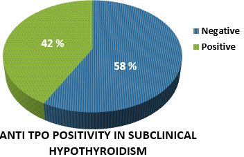
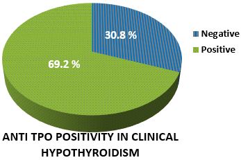
Results: 170 subjects with hypothyroidism were studied. Out of 50 subjects with subclinical hypothyroidism,64% were females. 70% of patients had a BMI of more than 25. Waist circumference was greater than 80 cm in 96% of patients. A family history of hypothyroidism was present in 16%. Anti-TPO antibody was positive in 42% of the subjects. Total cholesterol, LDL and triglyceride levels were higher in anti-TPO-positive subjects. Out of 120 subjects with clinical hypothyroidism,76.7% were females. 74.9% of patients had a BMI of more than 25. Waist circumference was more than 80 cms in 92% of patients. A family history of hypothyroidism was present in 24.2% of the subjects. Anti-TPO antibody was positive in 69.2% of the subjects. Total cholesterol, LDL and triglyceride levels were higher in anti-TPO-positive patients.

Conclusions: Prevalence of anti-TPO antibody was present in 42% of subclinical hypothyroidism patients. As anti-TPO positive patients progress to clinical hypothyroidism at a higher rate than negative patients, estimation of anti-TPO antibodies should be an integral part of an investigation of subclinical hypothyroidism. Prevalence of anti-TPO antibody was present in 69.2% of clinical hypothyroidism patients.

Keywords: Anti-thyroid peroxidase antibodies, Hypothyroidism, TSH

Corresponding Author How to Cite this Article To Browse
R Anil Kumar, Professor and HOD, , Karnataka Institute of Endocrinology and Research, Bengaluru, Karnataka, India.
Email:
Anil Kumar R, Lalitha R, Surekha B Shetty, Prevalence of Anti Thyroid Peroxidase Antibody Positivity in Subclinical and Clinical Hypothyroidism subjects attending a tertiary centre in South India. Int J Med Res Rev. 2024;12(4):109-110.
Available From
https://ijmrr.medresearch.in/index.php/ijmrr/article/view/1488

Manuscript Received Review Round 1 Review Round 2 Review Round 3 Accepted
2024-07-05 2024-07-11 2024-07-17 2024-07-23 2024-07-29
Conflict of Interest Funding Ethical Approval Plagiarism X-checker Note
Nil None yes 11.11

© 2024by Anil Kumar R, Lalitha R, Surekha B Shettyand Published by Siddharth Health Research and Social Welfare Society. This is an Open Access article licensed under a Creative Commons Attribution 4.0 International License https://creativecommons.org/licenses/by/4.0/ unported [CC BY 4.0].

Introduction

Autoimmune thyroid diseases (AITDs) are the most common human autoimmune diseases. They are organ-specific autoimmune diseases and include several inflammatory thyroid diseases with Graves’ disease (GD) and Hashimoto’s thyroiditis (HT) as the most frequent forms[1]. Autoimmune thyroid diseases are usually accompanied by the presence of anti-thyroid peroxidase (TPO), anti-thyroglobulin (Tg), and anti-thyroid-stimulating hormone receptor (TSHR) antibodies. Antibodies against thyroid antigens such as carbonic anhydrase 2, megalin, T3 and T4, sodium iodide symporter (NIS), and pendrin have also been detected, although rarely [2].

Thyroid autoimmunity could be initiated by genetic, environmental and endogenous factors. Exogenous and endogenous factors contribute to the risk of developing AITDs. Major exogenous causative factors are infections, intake of particular substances, and radiation, while endogenous factors are mainly gender and genetic disposition [3].

The most common antibodies frequently measured in serum in population surveys are the thyroid peroxidase antibody (TPOAb) andthyroglobulin antibody (TgAb). Thyroid Peroxidaseis a poorly glycosylated membrane-bound enzyme, responsible for iodine (I2) oxidation and iodination of tyrosyl residues of the Tg molecule [4].

It had been termed microsomal antigen based on its intracellular localization. Antibodies react against conformational epitopes at the surface of the molecules and against linear epitopes [5].

Polyclonal antibodies from healthy individuals and patients are directed against the same epitopes. Anti-TPO antibodies from healthy subjects don’t block TPO activity or interfere with the blocking activity of anti-TPO antibodies from AITD patients [6], while anti-TPO antibodies from AITD patients can fix complement, destroy thyrocytes, and act as competitive inhibitors of enzymatic activity [7].

Anti-thyroid peroxidase (anti-TPO) antibodies are more prevalent than anti-thyroglobulin (anti-Tg) antibodies and serve as stronger indicators of thyroid disease [8]. Anti-TPO antibodies are inductors of oxidative stress evidenced by decreased antioxidant potential, advanced glycosylation products and oxygen metabolites in the blood [9].

However, their contribution to thyroid damage compared to T cell and cytokine-mediated apoptosis is minor [10]. Notably, while anti-TPO antibodies may exhibit cytotoxic effects on thyrocytes in Hashimoto’s thyroiditis (HT), they do not play a well-established role in Graves’ disease (GD) [11].

Anti-TPO antibodies are present in approximately 90% ofHashimoto's thyroiditis, 75% of Graves' disease and 10–20% ofnodular goitreorthyroid carcinoma. Additionally, 10–15% of normal individuals can have high-level anti-TPO antibody titres[12], [13], [14].

Hypothyroidism may be either subclinical (SCH) or clinical. Subclinical hypothyroidism is characterized by a serum TSH above the upper reference limit in combination with a normal free thyroxine (T4). An elevated TSH, usually above 10 mIU/L, in combination with a low free T4characterizes clinical hypothyroidism.

The clinical importance of SCH is that it has a high rate of progression to clinical hypothyroidism, with an annual incidence of 2.6% in the absence of anti-TPO and 4.3% in their presence [15]. A 2022 Indian study showed that the rate of progression to clinical hypothyroidism was higher in the anti-TPO positive group compared to the anti-TPO negative group (P< 0.023; odds ratio [OR]: 4.58; 95% confidence interval [CI]: 1.14, 18.28) [16]. SCH patients with elevated anti-TPO have a higher conversion to clinical hypothyroidism than those without, and hence, it is recommended that anti-TPO measurement should be an integral part of the diagnosis of SCH. Evidence suggests that both serum TSH and anti-TPO analyses are essential in determining thyroid status, particularly for the diagnosis of patients suspected of SCH [17].

Indications for anti-TPO antibody measurement include the following:

1. To help confirm the diagnosis of Hashimoto's disease.
2. To help with the treatment decision in the patient with subclinical hypothyroidism.

In patients with autoimmune hypothyroidism (Hashimoto’sthyroiditis), anti-TPO antibodies usually persist in the body. Levelsmay reduce over time, but hardly ever normalize completely, even after medication has restored thyroid hormone levels to normal.


Studies have classified Anti-TPO antibody levels depending on their titers as 0 to 34 – normal, low >34 to 100, high >100 to 499 and very high >500 [18].

Based on these findings, it is recommended for TPOAb measurement to help confirm the diagnosis of Hashimoto's disease and to help with the treatment decision in the patient with subclinical hypothyroidism.With this background, we decided to conduct a study to know the prevalence of anti-TPO antibodies and their titers in subclinical and clinical hypothyroid subjects.

Materials and Methods

Subclinical hypothyroidism is characterized by a serum TSH above the upper reference limit in combination with a normal free thyroxine (T4). Clinical hypothyroidism is characterized by an elevated TSH, usually above 10 mIU/L, in combination with a subnormal free T4. If the antibody titres are more than 34 IU/ml such patients are considered as anti TPO antibody positive.

Inclusion Criteria:

1. Willing to participate in the study.
2. Patients in the age group of 18 to 60 years of age.
3. Patients with clinical and biochemical evidence of hypothyroidism.

Exclusion criteria:

1. Persons not willing to participate in the study.
2. Females in the gestational or postpartum period
3. Thyroid destruction (from radioactive iodine or surgery)
4. Medications causing thyroid dysfunction like amiodarone, lithium, anti-thyroid drugs

Statistical Analysis

Statistical Methods: Descriptive and inferential statistical analysis has been carried out in the present study. Results on continuous measurements are presented on Mean ± SD (Min-Max) and results on categorical measurements are presented in Number (%). Significance is assessed at a 5% level of significance. The following assumptions on data are made, Assumptions: 1 Dependent variables should be normally distributed, 2.Samples drawn from the population should be random, Cases of the samples should be independent.

Student t-test (two-tailed, independent) has been used to find the significance of study parameters on a continuous scale between two groups (Intergroup analysis) on metric parameters. Leven`s test for homogeneity of variance has been performed to assess the homogeneity of variance. A t-test is astatistical testthat is used to compare the means of two groups. It is often used inhypothesis testingto determine whether a process or treatment has an effect on the population of interest, or whether two groups are different from one another with the null hypothesis (H0) is that the true difference between these groups means is zero and the alternate hypothesis (Ha) is that the true difference is different from zero.

The Chi-square/ Fisher Exact test has been used to find the significance of study parameters on a categorical scale between two or more groups in a non-parametric setting for Qualitative data analysis. The Fisher Exact test is used when cell samples are very small.

Significant figures

+ Suggestive significance (P value: 0.05<P<0.10)

* Moderatesignificance (P value: 0.01<P £ 0.05)

** Strong significance (P value: P£0.01)

Statistical software: The Statistical software namely SPSS 22.0, and R environment ver.3.2.2 were used for the analysis of the data and Microsoft Word and Excel were used to generate graphs, tables etc.

Results

170 subjects with hypothyroidism were studied. 50 had subclinical hypothyroidism and 120 subjects had clinical hypothyroidism.

Out of 50 subjects with subclinical hypothyroidism, 64% were females. 70% of subjects had a BMI of more than 25. Waist circumference was more than 80 cms in 96% of the subjects. A family history of hypothyroidism was present in 16% of the subjects. Anti-TPO antibody was positive in 42% of the subjects. Total cholesterol, LDL and triglyceride levels were higher in anti-TPO-positive subjects. Anti-TPO antibody was positive in 21 subjects with subclinical hypothyroidism. 4 subjects had titers of 34 to 100, 13 had titers of 100 to 499 and 4 subjects had more than 500 titers.


Mean TSH was 6.99±1.76 in anti-TPO negative and 8.54±3.56 in TPO positive subjects and mean FT4 was 16.47±2.81 in anti-TPO negative and 15.38±2.54 in anti-TPO positive subjects. (Tables 1,3,5,7,9)

Out of 120 subjects with clinical hypothyroidism, 76.7% were females. 74.9% of subjects had a BMI of more than 25. Waist circumference was more than 80 cms in 92% of subjects. A family history of hypothyroidism was present in 24.2%. Anti-TPO antibody was positive in 69.2%. Total cholesterol, LDL and triglyceride levels were higher in anti-TPO-positive subjects. Anti-TPO antibody was positive in 83 subjects of clinical hypothyroidism. 11subjects had titers of 34 to 100, 56 had titers of 100 to 499 and 16 subjects had more than 500 titers. Mean TSH was 10.93±22.44 in anti-TPO negative and 17.78±25.09 in TPO positivesubjects. Mean FT4 was 16.43±3.86 in anti-TPO negative and 15.32±5.14 in anti-TPO positive subjects. (Table 2,4,6,8,10)

Table 1: Age in Years-frequency distribution of Subclinical hypothyroidism subjects studied

Age in YearsNo. of Patients%
21-30510.0
31-40714.0
41-501428.0
>502448.0
Total50100.0

    Mean ± SD: 47.94±12.04

Table 2: Age in Years-frequency distribution of Clinical hypothyroidism subjects studied

Age in YearsNo. of Patients%
<2032.5
20-301411.7
31-402319.2
41-503025.0
>505041.7
Total120100.0

    Mean ± SD: 46.65±14.07

Table 3: Body Mass Index (kg/m2)-frequency distribution of Subclinical hypothyroidism subjects studied

Body Mass Index (kg/m2)No. of Patients%
<18.500.0
18.5-24.91530.0
25.0-29.92346.0
>30.01224.0
Total50100.0

Table 4: Body Mass Index (kg/m2) - frequency distribution of Clinical hypothyroidism subjects studied

Body Mass Index (kg/m2)No. of Patients%
<18.543.3
18.5-24.92520.8
25.0-29.95041.7
>30.04134.2
Total120100.0

Table 5: WCR-frequency distribution of Subclinical hypothyroidism patients studied

WCRNo. of Patients%
<8024.0
80-1003978.0
>100918.0
Total50100.0

Table 6: WCR- frequency distribution of Clinical hypothyroidism subjects studied

WCRNo. of Patients%
<8097.5
80-1007865.0
>1003327.5
Total120100.0

Table 7: Comparison of clinical variables to anti-TPO positivity of Subclinical hypothyroidism subjects studied. 

VariablesANTI TPOTotalP Value
NEGATIVEPOSITIVE
AGE IN YEARS49.79±11.8845.38±12.0947.94±12.050.204
BODY MASS INDEX(KG/M2)28.04±7.0327.8±5.0327.94±6.210.895
WCR94.28±9.7894.06±10.8794.19±10.140.940
SBP138.97±21.99131.33±16.73135.76±20.130.189
DBP83.24±13.5678.43±11.1481.22±12.710.189

Table 8: Comparison of clinical variables to anti-TPO positivity of clinical hypothyroidism subjects studied.   

VariablesANTI TPOTotalP Value
NEGATIVEPOSITIVE
AGE IN YEARS52.92±12.7643.86±13.846.65±14.08<0.001**
BODY MASS INDEX(KG/M2)28.92±4.7928.15±8.728.39±7.690.613
WCR95.95±10.9893.47±12.1494.23±11.80.291
SBP131.62±18.66131.24±19.14131.36±18.910.919
DBP75.7±10.6778.84±11.3977.88±11.220.158

Table 9: Comparison of lipid parameters between anti TPO positive and negative Subclinical hypothyroidism subjects studied

VariablesANTI TPOTotalP Value
NEGATIVEPOSITIVE
TC180.93±49.96191.48±44.77185.36±47.670.446
HDL42.18±11.1537.49±8.6940.21±10.360.115
LDL113.78±43.66130.33±35.8120.73±40.990.161
TG169.72±76.37233.76±34.87196.62±21.710.296

Table 10: Comparison of lipid parameters between anti TPO positive and negative hypothyroidism subjects studied

VariablesANTI TPOTotalP Value
NEGATIVEPOSITIVE
TC168.92±34.15178.59±44.8175.61±41.90.245
HDL41.34±14.0740.27±9.5940.6±11.110.630
LDL106.92±32.28119.79±38.38115.82±36.960.078+
TG139.81±48.88157.17±88.03151.82±78.270.264

ijmrr_1488_01.JPG
ijmrr_1488_02.JPG

Discussion

Several studies have looked at the prevalence of thyroid antibodies in clinical and subclinical hypothyroidism.The prevalence of anti-TPO antibodies noted in a study by Jayashankar C.A, et al from Bangalore in patients with clinical and subclinical hypothyroidism was 80% and 50% respectively.

This study finding validates that the elevated anti-TPO levels could be correlated with autoimmune thyroid dysfunction. This study was a small study with 50 subjects compared to 170 subjects in our study. Female preponderance was seen in both studies [19].

Another study by Jeena et al(2013) has concluded the usefulness of anti-TPO antibody estimation in establishing the etiological diagnosis of autoimmune thyroid diseases. The study found that out of 47 hypothyroid subjects evaluated, 28 (60%) had an elevated TPO antibody titer [20]. Another Indian study by Mohanty et al has reported that among the 38 frank hypothyroid subjects, 76% had raised anti-TPO levels [17].

J.Dutta et al studied 74 cases where 30 cases had clinical hypothyroidism and 44 cases had subclinical hypothyroidism. 70.3% of cases of clinical hypothyroidism were anti-TPO positive and subclinical hypothyroidism had 51.5% anti-TPO positive. These results are almost similar to our study results [21].

Bjoroet alin a 20-year follow-up study, conducted among Norwegian inhabitants (94,009), have found that the positive anti-TPO levels correlated significantly with thyroid dysfunction and the prevalence of elevated TSH was nearly 10-fold higher in both females and males with positive anti-TPO when compared to anti-TPO-negative subjects [22].

The study by Silva et al, conducted on 89 Brazilian women, has noted elevated anti-TPO levels in around 90% of the patients with autoimmune thyroiditis [23].

Literature studies suggest a constant increase in the prevalence of anti-TPO-positive subjects in India. In a study conducted from 2007 through 2010 in Delhi, the percentage of TPO antibody-positive adults was found to be 13.3 % (24)

Similarly, an apparent rise in prevalence was noted in the southern part of India. The corresponding prevalence of anti-TPO positivity noted in two different studies conducted in Kerala and Chennai were 16.7% and 25.81 %.(25,26).

Conclusions

Prevalence of anti-TPO antibody was 42% in subclinical hypothyroidism subjects.


Since anti-TPO positive subjects progressing to clinical hypothyroidism is higher than negative subjects, estimation of anti-TPO antibodies should be an integral part of the investigation of subclinical hypothyroidism.

Prevalence of anti-TPO antibody was present in 69.2% of clinical hypothyroidism subjects. Anti-TPO antibody positivity indicates the autoimmune aetiology of hypothyroidism and helps in follow-up management of hypothyroidism. Anti-TPO antibody titers did not determine the severity of clinical hypothyroidism.

Abbreviations Used:

1. AITD- Autoimmune Thyroid Diseases.
2. TPO- Thyroid Peroxidase Antibody.
3. SCH- Subclinical Hypothyroidism.
4. HT-Hashimotos Thyroiditis.
5. GD- Graves Disease.
6. BMI- Body Mass Index.
7. SBP- Systolic Blood Pressure.
8. DBP- Diastolic Blood Pressure.
9. WCR- Waist Circumference.
10. TSH- Thyroid Stimulating Hormone.
11. TC - Total Cholesterol.
12. TG-Triglycerides.
13. LDL-Low-Density Lipoprotein.
14. HDL-High-Density Lipoprotein.

What is new in this study? Total cholesterol, LDL and triglyceride levels were higher in both anti-TPO positive subclinical and clinical hypothyroidism. Anti-TPO antibody titers did not determine the severity of clinical hypothyroidism.

Acknowledgement: Niharika S, and Swathi Gupta for the creation of Tables, Listing and Graphs(TLG) and Statistical Analysis Reporting(SAR) and Dr K.P.Suresh, PhD (Biostatistics) for reviewing methodology and results.

No potential conflicts of interest relevant to the article are reported.

Funding: Nil.

Permission from IRB:Yes.

References

1. McLeod DS, Cooper DS. The incidence and prevalence of thyroid autoimmunity. Endocrine. 2012 Oct; 42:252-65. [Crossref][PubMed][Google Scholar]

2. Marcocci C, Marino M. Thyroid directed antibodies. InWerner and Ingbar's the thyroid: a fundamental and clinical text 2005 (Vol. 9, pp. 360-372). Lippincott Williams &Wilkins [Crossref][PubMed][Google Scholar]

3. Fröhlich E, Wahl R. Thyroid autoimmunity: role of anti-thyroid antibodies in thyroid and extra-thyroidal diseases. Frontiers in immunology. 2017 May 9; 8:521. [Crossref][PubMed][Google Scholar]

4. McLachlan SM, Rapoport B. The molecular biology of thyroid peroxidase: cloning, expression and role as autoantigen in autoimmune thyroid disease. Endocrine reviews. 1992 May 1; 13(2):192-206. [Crossref][PubMed][Google Scholar]

5. Le Fourn V, Ferrand M, Franc JL. Endoproteolytic cleavage of human thyroperoxidase: role of the propeptide in the protein folding process. Journal of Biological Chemistry. 2005 Feb 11; 280(6):4568-77. [Crossref][PubMed][Google Scholar]

6. Kohno Y, Yamaguchi F, Saito K, Niimi H, Nishikawa T, Hosoya T. Anti‐thyroid peroxidase antibodies in sera from healthy subjects and from patients with chronic thyroiditis: differences in the ability to inhibit thyroid peroxidase activities. Clinical & Experimental Immunology. 1991 Sep; 85(3):459-63. [Crossref][PubMed][Google Scholar]

7. Kaczur V, Vereb G, Molnar I, Krajczar G, Kiss E, Farid NR, Balazs C. Effect of anti-thyroid peroxidase (TPO) antibodies on TPO activity measured by chemiluminescence assay. Clinical chemistry. 1997 Aug 1; 43(8):1392-6. [Crossref][PubMed][Google Scholar]

8. Balucan FS, Morshed SA, Davies TF. Thyroid autoantibodies in pregnancy: their role, regulation and clinical relevance. Journal of thyroid research. 2013; 2013(1):182472. [Crossref][PubMed][Google Scholar]

9. Ruggeri RM, Vicchio TM, Cristani M, Certo R, Caccamo D, Alibrandi A, Giovinazzo S, Saija A, Campennì A, Trimarchi F, Gangemi S. Oxidative stress and advanced glycation end products in Hashimoto's thyroiditis. Thyroid. 2016 Apr 1; 26(4):504-11. [Crossref][PubMed][Google Scholar]

10. Zaletel K, Gaberscek S. Hashimoto's thyroiditis: from genes to the disease. Current genomics. 2011 Dec 1; 12(8):576-88. [Crossref][PubMed][Google Scholar]


11. DeGroot LJ. Graves. disease and. the manifestations of thyrotoxicosis. [Crossref][PubMed][Google Scholar]

12. Werner SC, Ingbar SH, Braverman LE, Utiger RD, editors. Werner & Ingbar's the thyroid: a fundamental and clinical text. Lippincott Williams & Wilkins; 2005. . [Crossref][PubMed][Google Scholar]

13. Saravanan P, Dayan CM. Thyroid autoantibodies. Endocrinology and metabolism clinics of North America. 2001 Jun 1; 30(2):315-37. [Crossref][PubMed][Google Scholar]

14. Chardès T, Chapal N, Bresson D, Bès C, Giudicelli V, Lefranc MP, Péraldi-Roux S. The human anti-thyroid peroxidase autoantibody repertoire in Graves' and Hashimoto's autoimmune thyroid diseases. Immunogenetics. 2002 Jun; 54:141-57. [Crossref][PubMed][Google Scholar]

15. Fatourechi V. Subclinical hypothyroidism: an update for primary care physicians. InMayo Clinic Proceedings 2009 Jan 1 (Vol. 84, No. 1, pp. 65-71). Elsevier [Crossref][PubMed][Google Scholar]

16. Singh SK, Singh R, Singh SK, Iquebal MA, Jaiswal S, Rai PK. Risk of progression to overt hypothyroidism in Indian patients with subclinical hypothyroidism: A prospective observational study. Int. J. Adv. Med. 2022 Dec; 9:1183 [Crossref][PubMed][Google Scholar]

17. Mohanty S, Amruthlal W, Reddy GC, Kusumanjali G, Kanagasabapathy AS, Rao P. Diagnostic strategies for subclinical hypothyroidism. Indian journal of clinical biochemistry. 2008 Jul; 23:279-82. [Crossref][PubMed][Google Scholar]

18. Ehlers M, Jordan AL, Feldkamp J, Fritzen R, Quadbeck B, Haase M, Allelein S, Schmid C, Schott M. Anti-thyroperoxidase antibody levels> 500 IU/ml indicate a moderately increased risk for developing hypothyroidism in autoimmune thyroiditis. Hormone and Metabolic Research. 2016 Sep; 48(10):623-9. [Crossref][PubMed][Google Scholar]

19. Jayashankar CA, Avinash S, Shashidharan B, Sarathi V, Shruthi KR, Nikethan D, Harshavardhan J. The prevalence of anti-thyroid peroxidase antibodies in subclinical and clinical hypothyroid patients. Int J Res Med Sci. 2015 Jan 1; 3(12):3564-6. [Crossref][PubMed][Google Scholar]

20. Jeena EJ, Malathi M, Sudeep K. A hospital-based study of anti-TPO titer in patients with thyroid disease. Muller Journal of Medical Sciences and Research. 2013 Jul 1;4(2):74-7. [Crossref][PubMed][Google Scholar]

21. Dutta J, Jain S, Jain A, Jain S, Jain S. An association of anti-thyroid peroxidase antibodies. in clinical and subclinical hypothyroidism. . [Crossref][PubMed][Google Scholar]

22. Bjoro T, Holmen J, Kruger O, Midthjell K, Hunstad K, Schreiner T, Sandnes L, Brochmann H. Prevalence of thyroid disease, thyroid dysfunction and thyroid peroxidase antibodies in a large, unselected population. The Health Study of Nord-Trondelag (HUNT). European journal of endocrinology. 2000 Nov; 143(5):639-47 [Crossref][PubMed][Google Scholar]

23. Silva LM, Chavez J, Canalli MH, Zanetti CR. Determination of IgG subclasses and avidity of antithyroid peroxidase antibodies in patients with subclinical hypothyroidism–a comparison with patients with overt hypothyroidism. Hormone research. 2003 Jul 1; 59(3):118-24. [Crossref][PubMed][Google Scholar]

24. Unnikrishnan AG, Kalra S, Sahay RK, Bantwal G, John M, Tewari N. Prevalence of hypothyroidism in adults: An epidemiological study in eight cities of India. Indian journal of endocrinology and metabolism. 2013 Jul 11`; 17(4):647-52. [Crossref][PubMed][Google Scholar]

25. Marwaha RK, Tandon N, Ganie MA, Kanwar R, Sastry A, Garg MK, Bhadra K, Singh S. Status of thyroid function in Indian adults: two decades after universal salt iodization. J Assoc Physicians India. 2012 Apr 1; 60:32-6. [Crossref][PubMed][Google Scholar]

26. Menon U, Sundaram KR, Unnikrishnan AG, Jayakumar RV, Nair V, Kumar H. High prevalence of undetected thyroid disorders in an iodine sufficient adult south Indian population. Journal of the Indian Medical Association. 2009 Feb 1; 107(2):72-7. [Crossref][PubMed][Google Scholar]