E-ISSN:2320-8686
P-ISSN:2321-127X

Research Article

Postmenopausal Coronary

International Journal of Medical Research and Review

2024 Volume 12 Number 4 Jul-Aug
Publisherwww.medresearch.in

Differences Between Premenopausal And Postmenopausal Coronary Artery Disease (CAD) Risk Factors and Clinical Profile

MS H1*, Bhuiyan M2, Sowdagar M3, Siddique N4, Paul G5, Paul P6, Shakil S7, Khan M8, Sohan A9, Alam M10, Hoque M11
DOI:https://doi.org/10.17511/ijmrr .2024.i04.04

1* Hossain MS, Assistant Professor, Department Of Cardiology, Mymensingh Medical College Mymensingh Bangladesh, Bangladesh.

2 MAS Bhuiyan, Assistant Professor, Department Of Cardiology, Mymensingh Medical College Mymensingh, Bangladesh.

3 MNH Sowdagar, Associate Professor and Head, Department Of Cardiology, Border Guard Hospital Dhaka, Bangladesh.

4 NA Siddique, Assistant Professor, Department of Cardiology, Mymensingh Medical College Mymensingh, Bangladesh.

5 GK Paul, Associate Professor and Head, Department Of Cardiology, Mymensingh Medical College Mymensingh, Bangladesh.

6 PK Paul, Consultant, Department Of Cardiology, Mymensingh Medical College Hospital Mymensingh, Bangladesh.

7 SS Shakil, Senior Consultant, Department of Cardiology, Mymensingh Medical College Hospital Mymensingh, Bangladesh.

8 MTI Khan, Registrar, Department Of Cardiology, Mymensingh Medical College Hospital Mymensingh, Bangladesh.

9 AM Sohan, Associate Professor, Department Of Cardiology, Ahsania Medical College Uttara Dhaka, Bangladesh.

10 MM Alam, Associate Professor, Department of Cardiology, Dhaka National Medical College Dhaka, Bangladesh.

11 MS Hoque, Consultant, Department Of Cardiology, BSMMU Dhaka, Bangladesh.

Introduction: Coronary artery disease is the leading cause of mortality and morbidity of both men and women accounting for over one-third of total deaths. It has reached epidemic proportions among Indians. It accounts for 1 out of 3 womens deaths regardless of race or ethnicity. In women, the annual mortality rate from CAD is high.

Methods: A prospective observation study was conducted at the cardiology department, NICVD, Dhaka, Bangladesh from January to December 2019. A total of 200 female CAD patients were drawn from the cardiology department. A total of 50 premenopausal cases and 150 postmenopausal cases were enrolled according to their status at the time of diagnosis. All patients underwent percutaneous coronary intervention and were given conventional drug therapy for coronary artery disease during follow-up.

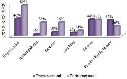
Results: Differences were compared between the 2 groups. Fewer premenopausal women suffered from hypertension (46% versus 82%, P< 0.001), type 2 diabetes (10% versus 36%, P = 0.001), and hyperlipidemia (4% versus 36%, P<0.001), but more had a positive family history of premature CAD (42% versus 26%, P = 0.03). The cumulative recurrence rate was 1.76 times higher in postmenopausal patients than in premenopausal patients. Clinical diagnosis (HR = 2.54, 95%CI: 1.21-4.85, P = 0.02) and type 2 diabetes (HR = 4.10, 95%CI: 2.37-8.83, P = 0.004) were two factors that influenced recurrence in premenopausal subjects, while the clinical diagnosis (HR = 1.93, 95%CI: 1.59-3.46, P = 0.03) and Gensini score (HR =1.20, 95% CI: 1.11-1.45, P = 0.02) were influencing factors in the postmenopausal patients.

Conclusion: Our study clarified the differences between postmenopausal and premenopausal women concerning risk factors, clinical symptoms, cardiovascular features, and recurrence rate, and provided a reference for further study on the mechanism and prognosis in postmenopausal or premenopausal CAD patients.

Keywords: Premenopausal, Postmenopausal, Coronary Artery Disease (CAD), Risk Factors

Corresponding Author How to Cite this Article To Browse
Hossain MS, Assistant Professor, Department Of Cardiology, Mymensingh Medical College Mymensingh Bangladesh, , , Bangladesh.
Email:
MS H, Bhuiyan M, Sowdagar M, Siddique N, Paul G, Paul P, Shakil S, Khan M, Sohan A, Alam M, Hoque M, Differences Between Premenopausal And Postmenopausal Coronary Artery Disease (CAD) Risk Factors and Clinical Profile. Int J Med Res Rev. 2024;12(4):116-117.
Available From
https://ijmrr.medresearch.in/index.php/ijmrr/article/view/1477

Manuscript Received Review Round 1 Review Round 2 Review Round 3 Accepted
2024-05-01 2024-05-07 2024-05-13 2024-05-19 2024-05-25
Conflict of Interest Funding Ethical Approval Plagiarism X-checker Note
Nil None yes 11.11

© 2024by MS H, Bhuiyan M, Sowdagar M, Siddique N, Paul G, Paul P, Shakil S, Khan M, Sohan A, Alam M, Hoque Mand Published by Siddharth Health Research and Social Welfare Society. This is an Open Access article licensed under a Creative Commons Attribution 4.0 International License https://creativecommons.org/licenses/by/4.0/ unported [CC BY 4.0].

Introduction

Coronary artery disease is the leading cause of mortality and morbidity in both men and women accounting for over one-third of total deaths [1]. It has reached epidemic proportions in Bangladesh. It accounts for 1 out of 3 women's deaths regardless of race or ethnicity [2]. In women, the annual mortality rate from CAD is high. The worldwide INTERHEART Study, [3]a large cohort study of more than 52000 individuals with myocardial infarction, have revealed that women have their first presentation of coronary heart disease approximately 10 years later than men, most commonly after menopause. Despite this delay in onset, mortality is increasing more rapidly among women than men [3]. In Bangladesh, it is the fourth most common cause of death and accounts for 10.68% of total deaths, of which males 12.47% and females 8.19% [4]. Epidemiological studies from various parts of Bangladesh have reported the rising trends and a high burden in the levels of conventional risk factors such as diabetes, hypertension and metabolic syndrome [5],[6]. Postmenopausal and premenopausal women differ in the symptoms, risk factors, disease characteristics, prognosis, and recurrence of CAD. However, due to the low incidence of CAD in premenopausal women, research on this particular population has not been sufficient. Symptomatic obstructive coronary artery disease is less common in premenopausal women than in men of comparable age and the clinical diagnosis is often difficult to establish [7]. Many risk factors, first documented in males, were lately found to affect females similarly. Women younger than 55 years have a worse prognosis after acute myocardial infarction than their male counterparts, with a greater incidence of recurring events and higher mortality [8]. Currently, the Indian subcontinent is experiencing a rapid increase in the rates of CAD [9],[10]. Modern women have professional and housewife responsibilities, consume excess fat and carbohydrates, smoke, do not exercise regularly and do not have enough time to rest. This situation leads to overweight, dyslipidemia, arterial hypertension, impaired glucose tolerance and diabetes mellitus. As a result, an increasing number of young women are now suffering from coronary artery disease, not only in Western and industrialized countries but also in Asian countries.

Women do not often participate in preventive studies and undergo less intensive and invasive evaluation and treatment of chest pain when compared to men. However, the rate of coronary death is twice higher in women than in men after acute coronary syndromes and revascularization procedures. Symptoms among younger women were atypical, but the onset of the disease was faster and more urgent.

Materials and Methods

A prospective observation study was conducted at the cardiology department, NICVD, Dhaka, Bangladesh from January to December 2019. A total of 200 female CAD patients were drawn from the cardiology department. The subjects were enrolled following the criterion of diameter stenosis ≥50% in left mean and ≥70% in LAD, LCX and ECA in at least one invasive coronary angiogram. Patients with pulmonary embolism, aortic aneurysm, congenital heart disease, rheumatic heart disease, myocarditis, or cardiomyopathy were excluded. A total of 50 premenopausal cases and 150 postmenopausal cases were enrolled according to their status at the time of diagnosis. All patients underwent percutaneous coronary intervention and were given conventional drug therapy for coronary artery disease during follow-up. Routine drug therapy included antiplatelet agents (aspirin), lipid-lowering agents, beta-blockers, and angiotensin-converting enzyme inhibitors/angiotensin II receptor antagonists according to the patient’s existing conditions. No patients were treated with hormone replacement therapy. This study was approved by the ethical committee of the institution.

Clinical data collection: The following clinical data were collected: 1) demographic data and traditional risk factors, such as age, hypertension, hyperlipidemia, type 2 diabetes, smoking, obesity, and a positive family history of premature CAD; 2) clinical classification of disease, such as stable angina and acute coronary syndrome (ACS) including unstable angina, ST-elevation myocardial infarction, and non-ST-elevation myocardial infarction; 3) clinical measurements including body mass index (BMI), blood pressure, blood biochemical variables, 4) the severity of CAD was defined using the angio- graphic Gensini scores according to coronary angiography with Judkins catheters.


Coronary angiography test results included lesion location, the number of vascular stenosis, and the severity of stenosis.

Clinical follow-up: All patients were contacted by telephone every 6 months until June 2020, and their medical records were followed regularly. The endpoint was the combined recurrence of major adverse cardiovascular events, including death, targeted vascular revascularization, heart failure, ACS, and transient ischemic attack (TIA)/stroke. Death and targeted vascular revascularization with percutaneous coronary intervention or coronary bypass surgery were confirmed by a review of the medical records. Heart failure was defined as dyspnea and/or edema that was accompanied by pulmonary congestion on a chest roentgenogram and left ventricular dysfunction on an echocardiogram. ACS was defined as a rise in cardiac troponin I with ischemic symptoms and/or characteristic electrocardiographic changes. TIA/stroke was defined as the presence of a new neurological deficit verified by either magnetic resonance imaging or computed tomography.

Data analysis: Statistical analyses were performed using SPSS version 17.0. Discrete variables were compared with the chi-square test or Fisher Exact test.

Results

Comparison of CAD traditional risk factors: The mean age of the 50 premenopausal cases was 46.70 ± 4.40 years, while the mean age of the 150 postmenopausal subjects was 67.62 ± 9.86 years. The differences in traditional risk factors between the two groups are summarized in Figure-1.

ijmrr_1477_01.JPG
Figure 1:
Comparison of risk factors between premenopausal and post-menopausal groups

Compared with the postmenopausal group, the prevalences of hypertension (46% versus 82%, χ2 = 19.48, P < 0.001) and hyperlipidemia (4% versus 36%, χ2 = 18.86, P < 0.001) were significantly lower. Furthermore, fewer premenopausal patients suffered from type 2 diabetes (10% versus 36%, χ2 = 11.48, P = 0.001).

Yet, there were more premenopausal patients with a positive family history of premature CAD (42% versus 26%, χ2 = 4.77, P = 0.03). There were no significant differences in smoking and obesity.

Symptom presentation and clinical diagnosis: Eight cases (16%) had typical angina before the onset in premenopausal CAD women, while the percentage was much higher in postmenopausal patients (82%) (χ2 = 90.35, P< 0.001).

Based on the clinical manifestation, CAD was divided into stable angina and acute coronary syndrome, the latter including unstable angina (UA), acute ST-segment elevation myocardial infarction (STEMI), and acute non-ST-segment elevation myocardial infarction (Non-STEMI). Table-1 shows the main onset of disease in premenopausal women was ACS, which was significantly more frequent than in the post-menopausal group (80% versus 20%, χ2 = 19.87, P < 0.001).

Comparison of clinical parameters: Comparison of the clinical and biochemical markers between the premenopausal and postmenopausal women revealed that the systolic pressure (140.48 ± 20.54 mmHg versus 126.73 ± 18.25 mmHg, t =4.51, P < 0.001) and diastolic pressure (78.96 ± 9.72 mmHg versus 72.28± 11.35 mmHg, t = 4.33 mmHg, P < 0.001), fasting blood glucose (6.78 ± 2.34 mmol/L versus 6.01 ± 2.48 mmol/L, t = 2.13, P = 0.003), total cholesterol (5.43 ± 1.24 mmol/L versus 4.97 ± 1.92 mmol/L, t = 2.11, P= 0.04) and low-density lipoprotein cholesterol (3.27 ± 1.08 mmol/L versus 2.90 ± 1.25 mmol/L, t = 2.16, P = 0.03) of postmenopausal patients were higher than those of the premenopausal women.

There were no differences in other blood lipids markers, BMI, blood coagulation markers, or hsCRP.

Disease recurrence and influence factors: Four patients dropped out of the study because they had changed their phone numbers or addresses for communication and did not return after discharge from the hospital.


A total of 96 cases of recurrence of major adverse cardiovascular events occurred: 6 cases of death, 66 cases of acute coronary syndrome, 4 cases of TIA/ stroke, 10 cases of targeted vessel revascularization and 18 cases of heart failure.

There were 15 patients in the premenopausal group and 81 in the postmenopausal group (20% versus 50%, χ2 = 11.52, P=0.001).

The cumulative recurrence rate was 1.76 times higher in postmenopausal patients than in premenopausal patients (15.94 per person-month versus 9.07 per person-month).

Table 1: Comparison of clinical diagnosis between premenopausal and postmenopausal groups (n=200)

Acute coronary syndrome (n %)Stable angina(n %)X2p-value
Premenopausal group40 (80.00%)10(20.00%)19.87<0.001**
Postmenopausal group71 (47.33%)79 (52.77%)

Table 2: Comparison of clinical measurements and biochemical variables (n=200)

Premenopausal group (n=50)Postmenopausal group (150)p-value
BMI (kg/m)226.92±5.1525.87±3.640.09
Blood pressure (mmHg)
Systolic pressure126.73±18.25140.48±20.54<0.001**
Diastolic pressure72.28±11.3578.96±9.72<0.001**
Blood lipids
TC (mmol/L)4.97±1.925.43±1.240.04*
TG (mmol/L)1.96±1.502.05±1.200.290.64
HDL-C (mmol/L)1.28±0.411.27±1.080.84
LDL-C (mmol/L)2.90±1.253.27±0.260.03*
apoAl (g/L)1.48±1.0061.35±0.260.13
Apo (g/L)0.88±0.241.00±0.620.16
LPA (mg/L)210.10±153.15231.42±188.550.44
FBG (mmol/L)6.01±2.486.78±2.340.03*
haCRP (mg/L)3.46±4.113.47±3.290.20
Blood coagulation
PT12.93±2.9513.43±1.940.14
APTT29.28±10.7526.84±17.150.31
INR1.02±0.230.98±0.170.15

Table 3: Coronary angiographic features of premenopausal and postmenopausal and postmenopausal cad women (n=200)

Lesion locationNumber of stenotic vesselsGensini score
LMLADLCXRCA
Premenopausal group0(0.00%)35(70%)8(16%)7(14%)1.28±0.5012.48±7.23
Premenopausal group2(1.33%)61(40.6%)45(30%)42(28%)2.43±0.8946.49±26.97
P<0.001**<0.001**<0.001**

Characteristics of coronary lesions: The number of stenotic vessels was significantly less in the premenopausal group compared with postmenopausal cases (1.28± 0.50 versus 2.43± 0.89, p<0.001). In the premenopausal group, the left anterior descending branch was the stenosis vessel most often involved (70%), followed by the left circumflex (16%) and right coronary artery (14%). However, there was more left circumflex stenosis (30%) and right coronary artery (28%) stenosis in the postmenopausal group. The location of the vessel stenosis was significantly different between the two groups (Fisher’s exact test, P < 0.001).

Gensini’s degree integral was applied to identify the severity of coronary narrowing. The Gensini scores of premenopausal patients were significantly lower than those of postmenopausal patients (10.48 ± 7.23 versus 56.49 ± 26.97, P < 0.001).

Table 4: Multivariate Cox regression of risk factors of recurrence (n=200)

Premenopausal patientsPostmenopausal patients
HR95.0%CIPHR95.0%CIP
Age1.020.87-1.210.801.040.98-1.030.74
Clinical diagnosis2.541.21-4.850.02*1.931.59-3.460.03*
Hypertension1.660.97-6.970.071.280.60-5.680.06
Hypertension2.190.09-56.420.641.390.89-2.170.15
Diabetes mellitus4.102.37-51.240.004*1.030.66-1.620.89
Smoking5.990.70-51.240.101.180.65-2.130.57
Obesity1.380.88-2.150.161.240.69-2.230.47
Number of stenosis vessels0.840.15-4.690.840.870.62-1.210.401
Gensini scire1.010.92-1.110.881.201.11-1.450.02*

Cox regression analysis was conducted to investigate the factors influencing recurrence (Table 4).


Age, clinical diagnosis, risk factors of CAD, number of stenotic vessels, and Gensini scores were independent variables. Clinical diagnosis (HR = 2.54, 95%CI: 1.21-4.85, P = 0.02) and type 2 diabetes (Hazard ratio (HR) = 4.10, 95%CI: 2.37-8.83, P = 0.004) were two factors that influenced recurrence in premenopausal sub-jects, while clinical diagnosis (HR = 1.93, 95%CI: 1.59-3.46, P = 0.03) and Gensini score (HR = 1.20, 95%CI: 1.11-1.45, P= 0.02) were factors in postmenopausal patients.

ijmrr_1477_02.JPG
Figure 2:
Kaplan-Meier analysis of non-recurrence in the 2 study groups indicates the cumulative rate of non-recurrence at different followups.

Discussion

CAD in women continues to be a major public health problem that represents a leading cause of death and disability. Women have varied presentations of coronary artery disease. In premenopausal women undergoing coronary angiography for suspected myocardial ischemia, disruption of the ovulatory cycle characterized by hypoestrogenemia seems to be associated with CAD. Recognition of prodromal symptoms was reported to be critical for preemptive coronary heart disease screening and effective diagnosis and treatment. It was important to recognize the symptoms which are associated with acute subsequent cardiac events. However, symptoms among younger women were atypical, and patients with silent myocardial ischemia usually had more extensive and severe disease. A previous study also indicated that young women were more likely to have CAD without chest pain [11]. Another study in a Canadian cohort of patients whose age was under 55 years with acute coronary syndrome supported this pattern [12].

Our study showed that 48% of postmenopausal cases were identified as ACS, compared to 80% of premenopausal patients identified as ACS. Premenopausal women had a rapid onset and no typical angina, but myocardial infarction typically occurred. Autopsy research has demonstrated that coronary artery lesions in young women contain less calcium and dense fibrous tissue than those of men and older women. Coronary atherosclerotic plaque of young patients was comprised mainly of fatty deposits, which were extremely easy to rupture and caused acute coronary thrombosis that led to acute cardiovascular disease.

Coronary angiography of the premenopausal patients showed the coronary artery stenosis was limited to a single vessel, and the left anterior descending artery was the most frequently affected vessel, while the involvement of the left circumflex and right coronary artery was significantly less. It reported that left anterior descending branch stenosis and Q-wave myocardial infarction were common in young women. Another recent study found that left anterior descending branch stenosis was more common in young people; 63.9% of patients in the ≤ 35 years old age group, 41.7% in the 35 to 55 age group, and 33.7% in the older than 55 age group exhibited atherosclerosis in the left anterior descending artery (p<0.01) [13].

At present, there is no authoritative statement about why LAD disease occurs more often in the premenopausal group. Taking the anatomical structure of the left anterior descending artery into account, it was most likely because pre-menopausal women are more active, and the left ventricle needs to consume more oxygen and nutrients. The left anterior descending artery is more easily involved as it is the main blood supply artery of the left ventricle, and it supports large areas of the ventricle. In addition, the Gensini scores of premenopausal patients were much lower, which meant the lesions were confined mostly to single or double blood vessels. A previous study reported age-related differences in percent luminal stenosis at the site of thrombus and the extent of coronary disease [14].

There were relatively few narrowed segments of coronary arteries in younger women compared with women over 50 years of age. This was probably because young women were more likely to have inflammation, coronary spasm, plaque erosion, or rupture.


The clinical manifestations of postmenopausal patients were more complex, and collateral circulation was easily formed as they had a longer disease duration and long-term progress of the disease course. Our study results suggest that angiography in premenopausal women might underestimate the severity of the disease in younger women.

These findings emphasize that premenopausal women and postmenopausal women who experience CAD may represent a heterogeneous group. Cardiovascular risk factors were highly valuable predictors of the presence and severity of CAD. The prevalence of hypertension, hyperlipidemia, and type 2 diabetes was significantly lower in the premenopausal group. In menopause, women have significant effects on lipid metabolism, which is mainly manifested by an increase in LDL-C and a decrease in HDL-C. However, there was no difference in other blood lipid markers, except for total cholesterol and LDL. This suggested that abnormal metabolism occurred early on in premature menopause CAD women.

In other words, it appeared to lessen the cardio-protective effect associated with age. Smoking was reported as the most prevalent risk factor which caused endothelial dysfunction, spontaneous platelet aggregation, coronary spasm, and adverse hemostatic effects. However, due to the low smoking rate among Chinese women [15], smoking was a less significant risk factor in this study.

Among all the 6 main risk factors, only the proportion of the family history of the premenopausal women was significantly higher, which suggests that females with a positive family history of premature CAD should be more concerned with the occurrence of CAD before menopause. The cumulative recurrence rate was higher in postmenopausal patients, which was partly attributable to their older age and partly because their coronary lesions were much more complex, even though the onset of the disease was faster and more urgent in premenopausal patients.

The premenopausal group had a lower recurrence rate within a relatively short period. In our study, the influencing factors for each group were not identical. However, the initial clinical diagnosis before follow-up was the influencing factor in both groups.

The risk of recurrence of cardiovascular events remains high after acute coronary syndrome. The description of other symptoms in the absence of typical angina pectoris was not identified, and we did not analyze it in our study. Secondly, the sample size was not large. It was because the incidence of CAD in pre-menopausal women was not particularly high, and all enrolled subjects underwent coronary angiography to compare the coronary artery lesions.

Conclusion

Our study clarified the differences between postmenopausal and premenopausal women concerning risk factors, clinical symptoms, cardiovascular features, and recurrence rate, and provided a reference for further study on the mechanism and prognosis in postmenopausal or premenopausal CAD patients.

Permission from Institutional research board: Yes

Conflict of interest: None

Funding: Nil

References

1. Kreatsoulas Catherine, Sloane Debi, Pogue Janic, Velianou James L. , Anand Sonia S. Referrals in acute coronary events for cardiac catheterization: The RACE CAR Trial. Can J Cardiol. 2010;8: e290–e296 [Crossref][PubMed][Google Scholar]

2. Thom T. , Hasse N. , Rosamond W. Heart disease and stroke statistics-2006 update: a report from the American Heart Association Statistics Committee and Stroke Statistics Subcommittee. Circulation. 2006;113: e85 [Crossref][PubMed][Google Scholar]

3. Yusuf S. , Hawken S. , Ounpuu S. Effect of potentially modifiable risk factors associated with myocardial infarction in 52 countries (the INTERHEART study): case-control study. Lancet. 2004; 364:937 [Crossref][PubMed][Google Scholar]

4. Bangladesh Bureau of Statistics. Health, family planning & social statistics. BBS, Dhaka. 2006; Tab. no. 13-20 [Crossref][PubMed][Google Scholar]

5. Gupta R. V. Trends in hypertension epidemiology in India. J Hum Hypertens. 2004; 18:73–78 [Crossref][PubMed][Google Scholar]


6. Prabhakaran D. , Chaturvedi V. , Shah P. Differences in the prevalence of metabolic syndrome in urban and rural India: a problem of urbanization. Chronic Illn. 2007; 3:8–19 [Crossref][PubMed][Google Scholar]

7. Reddy KS, Yusuf S. Emerging epidemic of cardiovascular disease in developing countries. Circulation. 1998; 97:596-601. [Crossref][PubMed][Google Scholar]

8. 5. Nair M, Prabhakaran D. Why do South Asians have high risk for CAD? Global Heart. 2012; 7:307-14. [Crossref][PubMed][Google Scholar]

9. Solimene MC. Coronary heart disease in women: a challenge for the 21st century. Clinics 2010; 65:99-106. . [Crossref][PubMed][Google Scholar]

10. 2. Mosca L, Benjamin EJ, Berra K, Bezanson JL, Dolor RJ, Lloyd-Jones DM, et al. Effectiveness-based guidelines for the prevention of cardiovascular disease in women—2011 update: a guideline from the American Heart Association. Circulation 2011;123: 1243–62. [Crossref][PubMed][Google Scholar]

11. Leifheit‑ Limson EC, D’Onofrio G, Daneshvar M, Geda M, Bueno H, Spertus JA, et al. Sex differences in cardiac risk factors, perceived risk, and health care provider discussion of risk and risk modification among young patients with acute myocardial infarction: The VIRGO study. J Am Coll Cardiol 2015; 66:1949‑ 57. . [Crossref][PubMed][Google Scholar]

12. Bettegowda S. Clinical profile of ischemic heart disease in women with special reference to the risk factors. Sch J App Med Sci 2014; 2:3020‑ 5. . [Crossref][PubMed][Google Scholar]

13. Yusuf S, Hawken S, Ounpuu S, Dans T, Avezum A, Lanas F, et al. Effect of potentially modifiable risk factors associated with myocardial infarction in 52 countries (the INTERHEART study): Case‑ control study. Lancet 2004; 364:937‑ 52. . [Crossref][PubMed][Google Scholar]

14. Oomman A, Sathyamurthy I, Ramachandran P, Verghese S, Subramanyan K, Kalarickal MS, et al. Profile Of female patients undergoing coronary angiogram at a tertiary centre. J Assoc Physicians India 2003; 51:16‑ 9. . [Crossref][PubMed][Google Scholar]

15. Wenger NK. Coronary heart disease: The female heart is vulnerable. Prog Cardiovasc Dis 2003; 46:199‑ 229. . [Crossref][PubMed][Google Scholar]