E-ISSN:2320-8686
P-ISSN:2321-127X

Research Article

Mycobacterium Tuberculosis

International Journal of Medical Research and Review

2024 Volume 10 Number 3 May-June
Publisherwww.medresearch.in

Pulmonary and Extrapulmonary Tuberculosis, An Inflammatory Disease Misdiagnosed as Cancer

Gheryani NA1*, Elkwafi RS2, Anbaig A3, Berras Ali RJ4, Elmatri H5
DOI:https://doi.org/10.17511/ijmrr.2024.i03.01

1* Nabeia Al Gheryani, Department of Pathology, Faculty of Medicine, University of Benghazi, Benghazi, Libya.

2 Raja S Elkwafi, Department of Pathology, Faculty of Medicine, University of Benghazi, Benghazi, Libya.

3 Amal Anbaig, Department of Pathology, Faculty of Medicine, University of Benghazi, Benghazi, Libya.

4 Rasheed J Berras Ali, Faculty of Medicine, University of Benghazi, Benghazi, Libya.

5 Haitham Elmatri, Department of Otolaryngology, Hospital of Benghazi, Benghazi, Libya.

Introduction: Tuberculosis is a serious infection associated with a high mortality rate if not treated. The clinical and imaging presentation are usually nonspecific and can mimic malignancy; therefore, extrapulmonary TB should be considered in the differential diagnosis of any suspicious extrapulmonary masses, particularly in immunocompromised patients.

Objective: This study aims to analyse 160 cases of pulmonary and extra-pulmonary tuberculosis correlate the pathological features with the clinical data and find the most common differentiation diagnosis of both pulmonary and extrapulmonary tuberculosis.

Methodology: A retrospective analysis of 160 cases of pulmonary and extra-pulmonary tuberculosis was performed. The age, gender, site, and symptoms of the lesion were collected, extrapulmonary sites were detected including rare and unusual sites and the differential diagnosis of these lesions was highlighted. The data were plotted in graphs and were analyzed.

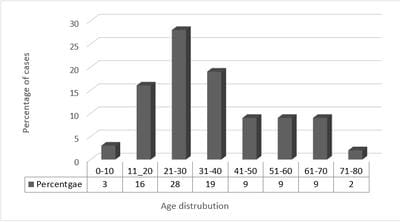
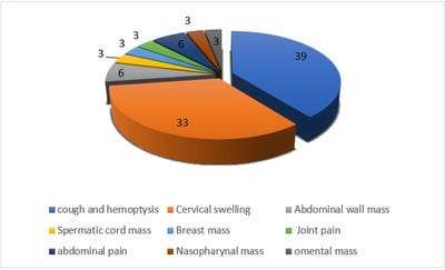
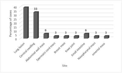
Result: In our study 63.6% were male and 36.4 were females. The majority of the patients were in the age group of 21-30 years followed by 31- 40 years, cough and hemoptysis were the most common symptoms followed by cervical swelling with 39% and 33% respectively. The most common sites were lung with (39%) and cervical swelling with (33%). The main differential diagnosis for many pulmonary and all extra-pulmonary tuberculosis was malignancy.

Conclusion: Tuberculosis is a serious young person's disease that can affect almost every organ and tissue of the body and should be considered in the differential diagnosis of suspicious masses, as tuberculosis is often misdiagnosed as cancer.

Keywords: Mycobacterium Tuberculosis, Cancer, Malignancy, Extrapulmonary

Corresponding Author How to Cite this Article To Browse
Nabeia Al Gheryani, Department of Pathology, Faculty of Medicine, University of Benghazi, Benghazi, , Libya.
Email:
Gheryani NA, Elkwafi RS, Anbaig A, Berras Ali RJ, Elmatri H, Pulmonary and Extrapulmonary Tuberculosis, An Inflammatory Disease Misdiagnosed as Cancer. Int J Med Res Rev. 2024;10(3):65-71.
Available From
https://ijmrr.medresearch.in/index.php/ijmrr/article/view/1473

Manuscript Received Review Round 1 Review Round 2 Review Round 3 Accepted
2024-02-27 2024-02-29 2024-03-07 2024-03-14 2024-03-21
Conflict of Interest Funding Ethical Approval Plagiarism X-checker Note
Nil Nil Yes 14.54

© 2024by Gheryani NA, Elkwafi RS, Anbaig A, Berras Ali RJ, Elmatri Hand Published by Siddharth Health Research and Social Welfare Society. This is an Open Access article licensed under a Creative Commons Attribution 4.0 International License https://creativecommons.org/licenses/by/4.0/ unported [CC BY 4.0].

Introduction

Tuberculosis is an old inflammatory disease with a high morbidity and mortality rate, primarily affects the lungs, but it can affect any organ [1]. According to World Health Organization (WHO), the approximate global incidence is ten million with 1.5 million deaths due to tuberculosis each year [2]. High-risk groups include immunosuppressed individuals, malnourished people, people of low socioeconomic status, prisoners, alcoholics, children, and elderly people [3]. Extra-pulmonary involvement occurs in more than 15% of immuno-competent patients and more than 50% of patients with HIV infection [4]. The most common extra-pulmonary sites are cervical lymph nodes, pleura, abdomen, genitourinary tract, skin, joints and bones, and meninges [5].

Tuberculous lymphadenitis is most common type of extrapulmonary tuberculosis [6] may arise from primary lung infection or via lymphatic dissemination from mediastinal lymph nodes [6][7], as a cervical mass, cervical tuberculosis may confused with a malignant tumour [6].

Tuberculosis of the CNS and spinal cord is associated with a high rate of morbidity and mortality and has been found in 20% of AIDS-related tuberculosis [8]. It is usually caused by hematogenous spread from primary focus [9], and clinical features are usually nonspecific, including headache, neck stiffness, fever, vomiting, and spinal cord pain or mass [10] which could be seen in other inflammatory diseases and brain tumour [11].

Tuberculosis of the breast is a rare form of extrapulmonary tuberculosis that affects multiparous and lactating women between 20 and 40years old. The infection arises from infected axillary lymph nodes or by direct spread from adjacent tissue [12]. It presents as a breast mass that mimics malignant tumours [13].

Abdominal tuberculosis either results from direct ingestion of infected lung secretions or infected dairy products and infected meats or as a complication of primary lung infection. Terminal ileum is the most common affected site in the gastrointestinal tract (form up to 90% of cases of gastrointestinal TB) and may associated with ascites and or omental mass which can sometimes be confused with peritoneal carcinomatosis [9] and the differential diagnosis

should also include Crohn's disease, and colorectal carcinoma [14]. Other common sites affected by tuberculosis are the renal system [15], liver [14] [16], muscle-skeletal [17], genital system [18], and rarely the heart [19]. Extrapulmonary TB represents a major diagnostic challenge as the condition is often misdiagnosed as cancer. Furthermore, the initial diagnosis of tuberculosis as a malignant lesion has been seen in organs considered a rare site of tuberculosis such as the thyroid, and pancreas [20], [21], [22].

Aim of work

This study aims to analyse 160 cases of pulmonary and extra-pulmonary tuberculosis correlate the pathological features with the clinical data and find the most common differentiation diagnosis of tuberculosis

Material and Methods

Study design: this study is a retrospective study performed on 160 cases of pulmonary and extrapulmonary tuberculosis. The data was collected from Tiba lab, a private histopathological laboratory in Benghazi, Libya from Jan 2017 to Dec 2022.

Inclusion criteria: all patients who were diagnosed with pulmonary or extrapulmonary disease

Data collection: the cases were divided into pulmonary and extrapulmonary lesions. The age, the sex and the site of the lesion were collected, and extrapulmonary sites were detected including rare and unusual sites such as breast, joint and spermatic cord. The symptoms were recorded for each patient

Statistical analysis: The obtained data were statistically analysed and were demonstrated as numbers and percentages using statistical graphs

Ethical consideration: The present study was performed according to the Ethical Guidelines for Clinical Research based on the declaration of Helsinki and verbal consent was obtained from all patients or the patient's family.

Results

Sex distribution of cases

Out of the total number of 160 patients, 63.6% were male and 36.4 were female (Figure 1)


ijmrr_1473_01.jpg
Figure 1
: The sex distribution.Out of 160 cases, 63.6% were male and 36.4 % were females

Age distribution of patients

The age of the patients ranged from 1 to 80 with a mean age of 33.09±3.75 (SD) years. The percentage of age distribution was as follows: the percentage of cases in the age group of 1-10 was 3%, whereas the age group 11-20 formed 16% of cases, 28% was seen in age group of 21-30, 6% was seen in the age group of 31-40, 9% was detected in the age group of 41-50, and the percentage for the age groups of 51-60, 61-70 and 71-80 were 9%, 9% and 6% respectively. The majority of the patients were in the age group of 21-30 years followed by 31-40 years, whereas the minimum percentage of cases was seen in patients older than 70 years, few cases were seen in the age group less than 10 years, the same number of cases were seen in patient aged from 41 to 70 years (Figure 2).

ijmrr_1473_02.jpg
Figure 2: The age distribution.The majority of the patients were in the age group of 21-30 years followed by 31-40 years, whereas the minimum percentage of cases was seen in patients older than 70 years, few cases were seen in the age group less than 10 years

The symptoms and complaints of the patients

In the study, all cases gave general symptoms such as fever, weight loss, fatigue, and night sweating.

Some specific symptoms were seen in some patients depending on the site of the infection. The patients who were suffering from pulmonary tuberculosis mainly presented with a cough that lasted more than 3 weeks associated with hemoptysis (blood in cough) or mucus. On the other hand, the patient who is suffering from extrapulmonary tuberculosis (such as tubercules infection in the lymph nodes, the bone and joint, the digestive system and reproductive system) presented with symptoms related to the site of the infection, such as cervical swelling, joint pain, abdominal pain, or mass in the reproductive organs. In this study, the symptoms were as follows, cough and hemoptysis were found in about 39% of cases followed by cervical lymphadenopathy in 33% of cases, whereas abdominal mass and joint pain were seen in 6% of cases, other symptoms such as mass in breast, mass spermatic cord and joint pain were seen in only 3% of cases (Figure 3).


ijmrr_1473_03.jpg
Figure 3:The percentage of symptoms and complaints of the patients.Cough and hemoptysis were the most common symptoms followed by cervical swelling, other symptoms such as breast mass, spermatic cord mass, joint pain and nasopharyngeal mass were only seen in a few cases

The sites of the lesion

In this study, most of the patients complained of pulmonary tuberculosis and the lung was the most affected site.

The most common extrapulmonary site was lymphoid system and most of the patients with extrapulmonary tuberculosis were presented with cervical lymphadenopathy. Some other rare sites were also recorded such as breast, spermatic cord, nasopharyngeal region, knee joint and abdominal wall (Figure 4).


ijmrr_1473_04.jpg
Figure 4: The sites of the lesion. The most common site was the lung followed by cervical swelling, other sites such as the small intestine, breast, spermatic cord, knee joint and nasopharyngeal region were also recorded

Discussion

Tuberculosis is a chronic infectious disease caused by Mycobacterium tuberculosis, and affects both males and females, lungs (pulmonary tuberculosis is the most common primary site followed by cervical lymph nodes as an extrapulmonary site [1] [5]

This study shows that the incidence of males is higher than females. This is in concordance with many research results [23] [24]. One explanation for the increase in cases in males is that they have greater health care than females who may not seek medical advice, especially in rural areas, also habits such as smoking and drinking alcohol may be one of the reasons for the increase in infection in males. Furthermore, the presence of males in gathering places such as cafes or sports activities, can facilitate the transmission and spread of tuberculosis bacteria [25].

Tuberculosis is a disease that affects all age groups, but it is most common in young people less than 30 years old and elderly above 70 years [26]. Yong age is a period where the person is more susceptible to bacteria, it is the period of work and age activity for both genders and the people who are most in contact with society, older age "over 70 years"

usually showed immunodeficiency status and physiological changes related with ageing, therefore, they are more susceptible to infection [27].

In this study, the most common symptom of tuberculosis in pulmonary tuberculosis is coughing and hemoptysis followed by cervical swelling.

Extrapulmonary tuberculosis such as tuberculosis of the breast, spermatic cord, abdominal wall, intestinal and nasopharynx usually presents with pain, masses and limitation of function as seen in sites such as joints and bone. This was the main pattern of symptoms of pulmonary and extrapulmonary tuberculosis in general [28].

Tuberculosis generally affects the lungs, but as extrapulmonary tuberculosis in lymph nodes, breast, spermatic cord, nasopharyngeal region, knee joint abdominal wall etc., it can create a diagnostic dilemma [21] [22] [29] [30] [31] [32]. Extrapulmonary tuberculosis was found in more than 50% of patients in this study, nearly all of them were referred to the histopathology lab for a suspicious mass and a malignancy that needed to be ruled out. In our study, all patients with extrapulmonary tuberculosis presented with nonspecific radiographic features and their symptoms were, in general, common with other diseases. For example, the patients with cervical tuberculosis were referred to a pathologist for FNAC to rule out malignant lymphoma in young patients and metastatic carcinoma in the old age group. On the other hand, patients with tuberculosis in the breast were presented with a hard cold painless mass gradually increasing in size and radiologically, the mass showed necrosis and was suspicious of malignancy. Spermatic cord tuberculosis was detected in young males as a hard suspicious mass misdiagnosed as para-testicular malignancy, the tuberculosis in the anterior abdominal wall was initially diagnosed as primary sarcoma or metastatic carcinoma, in addition, tuberculosis in musculoskeletal sites, and intestine was initially diagnosed as a malignant lesion for further workup. Although it is a curable disease, the delay in diagnosis of extrapulmonary tuberculosis may be associated with a high mortality rate [33]. The diagnostic workup for a breast, abdominopelvic and spinal mass and lymphadenopathy of indeterminate aetiology should include evaluation for extrapulmonary foci of Mycobacterium tuberculosis infection.

Patients with extrapulmonary tuberculosis and those with disseminated miliary tuberculosis have a high rate of treatment failure and a high risk of infection-associated death [34], which is often due to delayed diagnosis which leads to delay in the description of appropriate antituberculosis therapy. This is because of the misleading clinical picture.


Therefore, diagnosis of active tuberculosis requires a high level of suspicion because these infections are uncommon in healthy patients and the clinical and radiographic presentation may be quite variable [35]. So, tuberculosis should be considered in the differentiation diagnosis of nearly all malignant lesions in any organ.

Conclusion

Tuberculosis is a serious disease seen in young age group and can affect almost every organ and tissue of the body and should be considered in the differential diagnosis of many diseases presented with masses and vague symptoms, particularly in patients with immune system disorders. Considering tuberculosis as a differentiation diagnosis for malignant lesions can decrease the mortality rate associated with untreated tuberculosis and prevent unnecessary aggressive treatment such as mastectomy, oophorectomy, and splenectomy. Furthermore, the early detection of the lesion and treatment are the most critical factors in reducing morbidity associated with the disease.

Acknowledgement: None

What does this study add to existing knowledge?
Tuberculosis is a serious infection associated with a high mortality rate if not treated. Extrapulmonary tuberculosis constitutes 15–20% of all tuberculosis cases. if tuberculosis is not considered in the differential diagnosis it could lead to delays in diagnosis and treatment. This study highlights the importance of considering tuberculosis in the differential diagnosis of any suspicious extrapulmonary masses, particularly in immunocompromised patients to avoid mortality associated with delayed diagnosis

Author’s contribution:
Nabeia Al Gheryani: Concept, research design, recruitment of participants, data preparation, data analysis, manuscript preparation.
Haitham Elmatri: research design, manuscript preparation.
Rasheed J Berras Ali: manuscript review, supervision.
Raja S. Elkwafi: research design, manuscript review.
Amal Anbaig: data preparation, data analysis.

References

1. De la Rosa G, Jacobson K, Rolston K, Raad I, Kontoyiannis D, Safdar A. Mycobacterium tuberculosis at a comprehensive cancer centre: active disease in patients with underlying malignancy during 1990–2000. Clin Microbiol Infect. 2004;10:749–752. [Crossref][PubMed][Google Scholar]

2. Global Tuberculosis Report 2020. Geneva: World Health Organization; 2020. Licence: CC BY-NC-SA 3. 0 IGO. [Crossref][PubMed][Google Scholar]

3. Yang Z, Kong Y, Wilson F, et al. Identification of risk factors for extrapulmonary tuberculosis. Clin Infect Dis. 2004;38: 199–205. [Crossref][PubMed][Google Scholar]

4. Sharma SK, Mohan A. Extra-pulmonary tuberculosis. Indian J Med Res 2004; 120: 316-353. . [Crossref][PubMed][Google Scholar]

5. Heye T, Stoijkovic M, Kauczor H, Junghanss T, Hosch W. Extrapulmonary tuberculosis: radiological imaging of an almost forgotten transformation artist. RoFo 2011. 183(11):1019–1029. [Crossref][PubMed][Google Scholar]

6. Rodriguez-Takeuchi SY, Renjifo ME, Medina FJ. Extrapulmonary tuberculosis: pathophysiology and imaging findings. Radiographics. 2019; 39(7):2023–2037. [Crossref][PubMed][Google Scholar]

7. Sharma SK, Mohan A, Kohli M. Extrapulmonary tuberculosis. Expert Rev Respir Med. 2021;15(7):931–948. [Crossref][PubMed][Google Scholar]

8. Bernaerts A, Vanhoenacker F, Parizel P et al. Tuberculosis of the central nervous system: overview of neuroradiological findings. Eur Radiol. 2003;13(8):1876–1890. [Crossref][PubMed][Google Scholar]

9. Gambhir S, Ravina M, Rangan K, Dixit M, Barai S, Bomanji J. Imaging in extrapulmonary tuberculosis. Int J Infect Dis. 2017;56:237–247. [Crossref][PubMed][Google Scholar]

10. Natarajan A, Beena P, Devnikar AV, Mali S. A systemic review on tuberculosis. Indian J Tuberc. 2020;67:295–311. [Crossref][PubMed][Google Scholar]


11. Sardana V, Shringi P. Intramedullary tuberculoma of the spinal cord, clinical features & imaging: possibility of early diagnosis with imaging? Indian J Tuberc. 2019. 67(3):346–348. [Crossref][PubMed][Google Scholar]

12. Baykan AH, Sayiner HS, Inan I, Aydin E, Erturk SM. Primary breast tuberculosis: imaging findings of a rare disease. Insights Imaging. 2021;12(1):1–11. [Crossref][PubMed][Google Scholar]

13. Tewari M, Shukla H. Breast tuberculosis: diagnosis, clinical features & management. Indian J Med Res. 2005;122(2):103. [Crossref][PubMed][Google Scholar]

14. Ladumor H, Al-Mohannadi S, Ameerudeen FS, Ladumor S, Fadl S. TB or not TB: a comprehensive review of imaging manifestations of abdominal tuberculosis and its mimics. Clin Imaging. 2021;76:130–143. [Crossref][PubMed][Google Scholar]

15. Gibson MS, Puckett ML, Shelly ME. Renal tuberculosis. Radiographics. 2004;24(1):251–256. [Crossref][PubMed][Google Scholar]

16. Karaosmanoglu AD, Onur MR, Sahani DV, Tabari A, Karcaaltincaba M. Hepatobiliary tuberculosis: imaging findings. AJR Am J Roentgenol. 2016;207(4):694–704. [Crossref][PubMed][Google Scholar]

17. Leonard MK Jr, Blumberg HM (2017) Musculoskeletal tuberculosis. Microbiol. 2017. (9):5(2):5–2. [Crossref][PubMed][Google Scholar]

18. Naeem M, Zulfiqar M, Siddiqui MA et al. Imaging manifestations of genitourinary tuberculosis. Radiographics. 2021;41:1123–1143. [Crossref][PubMed][Google Scholar]

19. Cowley A, Dobson L, Kurian J, Saunderson C. Acute myocarditis secondary to cardiac tuberculosis: a case report. Echo Res Pract. 2017;4(3):K25. [Crossref][PubMed][Google Scholar]

20. Ortiz-Flores A, Gioia F, Montánez-Fernández L et al. Incidental finding of a primary thyroid tuberculosis. Oxford Med Case Rep. 2017;(6):omx022. [Crossref][PubMed][Google Scholar]

21. Al-Mulhim A, Zakaria H, Abdel hadi M, Al-Mulhim F, AlTamimi D, Wosornu L. Thyroid tuberculosis mimicking carcinoma: report of two cases. Surg Today. 2002;32:1064 –1067. [Crossref][PubMed][Google Scholar]

22. Wu C, Wang S, Kuo T. Pancreatic tuberculosis mimicking pancreatic head carcinoma: a case report and review of the literature. Infection. 1994;22:287–289. [Crossref][PubMed][Google Scholar]

23. Orgdorff MW, Nagelkerke NJ, Dye C, Nunn P. Gender and tuberculosis: a comparison of prevalence surveys with notification data to explore sex differences in case detection. Int J Tuberc Lung Dis. 2000;4:123–32. [Crossref][PubMed][Google Scholar]

24. Watkins RE, Plant AJ. Does smoking explain sex differences in the global tuberculosis epidemic? Epidemiology and Infection. 2006;134(2):333-339. . [Crossref][PubMed][Google Scholar]

25. de Waroux OlP, Cohuet S, Ndazima D, et al. Characteristics of human encounters and social mixing patterns relevant to infectious diseases spread by close contact: A survey in southwest Uganda. bioRxiv; 2017. DOI: 10. 1101/121665 [Crossref][PubMed][Google Scholar]

26. Dong, Z. , Wang, QQ. , Yu, SC. et al. Age–period–cohort analysis of pulmonary tuberculosis reported incidence, China, 2006–2020. Infect Dis Poverty. 2022;11, 85 [Crossref][PubMed][Google Scholar]

27. Teo, A. , Rahevar, K. , Morishita, F. et al. Tuberculosis in older adults: case studies from four countries with rapidly ageing populations in the western pacific region. BMC Public Health 2023;23, 370 [Crossref][PubMed][Google Scholar]

28. Ismail, Yasser. Pulmonary tuberculosis--a review of clinical features and diagnosis in 232 cases. The Medical journal of Malaysia. 2004;59:56-64. [Crossref][PubMed][Google Scholar]

29. Piura B, Rabinovich A, Leron E, Yanai-Inbar I, Mazor M. Peritoneal tuberculosis mimicking ovarian carcinoma with ascites and elevated serum CA-125: case report and review of the literature. Eur J Gynaecol Oncol. 2002;23:120–122. [Crossref][PubMed][Google Scholar]


30. Fukasawa H, Suzuki H, Kato A, et al. Tuberculous arthritis mimicking neoplasm in a hemodialysis patient. Am J Med Sci. 2001;322:373–375. [Crossref][PubMed][Google Scholar]

31. Rezeig M, Fashir B, Al-Suhaibani H, Al-Fadda M, Amin T, Eisa H. Pancreatic tuberculosis mimicking pancreatic carcinoma: four case reports and review of the literature. Dig Dis Sci. 1998;43:329–331. [Crossref][PubMed][Google Scholar]

32. Rolston K, Rodriguez S, Dholakia N, Whimbey E, Raad I. Pulmonary infections mimicking cancer: a retrospective, three-year review. Support Care Cancer. 1997;5:90 –93. [Crossref][PubMed][Google Scholar]

33. Jørstad MD, Aẞmus J, Marijani M, Sviland L, Mustafa T. Diagnostic delay in extrapulmonary tuberculosis and impact on patient morbidity: A study from Zanzibar. PLoS One. 2018 Sep 6;13(9). [Crossref][PubMed][Google Scholar]

34. Khan, A. H. , Sulaiman, S. A. S., Laghari, M. et al. Treatment outcomes and risk factors of extra-pulmonary tuberculosis in patients with co-morbidities. BMC Infect Dis. 2019;19, 691 [Crossref][PubMed][Google Scholar]

35. Chisale MRO, Sinyiza F, Kaseka P, Wu JST, Chimbatata C, Mbakaya BC, Kamudumuli PS, Kayira AB. Cancer obscures extrapulmonary tuberculosis (EPTB) at a tertiary hospital in Northern Malawi. Epidemiol Infect. 2020;(9):20;149:e3. [Crossref][PubMed][Google Scholar]