E-ISSN:2320-8686
P-ISSN:2321-127X

Review Article

Multicentric Carpotarsal Osteolysis

International Journal of Medical Research and Review

2023 Volume 11 Number 6 November-December
Publisherwww.medresearch.in

The general skeletal features of Multicentric Carpotarsal Osteolysis: what the radiologist needs to know

Albahkali S1*, Alsaiady M2
DOI: https://doi.org/10.17511/ijmrr.2023.i06.03

1* Salha M Albahkali, Resident, Radiology Department, King Saud Medical City, Riyadh, Saudi Arabia.

2 Moath Alsaiady, Pediatric Radiology Consultant, Radiology Department, King Saud Medical City, Riyadh, Saudi Arabia.

Purpose of review: Radiographic imaging is the first investigational step in detecting Multicentric carporarsal osteolysis, and the radiologist must be aware then familiar with the radiological features in order both to reach a prompt diagnosis and then to classify the patient’s condition at this important time for considering early management. Here we review the literature on this topic. Recent Findings: Patients with multicentric carpotarsal osteolysis (MCTO) are frequently diagnosed with juvenile idiopathic arthritis (JIA). Initially, their symptoms are arthritic and include pain, swelling and stiffness. Among the many published studies, some are case reports that describe the phenotypical manifestations with a brief discussion of the radiographic findings and subsequently of the genetic analysis which eventually leads to a definitive diagnosis. Some patients develop end-stage renal failure, and a delay in identifying this condition can lead to unfavorable progression of the disease. In addition, we found reports of joint inflammation based on imaging and pain relief with antirheumatics for osteolysis and bone destruction. Various studies discuss the radiological findings, including the disappearance of the carpal and tarsal bones. Others describe the genetic mutations, including MAFB, that are associated with the condition, and its possible management through the use of therapeutic drugs. A very recent description of serial radiographs taken from a young age suggests that dysfunctional bone formation can play a role in the skeletal phenotype of MCTO. Summary: The unique features of clinical presentation, atypical radiological findings, failure to obtain remission with medical treatment and consanguinity, should guide clinicians towards the diagnosis of this condition. We summarize the X-ray findings which are highly specific and can therefore differentiate with confidence between this condition and others. Further performance of gene.

Keywords: Multicentric carpo-tarsal osteolysis, Skeleton, MAFB, Nephropathy, Radiologist.

Corresponding Author How to Cite this Article To Browse
Salha M Albahkali, Resident, Radiology Department, King Saud Medical City, Riyadh, , Saudi Arabia.
Email:
Albahkali S, Alsaiady M, The general skeletal features of Multicentric Carpotarsal Osteolysis: what the radiologist needs to know. Int J Med Res Rev. 2023;11(6):146-158.
Available From
https://ijmrr.medresearch.in/index.php/ijmrr/article/view/1454

Manuscript Received Review Round 1 Review Round 2 Review Round 3 Accepted
2023-12-07 2023-12-09 2023-12-16 2023-12-23 2023-12-30
Conflict of Interest Funding Ethical Approval Plagiarism X-checker Note
Nil Nil Yes 18

© 2023by Albahkali S, Alsaiady Mand Published by Siddharth Health Research and Social Welfare Society. This is an Open Access article licensed under a Creative Commons Attribution 4.0 International License https://creativecommons.org/licenses/by/4.0/ unported [CC BY 4.0].

Introduction

The multicentric carpotarsal osteolysis (MCTO), termed also in old idiopathic multicentric osteolysis with renal failure is a rare skeletal disease, caused by a heterozygous mutation in the MAFB gene particularly (v-maf musculoaponeurotic fibrosarcoma oncogene ortholog B). Osteolysis of the carpal, metacarpal, and tarsal bones is the characteristic condition that causes anomalies in the extremities. It typically manifests in early childhood and causes discomfort and movement impairment in the bones. Most of the patients experienced renal disease at various ages, from months to years, exhibiting symptoms such as proteinuria and renal failure that necessitated kidney transplantation for treatment. [1]. Due to renal failure which can be deteriorate the patient condition and developing of subsequent complications, such as the trends in respiratory symptom patterns, X-ray findings

Figure 1, and recurrent admissions for hemodialysis. [2].

ijmrr_1454_01.JPG
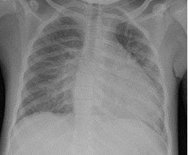
Figure 1: Nine-year-old presenting with skeletal deformity and renal osteodystrophy:

Anteroposterior radiograph of the chest demonstrating bilateral diffuse increased bilateral perihilar broncho-vascular lung markings and multiple and ill-defined patches of opacities in the left middle and lower lung zones, suggestive of multifocal subsegmental collapse, atelectatic consolidation and increased transverse diameter of the heart with an abnormal cardiothoracic ratio of 13.0/22.2 = 0.58% (as mild cardiomegaly).

(Radiology department, King Saud Medical City, Riyadh, SA).

The two main classifications of this disorder that have already been identified are with or without renal disease. The diagnosis is mostly based on the combination of radiological findings and clinical manifestations. It is an autosomal dominant disorder with a known gene abnormality. [3]. Patients with increasing bone loss, primarily affecting the tarsal and carpal bones, as well as kidney abnormalities, such as proteinuria, should be considered for this disorder.[4]. The disease can manifest in a variety of ways, with symmetric or asymmetric bone damage. After examining the literature, we discovered that numerous studies had an early clinical presentation that resembled the rheumatologic illness known as juvenile idiopathic arthritis.

This entity of unique skeletal phenotype may also be subtle craniofacial changes and the involvement of larger joints such as the knees and elbows. Due to the rareness of this condition, a diagnosis of MCTO is delayed on average by 3 to 4 years, with some adults not receiving a diagnosis of MCTO until after their child undergoes genetic testing. An earlier diagnosis of MCTO may enable screening for nephropathy and preventative measures to avoid nephrotoxic medications and preserve renal function for as long as possible. Also, the bone and joint destruction that progressively worsens over time can result in substantial debilitation and crippling of joints that are used for activities of daily living Figure 6. Early recognition of MCTO as a chronic condition may prompt sooner referrals to physical and occupational therapy to assist patients as they adapt to changes in their mobility and joint function [5]. Patients usually present with arthropathy, normal inflammatory markers, and atypical radiological features. It is a syndrome of non-inflammatory condition. When dealing with refractory disease and uncertain diagnosis, additional imaging and genetic testing can be extremely important in determining the cause. [6]. In the majority of cases, the renal disease appears later and quickly evolves to end-stage renal disease Figure 2 [7]. The ancient literature did not know the underlying cause of the genetic abnormalities of multicentric osteolysis linked to nephropathy. In 2007, Wenkert et al. The illness is either sporadic or occasionally autosomal dominant. It usually causes nephropathy and carpal-tarsal destruction, which can lead to renal failure.[8]. Furthermore, in some advanced current literature, they determine


the responsible genetic code refers to specific protein of the genetic mutations specifically [8,9]. Misdiagnosis of multicentric carpal tarsal osteolysis syndrome, an uncommon condition that is frequently mistaken for juvenile idiopathic arthritis, can also be a diagnostic trap. 

ijmrr_1454_02.JPG
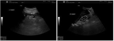
Figure 2: 9 years old girl presnted with ESRD, renal ultrasound Findings.

(A) Grayscale Ultrasound images of the kidneys showed, both kidneys are small but with preserved cortical medullary differentiation and no stones or hydronephrosis (Radiology department, King Saud Medical City, Riyadh, SA).

Most recently, Wu et al, 2021, they reported two Chinese boys with comparable ages displayed completely different clinical presentations based on when the disease started and how long it took to progress. Following the two patients' incorrect diagnosis of juvenile idiopathic arthritis and poor response to treatment for rheumatologic diseases, genetic studies were ultimately used to guide the diagnosis of MCTO [10]. Mehawej et al., 2013, reported eight patients with multicentric carpo-tarsal osteolysis from six families. They found that all eight patients had MAFB mutations, of the eight patients, only six had kidney disease, indicating that the condition is also heritable and supporting our findings regarding the underlying clinical variability of this condition. [7]. There have been reports of multicentric carpo-tarsal osteolysis in both inherited and sporadic cases. Mumm et al., 2014. They identified five different heterozygous missense defects among eight probands from their genetic study of nine multicentric carpo-tarsal osteolysis patients. Therefore, the mutations in both parents' multicentric carpo-tarsal osteolysis features for the seven patients thought to have developed spontaneously. As a result, it appeared that MAFB mutation penetrance was widespread. This also led us to believe that the disease's mode of occurrence is not caused by all MAFB mutations; rather, a small number of specific MAFB mutations cause multicentric carpo-tarsal osteolysis [11].

Zankl et al, 2012, tried to determine the underlying genetic cause, they found MAFB encodes a responsible transcription factor that regulates RANKL-induced osteoclast-genesis negatively which is important for renal development and gives the way for therapeutic approaches as well as proves the concept of bone and kidney normal development. MAFB is also known to play a role in osteoclastogenesis, making this gene highly responsible for underlying bone deformity [12].

Recent research by R. Regev et al., 2021, revealed that one patient received treatment with denosumab, a human monoclonal RANKL inhibitor, which stopped further osteolysis and stabilized the osteoporosis process. The high bone turnover and osteoporosis are consistent with a high serum level of RANKL. The process of osteoporosis is suggested to be more tolerable by denosumab, although further studies are necessary to cover the dose, frequency, and effectiveness extent [13]. Multidisciplinary care including a pediatrician, nephrologist, radiologist, orthopedic surgeons, and geneticists are important parts of patient care [14]. Eventually, we are considering the treatment of this disease is variably differentiating according to the patient’s situation inheritably then clinically, hemodynamically, and surgically.

Rationale: We encountered, to the best of our knowledge, the first Saudi female child case of multicentric carpal tarsal osteolysis syndrome (MCTO), which unfortunately resulted in end-stage renal failure. The diagnosis of MCTO was confirmed by genetic testing for the MAFB mutation, and related radiological findings were generally consistent with this diagnosis in the current literature. Here, our attention is focused on the skeletal features that radiologists need to be aware of in order to become more familiar with this entity and to make an early image-based diagnosis.

Purpose

A review of the literature suggests that prospective studies are needed to clarify the generalized skeletal features. Here we are more focused on the skeletal radiographic features, which can bridge a lack of knowledge about this rare entity and encourage radiologists and clinicians to build up an image-based background that can help in the early diagnosis of this condition.


Perspectives on Radiological Features of MCTO 

Skeletal Survey: Bilateral anteroposterior (AP) and posteroanterior (PA) projections of the hands, forearms, humerus, foot, leg, femur, pelvis, spine, and skull are commonly included in a standard skeletal scan performed with a conventional x-ray. Bilateral AP and PA images of the wrist, elbow, shoulder, ankle, knee, hip, and sacroiliac joints are included in a joint survey Figure 3, [15].

ijmrr_1454_03.JPG
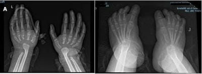
Figure 3. Nine-year-old girl presenting with ESRD, radiological X-ray findings:

(A) Anteroposterior view of the hands and dorsoplantar view of the feet showed significant bilateral osteolysis or absence of carpal and tarsal bones, with erosions and tapered proximal ends of the metacarpal and metatarsal bones. Findings are typical for multicentric carpo-tarsal osteolysis (Radiology department, King Saud Medical City, Riyadh, SA).

ijmrr_1454_04.JPG
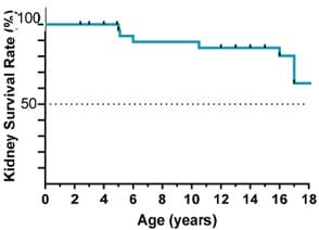
Figure 4: Kidney failure-free survival until adulthood.

In 2022, Drovandi et al. and his associates reported that 53 subjects had bone disease symptoms, with the median age of clinical manifestation being 2 years old. Rheumatological disorders were misdiagnosed in 30% of patients; all patients had involvement of the upper limbs, and about 70% had involvement of the lower limbs. In descending order of frequency, the disease affected the

following joints/sites: knees, tarsal bones, elbows, fingers/toes, and carpal bones. Three patients had Arnold-Chiari syndrome or cervical deformity, and one-third of patients had scoliosis. He also reviewed retrospectively the survival rate of patients they get affected with renal function Figure 4 [16].

Fig (4) Drovandi et al., 2022., Through retrospective analysis and a thorough revision of the literature for 54 patients diagnosed with MCTO. As a result, the median age at the last observation was 15 years, and the median follow-up from the time of kidney disease diagnosis was 9 years. This indicates that 63% of the observed patients reach adulthood at the age of 18 years, maintaining their kidney functions. (Figure 2) [17].

Peeters et al., 2011, reported significant radiographic findings that are fundamentally known to allow the radiologist to diagnose the patient based on plain radiographs, which is crucial for disease diagnosis. Osteolysis of the tarsal and carpal bones is the defining feature. In the early stages of the disease, the affected bone gradually lost its normal contours due to demineralization. Subsequently, fragmentations of the tarsal and carpal bones, sclerotic remnants, and additional bone resorption led to bone collapse. Last but not least, nearby tubular bones experiencing partial resorption showed signs of tapering at the proximal ends of the metacarpals and metatarsals, which could be described as a "sucked candy" appearance Figure 7. Patients with severe disease developed deformities in the interphalangeal, metatarsal, and metacarpal joints. The epiphyseal centers of the elbows and shoulders may undergo irregular delineation as well [3]. The carpal, metacarpal, and tarsal bones are the primary affected areas of the bony skeleton, though some cases have been reported involving other bones as well. According to Chen et al., 2022, the first Brazilian family to be diagnosed with MCTO demonstrated progressive osteolysis of the tarsal and carpal bones, confirming the disease's natural course. Due to the progressive nature of MCTO's symptoms, it's critical to diagnose the condition early in order to prevent unnecessary testing and treatments and to ensure that the right follow-up is provided on time. [17]. Rheumatologists or orthopedic surgeons are typically the first to identify the phenotypic features of multicentric carpo-tarsal osteolysis syndrome in a patient presenting with skeletal bone deformity or joint pain. Typically, the family's


primary concern is the clinical skeletal bone deformity. Therefore, in the context of a rheumatologic workup, the identification of typical bone osteolysis on radiographs is a negative, and the most crucial step in determining the appropriate time to make a diagnosis is an early referral to a clinical pediatric geneticist.

Bahavani et al, American Journal of Medical Genetics, Part A, 2016.

ijmrr_1454_05.JPG

Figure 5: Children's hand radiographs with When a disease is advanced, osteoporosis becomes more noticeable. Phalanges and metacarpals enlarge and change from their typical shape. Early in the disease, well-formed carpals frequently experience nearly total lysis. Carpal osteolysis varies in severity and progression; some people (C and D) exhibit early complete osteolysis, while other people (H and J) may experience it later. Radiographically, this condition is characterized by lysis of the tarsal and carpal bones. Other hand and foot bones, such as the metacarpals, metatarsals, and phalanges, are frequently affected as well. These bones have poor modeling, giving the impression that they are rounded and uneven. Bone cortices seem slender. In older children, the entire skeletal system exhibits osteoporosis or osteopenia [18].

The hand could be a crucial diagnostic tool for skeletal congenital abnormalities. Some authors suggest that the first step to successful characterization is a systematic examination of the radiologic semiology. After that, in order to get a more precise diagnosis, each case should be discussed with the referring pediatrician and clinical geneticist. The radiologist should also be knowledgeable about the key characteristics of the majority of common inherited bone diseases. [19]. In 2004, Faber M. et al. described a specific appearance in X-ray films of a case that showed

how bone destruction developed over time, with the carpal bones disappearing and the carpus shortening as a result, and the metacarpals developing a "sucked candy" appearance [20]. Thus, one of the primary MCTO criteria is radiological characteristic. Therefore, it's critical to recognize that this disorder depends on imaging in order to prevent pointless tests and treatments.

Ishaq, T., et al, BMC Musculoskeletal Disorders, 2023.

ijmrr_1454_06.JPG

Figure 6: Development of skeletal abnormalities in patients from Family 2 as they become adults. A, B, and C radiographs of Patient 1 at 20, 28, and 36 years old, demonstrating progressive deformity, osteolysis, and severe scoliosis. D, E, F, and G In addition to a vertebral compression fracture in the spine, radiographs of Patient 2 taken at ages 30 and 32 reveal widespread osteopenia, osteolysis, and osteoarthritis in the knee and hip joints. [21]. 

In 2017, Lazarus, S. et al. Categorize the condition according to site-specific bone abnormalities. The disease is thought to be caused by excessive osteolysis, as evidenced by the progressive destruction of carpal and tarsal bones as well as the epiphyses of numerous long bones that are visible on serial radiographs Figure 12. We refer to these three syndromes as carpotarsal osteolysis disorders (CTODs) because carpotarsal osteolysis is the predominant feature that unites them all (MCTO, Multicenteric osteolysis, nodulosis and arthropathy (MONA), Winchester syndrome. Another frequent occurrence is contractures throughout the affected joints. [22]. In its later stages, the illness may cause additional musculoskeletal issues, like scoliosis. [23]. Reviewing the literatures most bones are affected are the carpal bones in the skeleton, thus far, Li, Zhuang, and others 2017. According to the reported course of the carpal bone destruction,


the index patient had all of the carpal bones at age 5, but there were noticeable changes to their location and orientation. The carpal bones exhibited partial destruction and erosions at the age of ten. The ulna and radius epiphyses, as well as the carpal bones, had nearly entirely resorbed by the time the child was 13 years old. The left hand's ulnar deviation and progressive thinning and sclerosis of the proximal ends of the metacarpals were observed at the age of sixteen. At nine years old, multilocular erosions were seen in the tarsal bones. They did not resorb like the carpal bones did, but the erosions caused osteoarthritis. [24]. In another side, there is no essential role of interventional radiology to diagnose these cases, through review of literature, Thomas et al reported the intraarticular biopsies revealed no evidence of synovitis, and William m et al 1980, they reported pathological features of nephropathy as the glomerular and interstitial pathology was nonspecific and could be consistent with primary vascular disease, thus far the renal biopsy will not add a point through diagnostic work up.

Klein, C., et al, Skeletal Radiol 2018.

ijmrr_1454_07.JPG

Figure 7: Radiographs of patient 1. a The patient's left hand displaying carpal osteolysis at that age. b At 16 years old, the left hand exhibits ulnar deviation, a short first metacarpal, and carpal osteolysis. c Left hand at age 22 displaying the ulna extremity and metacarpals as if they were a sucked candy. d, e At 12 years old, plain and lateral view radiographs of the left elbow demonstrate total osteolysis of the trochlea and enlarged soft tissues. f, g Plain and lateral view the left forearm at age 22, displaying an ulnar deviation of the wrist, anteromedial dislocation of the olecranon, and total osteolysis of the humeral distal epiphysis. h Partial tarsal osteolysis was visible on lateral view radiographs of the foot at age 14. [15].

According to certain authors, radiographic evidence of the pathologic osseous process's destructive nature has been observed in the capitellum and trochlea. Additionally, a dysplastic olecranon and coracoid process have been noted, which can result in adult radial head and ulna dislocations Figure 11 [15]. A close differential diagnosis of MCTO is multicentric osteolysis nodulosis and arthropathy (MONA), which has osteolysis (25). Tyler T et al 2018., they but an old classification of this disease, Type 1 : hereditary multicentric osteolysis with dominant transmission. Type 2: hereditary multicentric osteolysis with recessive transmission. Type 3: non-hereditary multicentric osteolysis with nephropathy. Type 4: Gorham’s massive osteolysis.Type 5: the Winchester syndrome. Osteolysis in the hands and feet, osteopenia, arthropathy, and subcutaneous nodules on the palms and soles are the hallmarks of MONA. Heart problems, corneal opacities, and coarse facial features are possible extra features. Similar symptoms to MONA are present in Winchester syndrome, with the exception of subcutaneous nodules. Mutations in matrix metalloproteinases (MMP) that are recessive cause MONA and Winchester syndrome [26]. When the response to therapy or the inflammatory parameters are not appropriate, mimics of JIA such as Farber's MCTO should be actively investigated. Despite being a rare condition, MCTO can be identified with straightforward radiography, siblings must be screened, and bisphosphonates can be tested in order to lessen skeletal complications. (?). A few writers stated to investigate the impact of combination therapy (DMARDs and bisphosphonates) on the bones of the wrists and ankles, serial radiographs of the wrists and ankles were taken. Regretfully, the wrist osteolysis persisted. The already small dystrophic carpal bones either shrank further or vanished. She was down to one tiny carpal bone by the time she was 4 years 9 months old. Additionally, there was a noticeable narrowing of the wrist joint space and visible proximal metacarpal erosions. Remarkably, following the initiation of bisphosphonate treatment, the weight-bearing bones of the mid- and hindfoot seemed to stay stable or slightly increased in size. [27].

Through review of the available literature, we try to summarize the skeletal features involving both upper and lower limbs radiographs,


table1, as the following: cortical thinning with erosion, eroded, tapered and crenated appearance, progressive osteolysis, and deformity it can appear with loss of the joint space. Subchondral destruction as well as generalized osteopenia. Destructive or dysplastic alterations, progressive erosive changes, flattening, shortening, and penciling of carpal or metacarpal bases which can lead to pencil-in cup deformity. Gradually resorbing of carpometacarpal joints and disappearing then can lead to absence of the carpal bones, sometimes the ossification center also can be destroyed, soft tissue swollen and dislocations, in MRI reported findings were, tenosynovitis with joint effusion and osteoarthritis in advanced cases.

Table1: Summary of generalized skeleton features with multicentric carpo-tarsal osteolysis with or without nephropathy.

DiagnosisRadiographyPhenotype of the patientAge Study
Radiographs, and genetic testingCortical thinning with erosion and osteolysis of the middle and distal phalanges, deformity of the interphalangeal and metacarpophalangeal joins, decreased space of interphalangeal joints, and generalized osteopenia.It was found that the wrist and hand joints had both range of motion restriction and fixation.A 5 years and 9 months old girlShakiba M et al, 2023. [32]
Radiographs, and genetic testingOsteolysis of the scaphoid, cuneiform, and proximal phalanges of both ankles, as well as the thumbs. The arrangement of the carpal bones was unusual.Manifested as increasing wrist edema, pain, and restricted mobility.A 6-years-old girlLenka D et al, 2023.[22]
Radiographs, and genetic testingDestructive alterations to the carpal bones, which cause the wrists to shorten and the metacarpals to overlap over the distal radius. The navicular and cuneiform bones are the main targets of progressive erosive change and destruction.Absence of renal impairment, clouding of the cornea, or other manifestations (e.g., skin deposit).12 years old childFurness, L et al, 2022. [6]
Radiographs, and genetic testingThe distal end of the metacarpal bones shows progressive osteolysis, while the proximal end shows no carpal bones. And the phalanges remain relatively wellpreserved.Renal failure combined with craniofacial abnormalities.3, 8, and 10 years old.Zankl A. et al, 2012 [12]
Clinical and Radiographs imaging.Right-hand revealed flattening of the os hamatum and shortening of the carpus, along with the loss of recognition in the os capitatum. There was penciling on the third and fourth metacarpal bases.The mother's face is triangular, with small mouths, noses, and chins; they also have orbit hypoplasia, slight exophthalmos, and maxillary hypoplasia.Two patient at 1 year,two patients at 2 yearsand one at 3 years.Faber M et al, 2004 [20]
Radiographs imaging.Prominent osteolysis was observed in the hand, elbow, and ankle joints.Small forehead and hypotelorism; clubfoot was also noted, along with a shortening of the fingers on both hands and upper limbs.20-year-old. Scoliosis was apparent sinceshe was 12 years oldMiyazaki, K et al 2018.[23]
Radiographs, and genetic testing.The majority of the other bones are still in place; however the carpal bones were all present for a while before gradually resorbing and disappearing altogether at the age of sixteen. The only affected bones are the metacarpals where they meet the carpal bones.Her right wrist hurt when she was four years old.20 years old.Zhuang, Lei et al 2017. [24]
Radiographs imaging.Dysplastic ulna and distal radius, absent carpal bones, and a dysplastic base of the metacarpals. Absent navicular, medial, and lateral cuneiform.Swelling and constriction in the left ankle and right wrist joints.3 years oldAbhishek S. et al, 2018.[31]
Radiographs, and genetic testing.significant erosion of the proximal metacarpal and carpal bones. Deformities of the proximal ulnar and radius, as well as the distal right humerus; additionally, the ossification center is destroyed, the right elbow joint's space is narrowed, and the soft tissue surrounding the elbow joint is swollen.A painful, swollen right wrist with restricted passive and active wrist flexion and extension12 years old boy.Wang, Li et al 2021. [7]
Radiographs imaging.Partial and progressive carpal osteolysis radial head and ulna dislocation. Osteolysis linked to the pes cavus and navicular deformity, as well as subchondral destruction of the talus.Bilateral wrist pain, bilateral pes cavus and irreducible claw toes, cervicothoracic scoliosis, walking difficulties with an irreducible left knee flexion and valgus deformity24 years old maleKlein C et al, 2018.[15]
Radiographs, MRI, and genetic testing.His entire proximal row of carpal bones was lost in the center. An MRI of the right wrist revealed extensor tenosynovitis, lateral flexor tenosynovitis, and pan-carpal synovitis with a considerable joint effusion.Symptoms of arthritis. Later in life, he experienced wrist pain that limited his range of motion and caused diffuse swelling.13 months old boyRegev R, et al, 2021. [14]
Radiographs, and genetic testing.Dysplastic alterations to the tarsalia and irregular, partially absent carpalia.Wrist swelling in his right hand suggests that juvenile rheumatoid arthritis may be developing.19 years old maleDworschak, G et al 2013. [9]
Radiographs, and genetic testing.Narrowing of the right wrist joint and bony erosion of the right scaphoid, trapezium, and triquetral bones as well as the distal end of the radius.Multiple proximal interphalangeal joints as well as swelling and tenderness in the right carpal and metacarpal joints.12 years old female patientPark, P et al, 2018. [4]
Radiographs, and genetic testing.The carpal bones destroyed. Osteolysis of the proximal end of the metacarpal bones, dislocation of both elbow joints, and absence of carpal bones. Her wrists, ankles, and face were the first areas where the symptoms were noticed, along with an unusual gait brought on by decreased joint mobility.13 years old girlStajkovska, A et al 2018. [10]
Radiographs, and genetic testingDistal ends of the ulna and radial bones, osteolytic lesions of the metacarpal, and absence of the carpal bones. Severe cortical thinning, distal end of fibula, osteolytic lesions of the metatarsal bones, and absence of tarsal bones.Deformities of upper and lower extremities.14 years old girlChoochuen, P et al, 2018. [34]
Radiographs, and genetic testing.The mother's radiographs at age 33 showed marked osteolysis, dysplastic changes, and narrowed joint space of the tarsal bones. Ulnar deviation of the wrists, absent carpal bones, and eroded and tapered proximal metacarpal bones.Dysmorphic features and developmental delay.7 years old boy.Upadia, J., et al 2018. [1]
Radiographs, and genetic testingThe distinctive appearance of "sucked candy" is caused by the resorption of metacarpals, which results in the absence or crenated appearance of the carpal bones. Deformities of variable severity, bone destruction in the tarsal and metatarsals, and Cavo varus deformities. Joint instability, wrist effusions, proximal and metacarpophalangeal joint limited motion, distal interphalangeal joint deformity, and foot deformity.6 years old boy.Wenkert D, et al 2007.[8]

Characterization of the joint disease:

Compared to the wrists and ankles, the larger joints such as the elbows and knees are less commonly affected. An improved comprehension of the natural history of the MCTO arthritic presentation (5). The Main findings were noted in the affected large joints, widespread osteopenia, osteolysis, osteoarthritis in the shoulder, knee, and hip joints in advanced stages Figure 8. The small joints are usually affected by osteolysis as the early and main findings. In advanced cases can developing of other associated inflammatory manifestations. However, the joint disease with inflammatory changes is recurrent. Some authors reported that these changes were resolved by medical treatment and appeared after discontinuation of those medications, Figure 13.

ijmrr_1454_08.JPG
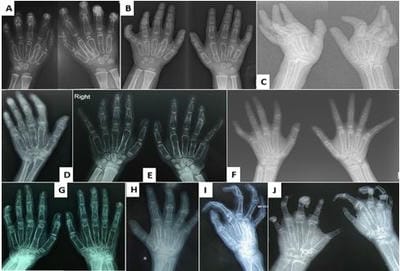
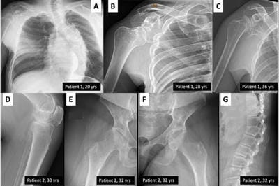
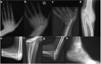
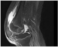
Figure 8: Nine-year-old known case of end stage renal disease on automated peritoneal dialysis, presenting with left knee swelling:

Lateral view of the left knee image T2 fat sat MRI sequence demonstrates narrowing of the joints associated with irregular thinning of normal cartilage. A moderate suprapatellar joint effusion was noted, associated with enhancement in post contrast images, and worrisome for underlying inflammatory process (Radiology department, King Saud Medical City, Riyadh)

A case of an adolescent patient with MTCO who underwent complex hip replacement surgery and showed successful, problem-free healing was documented by Sun et al. in 2016. In order to manage multicentric carpo-tarsal osteolysis, one must first restore function for the affected individual.

This requires prompt referral for orthopedic surgery, as medical therapy or surgical interventions are difficult because of the anatomical bone differentiation. [28].

According to research by Nishikomori et al. in Pediatric Rheumatology (2015), tocilizumab significantly reduced the articular pain of a multicentric carpo-tarsal osteolysis patient who had a mutation in MAFB.

This suggests that tocilizumab may be a useful treatment for MCTO patients' articular pain. [29]. In 2023, Melissa A. et al. The radiography of their patient revealed degenerative changes in the carpal bones, which are suggestive of a chronic illness. Every other joint functioned normally. The joint disease can be worsened over time. Lead to involve wrist, hand, many fingers, and elbows which are manifested by decreased range of motion, and the magnetic resonance imaging (MRI) will reveal that the articular surfaces of joints are gradually being destroyed Figure 9 [30].

Lerman, M. A. et al, . JBMR Plus, 2023.

ijmrr_1454_09.JPG

Figure 9: Images of the left wrist. (A). The left wrist's frontal radiographs show numerous erosions in the carpus, proximal metacarpals, and distal radius along with progressive carpal fusion (*) and loss of joint space. Multiple growth recovery lines and overgrown distal radius and ulna epiphyses are visible posttreatment.

(B) T1 signal-weighted MR images of the left wrist show multiple erosions in the ulna, carpals, metacarpal bases, and radius (thin black arrows and thick black arrows, respectively) (white arrow). The lunate and triquetrum have united by 2017. There won't be much carpal joint space left by 2020. [30].

Lerman, M. A. et al, . JBMR Plus, 2023.


ijmrr_1454_10.JPG
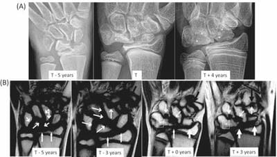
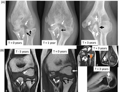
Figure 10: Images of the left elbow. (A). The left elbow's frontal radiographs show several progressive erosions in the ulna, humerus, and radius (black and white arrows, respectively), along with a progressive deformity of the distal humerus and overgrowth of the subluxed radial head. (B) (a,b,c,d) T1 signal-weighted MR images of the left elbow show several progressive erosions in the coronal plane (a, b) of the radius and ulna (orange arrows) and humerus (blue arrows). By 2020, acquisition in standard planes was impossible for the patient due to a fixed flexion deformity (c, d). (e) Radio-capitellar subluxation and synovitis (arrow) are visible in the post-contrast T1 signal-weighted image with fat saturation.
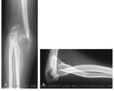
The elbow joint specifically can be affected in various degrees; initially by progressive erosions, destroying of the ossification center, then elbow joint's space narrowing, radio-capitellar subluxation, synovitis, and the soft tissue surrounding the elbow joint can affected by swelling Figure 10.

Wenkert, D. et al, Clinical orthopaedics and related research, 2007.ijmrr_1454_11.JPGFigure 11:

A, and B. Bone loss and dislocation at the elbows.

Ishaq, T., et al, BMC Musculoskeletal Disorders, 2023.

ijmrr_1454_12.JPG

Figure 12: Family 1 and Family 2 radiographs. Radiographs of the patient at age 25 and age 26 show increased osteolysis, poor carpal bone visualization, cortical thinning of the metacarpal bones, and medullary expansion due to advanced osteopenia. The patient's wrist is deformed, the distal radioulnar joint has cortical thinning, and the metacarpals are bullet-shaped.(A) The same patient's 26-year-old radiograph, seen in vertical view, displays the left hand's ulna and radius protrusion (D) The same patient's age-32 radiograph displays complete osteolysis of the carpal bones as well as an aberrant metacarpal bone shape. Additionally evident is pencil-in cup deformity caused by osteolysis in the majority of the proximal and distal interphalyngeal joints. (E) A knee radiograph taken at age 25 reveals a noticeable narrowing of the knee's medial joint space (F) The knee displayed increasing osteolysis and space narrowing at the age of 26 (G). The surfaces of the left knee joint have been destroyed at the age of thirty-two. Due to ongoing osteolysis, the joint is locked, unable to move, and exhibiting early osteoarthritic changes (H). Hip joint exhibiting generalized osteopenia osteonecrosis and periarticular osteopenia at age thirty-two. The advanced protrusion of the acetabula bilaterally is completely destroyed, resulting in a deformed pelvic shape. The left femur's Shepherd Crock deformity is evident in (I) Tarsal bone coalition and deformity. (J, K) Radiographs of the 13-year-old male patient. Images display the extremely thin fibula bowing and severe generalized demineralization. Osteolytic alterations and joint surface destruction are seen in the knee and ankle joints. The proximal femurs exhibit similar osteopenic characteristics, with


thin femoral necks and irregular acetabuli and femoral epiphyses. (21).

Thomas and colleagues (2006). It usually starts in childhood and affects the carpal bones, tarsal bones, and other joints to varying degrees. An MRI was conducted on his wrist to check for any signs of an active medical condition. This revealed that neither wrist's carpal bones remained visible. The base of the metacarpals, the ulna, and the distal radius all tapered to a point. Along the distal shaft of the ulna, a whorled-like mass of intermediate signal extended along the carpal area. Furthermore, there was evidence of active inflammation around the distal ulna on T2* images, indicating a high signal. [31]. In 2023, Trinkino, B. et al. Their child patient had inflammation on imaging studies multiple times, and they showed a clinical improvement in joint symptoms after anti-rheumatic therapy. They also reported the association between joint symptoms and inflammation on imaging studies with longitudinal follow-up over 3-1/2 years. Antirheumatic medications may be helpful in the acute treatment of joint pain in certain patients, and joint inflammation may be a common but poorly understood feature of MCTO. Furthermore, the early recording of radiographs of the wrist and ankle revealed that the carpal and tarsal bones were deformed from an early age or did not develop at all. This observation suggests that a component of dysfunctional chondrocytes or bone-forming cells prevents the carpal and tarsal bones from ossifying normally in MCTO. It also explains why antiresorptive agents have not been very effective in stopping the progression of bone disease, and early radiographs suggest that MCTO's skeletal phenotype may be related to dysfunctional bone formation. [27]. Despite the data available until now on continuous or recurrent joint disease, continued research into the joint inflammation is likely to be fruitful. Taking everything into account, plain X-rays play an important role from the start and basically offer a diagnosis based on the atypical generalized skeletal features. The initial chest X-ray is not important unless the patient is developing end stage renal disease. Imaging modalities can be put to further use in patient follow up, and in symptomatic patients in the assessment of joint pain, swelling, stiffness and reduced range of motion. MRI is also useful in assessing joint disease and in evaluating the process of inflammation.

Ultrasound imaging modality gives no particular value in joint disease itself, but is useful for renal assessment, either in related symptomatic or asymptomatic patients. There are no studies that have mentioned a role for computed tomography imaging (CT) in the diagnosis or follow up of these cases. Moreover, bone turnover markers and bone mineral density (BMD), achieved by dual-energy X-ray absorptiometry (DXA), have been reported in the literature for determining the effect of pharmacological treatment, but not for diagnostic use.

Trinkino, B., et al, Bone Reports, 2023.

ijmrr_1454_13.JPG
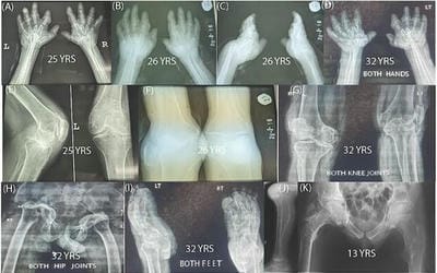
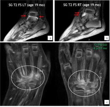
Figure 13: Joint inflammation on MRI. A, B: Presence of joint effusions and synovitis in bilateral ankle compartments at age 19 months. These images are prior to initiation of treatment with anti-rheumatic agents. C: Redemonstration of diffuse inflammation in bilateral wrists after discontinuation of methotrexate therapy (32).

Conclusions

1. The unique features of clinical presentation, atypical radiological findings, failure to obtain remission on medical treatment and consanguinity can guide clinicians towards a diagnosis of this entity. We encourage periodic follow-up of similar cases if they have a developmental delay, with renal function monitoring to avoid renal failure. We have summarized the X-ray findings, which are highly specific, and which confidently differentiate the condition from others. Performance of genetic tests is confirmative.


2. The top differential diagnoses of this entity include MONA and JIA, Winchester syndrome and Farber's disease.

3. Joint disease is not conclusive, since progressive osteolysis with erosions leads to a loss of bone over time. In occasional cases there are acute rheumatological manifestations characterized as inflammatory, which can be stabilized by antirheumatics. However, these do not prevent further progression of osteolysis. The nature of the inflammation in the chronic state is not well understood, and more research on this point is needed.

4. Presentation of an abnormal skeletal phenotype, developmental delay or facial dysmorphic features in a child or a young patient should raise suspicion of this condition. A skeletal survey is then mandatory for diagnosis and for further syndromic work up.

5. The radiological criteria for diagnosis of this condition are based on X-ray at a young age. Ultrasonography is used only for renal assessment and has no specific value in the joint manifestations of the condition. MRI plays a role in the case of rheumatological clinical manifestations for the assessment of joint disease. CT has no useful role in diagnostic work up, while nuclear medicine is used only for pharmacological therapeutic trials. Interventional radiology biopsies for either renal or affected synovial joint have no diagnostic value in this disease.

List of abbreviations:

MCTO: Multicentric carpotarsal osteolysis syndrome.

JIA: Juvenile idiopathic arthritis.

MONA: Multicenteric osteolysis nodulosis arthropathy.

MR: magnetic resonance.

Consent for publication: Informed consent for publication of primary accompanying images was obtained from the patient mother. Other images' license were taken from the publishing research center website, soft copies are available for review by the Editor-in-Chief of this journal.

Declarations:

Ethics approval and consent to participate: Application for ethical approval was done from

the institutional review board office, King Saud University, Riyadh, Saudi Arabia.

Consent for publication: Informed consent for publication of primary accompanying images was obtained from the patient mother. Other images' license were taken from the publishing research center website, soft copies are available for review by the Editor-in-Chief of this journal.

Availability of supporting data: The data used during the current study are not publicly available due to patient privacy but are available from the corresponding author upon reasonable request.

Competing interest: The authors declare that they have no conflict of interest.

Funding: This research received no specific grant from any funding agency.

Copyright: The general skeletal features of Multicentric Carpotarsal Osteolysis: what the radiologist needs to know by Salha Albahkali is marked with CC0 1.0. To view a copy of this license, visit http://creativecommons.org/publicdomain/zero/1.0.

Authors' contributions: Dr.Salha and Dr.Mamdouh was involved in review for the manuscript. Dr.Salha acted as a person who starting review of the literatures, write this manuscript, offered guidance on publication. Dr.Moath is the consultant who provided the primary case images with report, giving the idea.

References

1. Upadia J, Gomes A, Weiser P, Descartes M. A Familial Case of Multicentric Carpotarsal Osteolysis Syndrome and Treatment Outcome. J Pediatr Genet. 2018 Dec;7(4):174-179. doi: 10.1055/s-0038-1657760. Epub 2018 Jun 16. PMID: 30430035; PMCID: PMC6234043.

2. Shaik L, Thotamgari SR, Kowtha P, Ranjha S, Shah RN, Kaur P, Subramani R, Katta RR, Kalaiger AM, Singh R. A Spectrum of Pulmonary Complications Occurring in End-Stage Renal Disease Patients on Maintenance Hemodialysis. Cureus. 2021 Jun 3;13(6):e15426. doi: 10.7759/cureus.15426. PMID: 34249571; PMCID: PMC8254517.

3. Peeters, J., Vanhoenacker, F. M., Mortier, G., & Parizel, P. M. Multicentric carpal-tarsal osteolysis.


Journal of the Belgian Society of Radiology, 2011;94(4), 218-218.‏

4. Park PG, Kim KH, Hyun HS, et al. Three cases of multicentric carpotarsal osteolysis syndrome: a case series. BMC Med Genet. 2018;19(1):164. Published 2018 Sep 12. doi:10.1186/s12881-018-0682-x

5. Ma NS, Mumm S, Takahashi S, Levine MA. Multicentric Carpotarsal Osteolysis: a Contemporary Perspective on the Unique Skeletal Phenotype. Curr Osteoporos Rep. 2023;21(1):85-94. doi:10.1007/s11914-022-00762-7

6. Furness L, Riley P, Wright N, et al. Monogenic disorders as mimics of juvenile idiopathic arthritis. Pediatr Rheumatol Online J. 2022;20(1):44. Published 2022 Jun 18. doi:10.1186/s12969-022-00700-y

7 Mehawej C, Courcet JB, Baujat G, et al. The identification of MAFB mutations in eight patients with multicentric carpo-tarsal osteolysis supports genetic homogeneity but clinical variability. Am J Med Genet A. 2013;161A(12):3023-3029. doi:10.1002/ajmg.a.36151

8. Wenkert D, Mumm S, Wiegand SM, McAlister WH, Whyte MP. Absence of MMP2 mutation in idiopathic multicentric osteolysis with nephropathy. Clin Orthop Relat Res. 2007;462:80-86. doi:10.1097/BLO.0b013e3180d09db8

9. Dworschak GC, Draaken M, Hilger A, Born M, Reutter H, Ludwig M. An incompletely penetrant novel MAFB (p.Ser56Phe) variant in autosomal dominant multicentric carpotarsal osteolysis syndrome. Int J Mol Med. 2013;32(1):174-178. doi:10.3892/ijmm.2013.1373

10. Wu J, Wang L, Xu Y, et al. Multicentric Carpo-Tarsal Osteolysis Syndrome Mimicking Juvenile Idiopathic Arthritis: Two Case Reports and Review of the Literature. Front Pediatr. 2021;9:745812. Published 2021 Oct 15. doi:10.3389/fped.2021.745812
‏‏
11. Mumm S, Huskey M, Duan S, et al. Multicentric carpotarsal osteolysis syndrome is caused by only a few domain-specific mutations in MAFB, a negative regulator of RANKL-induced osteoclastogenesis. Am J Med Genet A. 2014;164A(9):2287-2293. doi:10.1002/ajmg.a.36641.

12. Zankl, A., Duncan, E. L., Leo, P. J., Clark,

G. R., Glazov, E. A., Addor, M. C., ... & Brown, M. A. (2012). Multicentric carpotarsal osteolysis is caused by mutations clustering in the amino-terminal transcriptional activation domain of MAFB. The American Journal of Human Genetics, 90(3), 494-501.‏‏

13. Regev R, Sochett EB, Elia Y, et al. Multicentric carpotarsal osteolysis syndrome (MCTO) with generalized high bone turnover and high serum RANKL: Response to denosumab. Bone Rep. 2021;14:100747. Published 2021 Jan 8. doi:10.1016/j.bonr.2021.100747

14. Klein C, Bellity J, Finidori G, Glorion C, Pannier S. Multicentric carpotarsal osteolysis syndrome: long-term follow-up of three patients. Skeletal Radiol. 2018;47(7):1015-1019. doi:10.1007/s00256-018-2874-z

15. Mudgal P, Murphy A, Weerakkody Y, et al. Skeletal survey. Reference article, Radiopaedia.org (Accessed on 19 Nov 2023) https://doi.org/10.53347/rID-28346

16. Drovandi S, Lugani F, Boyer O, La Porta E, Giordano P, Hummel A, Knebelmann B, Cornet J, Baujat G, Lipska-Ziętkiewicz BS, Ghiggeri GM, Caridi G, Angeletti A. Multicentric Carpotarsal Osteolysis Syndrome Associated Nephropathy: Novel Variants of MAFB Gene and Literature Review. J Clin Med. 2022 Jul 29;11(15):4423. doi: 10.3390/jcm11154423. PMID: 35956038; PMCID: PMC9369440.

17. 17. Chen, K., Zamariolli, M., Soares, M. D. F. D. F., Meloni, V. A., & Melaragno, M. I. Multicentric carpotarsal osteolysis syndrome in a mother and daughter with a MAFB missense variant and natural history of the disease. Molecular Syndromology, 2022;13(1), 50-55.

18. Bhavani GS, Shah H, Shukla A, et al. Clinical and mutation profile of multicentric osteolysis nodulosis and arthropathy. Am J Med Genet A. 2016;170A(2):410-417. doi:10.1002/ajmg.a.37447

19. Vanhoenacker FM, Simoni P, Bosmans F, Boutry N. Congenital and Developmental Abnormalities of the Hand and Wrist. Semin Musculoskelet Radiol. 2021;25(2):260-271. doi:10.1055/s-0040-1722613

20. Faber MR, Verlaak R, Fiselier TJ, Hamel BC, Franssen MJ, Gerrits GP. Inherited multicentric


osteolysis with carpal-tarsal localisation mimicking juvenile idiopathic arthritis. Eur J Pediatr. 2004;163(10):612-618. doi:10.1007/s00431-004-1502-1

21. Ishaq T, Loid P, Ishaq HA, Seo GH, Mäkitie O, Naz S. Clinical, radiographic and molecular characterization of two unrelated families with multicentric osteolysis, nodulosis, and arthropathy. BMC Musculoskelet Disord. 2023;24(1):735. Published 2023 Sep 14. doi:10.1186/s12891-023-06856-2

22. Lazarus S, Tseng HW, Lawrence F, Woodruff MA, Duncan EL, Pettit AR. Characterization of Normal Murine Carpal Bone Development Prompts Re-Evaluation of Pathologic Osteolysis as the Cause of Human Carpal-Tarsal Osteolysis Disorders. Am J Pathol. 2017;187(9):1923-1934. doi:10.1016/j.ajpath.2017.05.007

23. Miyazaki K, Komatsubara S, Uno K, Fujihara R, Yamamoto T. A CARE-compliant article: A case report of scoliosis complicated with multicentric carpotarsal osteolysis. Medicine (Baltimore). 2019;98(48):e17828. doi:10.1097/MD.0000000000017828

24. Zhuang L, Adler S, Aeberli D, Villiger PM, Trueb B. Identification of a MAFB mutation in a patient with multicentric carpotarsal osteolysis. Swiss Med Wkly. 2017;147:w14529. Published 2017 Oct 27. doi:10.4414/smw.2017.14529

25. Aradhya, A. S., Suri, D., & Prakash, A. Multicentric Carpotarsal Osteolysis Mimicking Juvenile Idiopathic Arthritis. INTERNATIONAL JOURNAL OF PEDIATRICS-MASHHAD, 2018; 6(11), 8605-8609.‏

26. Whyte, M. P., Murphy, W. A., Kleerekoper, M., Teitelbaum, S. L., & Avioli, L. V. Idiopathic multicentric osteolysis. Arthritis & Rheumatism: Official Journal of the American College of Rheumatology, 1978;21(3), 367-376.‏

27. Trinkino B, Ma NS. Treatment of a young child with multicentric carpotarsal osteolysis exhibiting joint inflammation and dysfunctional bone formation. Bone Rep. 2023;19:101701. Published 2023 Jul 19. doi:10.1016/j.bonr.2023.101701

28. Sun K, Barlow B, Malik F, Inglis A, Figgie M, Goodman S. Total Hip Arthroplasty in a Patient with Multicentric Carpotarsal Osteolysis:

a Case Report. HSS J. 2016;12(2):177-181. doi:10.1007/s11420-015-9478-0

29. Nishikomori R, Kawai T, Toshiyuki K, Oda H, Yasumi T, Izawa K, Ohara O, Heike T. Remarkable improvement of articular pain by biologics in a Multicentric carpotarsal osteolysis patient with a mutation of MAFB gene. Pediatr Rheumatol Online J. 2015 Sep 28;13(Suppl 1):P152. doi: 10.1186/1546-0096-13-S1-P152. PMCID: PMC4599764.

30. Lerman MA, Francavilla M, Waqar-Cowles L, Levine MA. Denosumab Treatment Does Not Halt Progression of Bone Lesions in Multicentric Carpotarsal Osteolysis Syndrome. JBMR Plus. 2023 Mar 9;7(5):e10729. doi: 10.1002/jbm4.10729. PMID: 37197321; PMCID: PMC10184019.

31. Thomas, C., Bisset, A., Sampson, M., & Armstrong, R. (2006). Multicentric carpal/tarsal osteolysis: Imaging review and 25-year follow-up. Clinical Radiology, 61(10), 892-895. https://doi.org/10.1016/j.crad.2006.03.018

32. Shakiba, M., & Alaei, F. (2023). A novel gene mutation for multicentric osteolysis nodulosis and arthropathy: Case report and review of literature. Heliyon, 9(4).‏

33. Dusatkova, L., Potuznikova, K., Zemkova, D., Marik, I., Rozenkova, K., Lebl, J., ... & Sumnik, Z. Growth characteristics of a girl with multicentric carpo-tarsal osteolysis caused by novel mutation in the MAFB gene. Bone & Mineral, 2016; 136, P1.‏

34. Choochuen P, Rojneuangnit K, Khetkham T, Khositseth S. The First Report of Multicentric Carpotarsal Osteolysis Syndrome Caused by MAFB Mutation in Asian. Case Rep Med. 2018;2018:6783957. Published 2018 Sep 16. doi:10.1155/2018/6783957

35. Bennett WM, Houghton DC, Beals RC. Nephropathy of idiopathic multicentric osteolysis. Nephron. 1980;25(3):134-138. doi:10.1159/000181769