E-ISSN:2320-8686
P-ISSN:2321-127X

Research Article

Abnormal Uterine Bleeding

International Journal of Medical Research and Review

2023 Volume 11 Number 1 January February
Publisherwww.medresearch.in

Histopathological Changes in Endometrial Biopsies of Patients with Abnormal Uterine Bleeding

M.Al-Shikey A.1, A.Abouzrig G.2, A.Al-Gheryani N.3, O.Omar G.4, A. Arhoma A.5, I.Ali I.6*
DOI: https://doi.org/10.17511/ijmrr.2023.i01.02

1 Ainour M.Al-Shikey, Assistant Lecturer, Department of Histology, University of Benghazi, Benghazi, Libya.

2 Ghazala A.Abouzrig, Assistant Lecturer, Department of Histology, University of Benghazi, Benghazi, Libya.

3 Nabeia A.Al-Gheryani, Associate Professor, Department of Pathology, University of Benghazi, Benghazi, Libya.

4 Ghazala O.Omar, Associate Professor, Department of Pharmacology, University of Benghazi, Benghazi, Libya.

5 Amal A. Arhoma, Lecturer, Department of Histology, University of Benghazi, Benghazi, Libya.

6* Iman I.Ali, Professor, Department of Histology, University of Benghazi, Benghazi, Libya.

Introduction: Abnormal uterine bleeding (AUB) is one of the commonest complaints leading to endometrial sampling by endometrial biopsy or curettage. Histopathological analysis revealed various patterns ranging from normal endometrium to malignancy and histopathological examination helps in the diagnosis of these diseases presenting with abnormal uterine bleeding. Objective: To assess the causes of AUB in reproductive, perimenopausal and menopausal women. Methodology: This is a retrospective study that includes a total of 375patients’specimens of endometrial biopsies which were clinically diagnosed as AUB in the department of pathology, faculty of medicine, the university of Benghazi from January 2009 to March 2010. The age of the patient ranged from 20 -80 years, and the mean age was (47.38yr). The patient was categorized into 3 groups with 198 cases in the perimenopausal age group,103 cases in the postmenopausal age group and only 74 cases in the reproductive age group. Results: in this study, the prevalence of non-neoplastic endometrial change was commonly seen in the perimenopausal and reproductive age groups (58.2%), (26.8%) respectively, whereas few cases in the postmenopausal age group (15%). The neoplastic endometrial changes (benign, premalignant, malignant) were commonly seen among the perimenopausal age group, followed by the postmenopausal age group, while neoplasia was rare in the reproductive age group. Conclusion: The causes of AUB depend on the age of the patient. In the reproductive age group, AUB was due to hormonal imbalance, while in perimenopausal and postmenopausal women it was generally due to hyperplasia and malignancy.

Keywords: Abnormal uterine bleeding, Menopause, Endometrial hyperplasia, endometrial carcinoma

Corresponding Author How to Cite this Article To Browse
Iman I.Ali, Professor, Department of Histology, University of Benghazi, Benghazi, , Libya.
Email:
Ainour M.Al-Shikey, Ghazala A.Abouzrig, Nabeia A.Al-Gheryani, Ghazala O.Omar, Amal A. Arhoma, Iman I.Ali, Histopathological Changes in Endometrial Biopsies of Patients with Abnormal Uterine Bleeding. Int J Med Res Rev. 2023;11(1):10-17.
Available From
https://ijmrr.medresearch.in/index.php/ijmrr/article/view/1415

Manuscript Received Review Round 1 Review Round 2 Review Round 3 Accepted
2023-02-04 2023-02-07 2023-02-14 2023-02-21 2023-02-28
Conflict of Interest Funding Ethical Approval Plagiarism X-checker Note
Nil Nil Yes 18%

© 2023by Ainour M.Al-Shikey, Ghazala A.Abouzrig, Nabeia A.Al-Gheryani, Ghazala O.Omar, Amal A. Arhoma, Iman I.Aliand Published by Siddharth Health Research and Social Welfare Society. This is an Open Access article licensed under a Creative Commons Attribution 4.0 International License https://creativecommons.org/licenses/by/4.0/ unported [CC BY 4.0].

Introduction

The uterus is a hollow organ, normally about the size and shape of a medium-sized pear, located between the bladder and the rectum. It functions to nourish and house a fertilized egg until the fetus, or offspring is ready to be delivered [1]. Abnormal uterine bleeding can occur any time between menarche and menopause, and in both ovulatory and anovulatory cycles. AUB is one of the most common causes in patients undergoing hysterectomy [2].

Abnormal uterine bleeding (AUB) is defined as the bleeding pattern that differs in frequency, duration and amount from a pattern observed during a normal menstrual cycle or after menopause [3]. Abnormal uterine bleeding is one of the commonest complaints leading to endometrial sampling by endometrial biopsy or curettage [4].

Histopathological examination of the endometrial biopsies and curetting revealed various patterns ranging from physiological to pathological lesions of the endometrium[5]. Proliferative and secretory endometrium were the two most common histopathological patterns which were seen in all three age groups[6]. A disordered proliferative pattern lies at one end of the spectrum of proliferative lesions of the endometrium that includes carcinoma at the other end with intervening stages of hyperplasia[7]. Atrophic endometrium is also one of the causes of AUB and was most common in postmenopausal women[5]. The exact cause of bleeding in atrophic endometrium is not known, it is thought to be due to anatomic vascular variations or local abnormal defective local homeostatic mechanisms [7]. Weakly proliferative endometrium was seen as common in the reproductive and perimenopausal age groups. It represents an intermediate point between profound atrophy of total estrogens deprivation and the normal proliferative phase response to cyclic estrogens production[8].

Endometrial hyperplasia is a precursor of endometrial cancer. It is more commonly seen during the perimenopausal period[10]. Hyperplasia is classified as simple or complex based on the absence or presence of architectural abnormalities such as glandular complexity and crowding. They are further designated as atypical if they demonstrate nuclear atypia [5].

Material and Methods

Study design: The current study was a retrospective study conducted from archives of the histopathology register, of the department of pathology, faculty of medicine, university of Benghazi-Libya. The study period was from January 2009 to March 2010.

Inclusion criteria: any patient with AUB from 18 to 80 years was included in this study.

Data collection: A total number of 375 specimens of endometria curettage and biopsy. The formalin-fixed samples were routinely processed and 4-5µ thick sections were cut from paraffin blocks. The sections were stained by routine hematoxylins and eosin stains. The patient was categorized into three age groups Reproductive& Perimenopausal and Postmenopausal age group. The data was classified into non-neoplastic endometrial changes and neoplastic endometrial changes. Demographic data were examined regarding the common endometrial changes according to age group and histopathological findings in different endometrial changes.

Statistical analysis: The entire data were evaluated statistically by using the SPSS statistical package version 18, independent t-test was used to correlate the mean age between patients with non-neoplastic and neoplastic endometrial changes. A P-value of less than 0.05 was considered statistically significant.

Ethical consideration: The present study was performed according to the Ethical Guidelines for Clinical Research based on the declaration of Helsinki and verbal consent was obtained from all patients or the patient's family.

Results

This study includes a total of 375 cases which were clinically diagnosed as AUB, the age of the patient ranged from (20 -80 years), and the mean age was (38-47yr). The patient was categorized into 3 groups with the maximum number of cases observed in the perimenopausal age group198 case (52.8%), followed by the postmenopausal age group with 103 cases (27.5%) and minimal in the reproductive age group with 74 cases (19.7%) (Figure1).


Out of 375 cases observed that 222 cases (59.2%) of neoplastic endometrial changes, while 153 cases (40.8%), were noted non-neoplastic endometrial changes, the large number of cases presented with a neoplastic endometrial change in this study was seen in perimenopausal age group 109 cases ( 49.1%), followed by postmenopausal age group 80 case (36%), and the least age group affected were noted among reproductive age group 33case (14.9%), while non-neoplastic endometrial change commonly seen in perimenopausal and reproductive age group.89 cases (58.2%) & 41case (26.8%) respectively, and 23 cases in the postmenopausal group(15%)(Figure 2).

ijmrr_1415_01.JPG
Figure 1: Distribution of patients according to age groups (n= 375)

ijmrr_1415_02.JPG
Figure 2: Age distribution of patients with neoplastic & non-neoplastic endometrial lesions


Non-Neoplastic Endometrial changes 

The most common histological finding among non-neoplastic endometrial changes was normal endometrial pattern accounted118case (77.12%), followed by a disorder of proliferative 28 cases (18.30%) and the least number of cases accounted for hormonal change only7 cases (4.58%)

Among normal endometrial changes,64 cases (54.24%) had a secretory phase, 34 cases (28.82%) had proliferative phases, and only 20 cases (16.94%) had an atrophic phase.

The incidence of the secretory phase was 59.4%, 35.9% and 4.7 % in the perimenopausal, reproductive and postmenopausal age groups respectively, whereas the proliferative phase was common among the perimenopausal age group 73.5%, and accounted for 17.6% in reproductive and8.8% postmenopausal age group respectively. atrophic endometrial changes were noted as more common in the older age groups as the postmenopausal age group 75%, followed by the perimenopausal age group 25%, with no case reported among the reproductive age group, P V =0.00 Highly significant (Figure 3)

ijmrr_1415_03.JPG

Figure 3: Age distribution of Normal endometrial pattern (n= 118).

Neoplastic endometrial changes

The most common histopathological finding was benign neoplastic endometrial changes in 143 cases out of 222 cases ( 64.43%), followed by premalignant neoplastic endometrial changes in 57 cases ( 25.67%).


Whereas malignant neoplastic endometrial changes account for 22 cases (9.90%) The benign endometrial changes, among these 112 cases had simple hyperplasia and 31 cases had endometrium polyps in the current study observed that simple hyperplasia was more common in the elderly patient so that perimenopausal age group accounted 63 cases (56.3%), postmenopausal 26 cases (23.2%) and reproductive .age group 23 case (20.5%) .while the endometrial polyp are more common in postmenopausal patient 21 cases (67.7%), while others age group were less common such as perimenopause and reproductive age group 7 case (22.6%) & 3 cases (9.7%) respectively, P value=0.000 (Highly Significant)( Figure 4).

ijmrr_1415_04.JPG
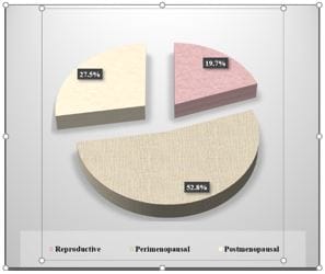
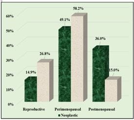
Figure 4: Age distribution of Benign endometrial lesion (n=143)

The next most common neoplastic endometrial change was premalignant changes were observed that42 cases (73.68%), had hyperplasia with atypia and15 cases(26.32%), were noted hyperplasia without Atypia, Twenty-two cases of malignant neoplastic endometrial changes were seen in the current study,19 cases (86.36%) of endometrial carcinoma, and only 3 cases (13.64%) of sarcoma.

The current study observed that the perimenopausal age group most commonly had benign endometrial changes ,premalignant endometrial changes, and malignant endometrial changes 70/143 case (49%), 28/57 case (49.1%) and 11/22 case (50%) respectively and second common age group affected in this study were postmenopausal age group had benign endometrial changes were 47/143 case (32.9%) while premalignant endometrial changes were 23/57 case (40.4%) and malignant were observed in 10/22 case (45.5%)

The least age group affected with neoplastic endometrial changes ,were noted in reproductive age group had benign endometrial changes 26/143 case (18.2%),premalignant 6/57 case (10.5%) and malignant only 1/22 case (4.5%) ,results was highly significant ( Figure 5).Malignant neoplastic endometrial changes, were noted more common among elderly age group such as ,endometrial carcinoma more common in postmenopausal age group 10 case (52.6%) ,followed by perimenopausal age group 8 case (42.1%),while uncommon in younger age group such as reproductive age group was only one case (5.3%).Regarding sarcoma were observed only 3 case (100%) in perimenopause age group only and P value = 0.044 results was highly significant. Figure 6.

ijmrr_1415_05.JPG

Figure 5: Age distribution of Neoplastic endometrial lesion

ijmrr_1415_06.JPG

Figure 6: Distribution of patients according to Malignant endometrial lesion (n=22).


ijmrr_1415_07.JPG
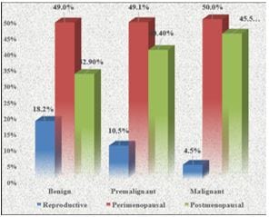
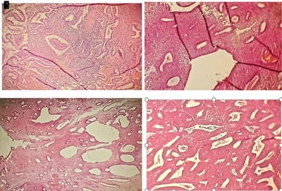
Figure 7: A. Section of secretory endometrium, showed glands with vacuolated cells (H&EX100).B. Section of simple cystic endometrial hyperplasia, showed a thick active stroma and hyperplastic glands with focal cystic changes (H&EX100).C. Section of Endometrial polyp, showed of dilated irregular glands, with fibrous stroma and thick wall blood vessels (H&E X100).D. Section of Endometrial hyperplasia with Atypia showed compact hyperplastic glands and thick active stroma (H&EX100)

Discussion

The endometrium is a hormonally sensitive, and responsive tissue which constantly changes the active reproductive life[11]. The most common indication of endometrial sampling is the workup of abnormal uterine bleeding[12]. A total number of 375 endometrial biopsies were presented with AUB. age of patient ranged from20-80yr, the main age was 47.38yr.However, in this study were observed the perimenopausal age group, accounted for 198 cases (52.8%), followed by 103case (27.5%) in was postmenopausal age group, and only 74 cases (19.7%) in was reproductive age group, these findings were similar to study done by[13]. It observed that the mean age was 45.38yr, with a minimum and maximum age of 28 and 70yr respectively, and the most common cases were in the perimenopausal age group In the current study were observed that 222 cases (59.25%) of neoplastic endometrial changes, while 153case (40.8%) were non-neoplastic endometrial changes. The incidence of neoplastic endometrial changes was higher than non-neoplastic could be due to the number of cases selected and the duration of the study. The incidence of non-neoplastic endometrial changes was common in the perimenopausal age group in 98 cases (58.2%), followed by the

reproductive age group in 41 cases (26.8%), and less frequently was seen in the postmenopausal age group. While common neoplastic endometrial changes were observed among the perimenopausal age group 109 cases (49.1%), followed by the postmenopausal age group 80 cases (36%), and the reproductive age group 33 cases (14.9%). A study was done by [11]. Where observed that the age of patients ranged from 21-78 years, with a mean age group was 49.5yr, and the highest incidence of AUB was found in 40-49yr of age. All these findings are in agreement with the current study[13]. Who observed that the prevalence of AUB increases with increasing age peaking before menopause could be that perimenopausal women have anovulatory cycles leading to AUB. A study was done by [11]. Who observed that the prevalence of AUB increase with increasing parity, but we do not include the parity in this study because of no information from sources? According to non-neoplastic results in the cases enrolled in the current study, the majority of them had normal endometrial changes,118case (77.12%) it was found that 64 cases (54.23%), had secretary endometrial changes,34 cases (28.81%) had proliferative endometrial changes, and 20case (16.94%) had atrophic endometrial changes. A similar finding was seen in a study done by[9],[14]. The secretory endometrial changes were founded in a higher number of cases (30.8%) and (35.4%) respectively.

A study by[15,9] and [16]. Were all observed proliferative endometrial changes more common than secretory endometrial changes (41.88%) (31%) and (75%) respectively, all these finding disagreements with the current results The secretory endometrial changes and proliferative endometrial changes were seen in the current study, more common among perimenopausal age group (59.4%), (73.5%) respectively, followed by reproductive age group(35.9%), (17.6%). However, a few cases were observed among the postmenopausal age group (4.7%), (and 8.8%) respectively. A study done by [11] found that most cases of proliferative endometrial change were among the perimenopausal age group, this finding was in agreement with the current study Moreover atrophic endometrial changes were seen, among the postmenopausal age group 15 cases (75%), followed by perimenopausal age group 5cases (25%), These finding in concordance


with most of the previous studies, and it is the most common cause of bleeding in postmenopausal age group[17] might be because of thin-walled veins, superficial to the expanding cystic gland, make the vessels vulnerable to injury and lead to excessive uterine bleeding[11]. The second common pattern of non-neoplastic endometrial changes was a disorder of proliferative 28 cases out of a total of 153 cases of non-neoplastic endometrial changes, the most common cases among the perimenopausal age group, (67.9%), followed by the reproductive age group was (28.6%), and only one case (3.6%) among the postmenopausal age group. A study done by[18] observed that the incidence of disorder proliferative changes is more common among the reproductive age group 20%, followed by the perimenopausal age group, while few numbers in the postmenopausal age group, the last finding similar to the current finding. However present results according to the disorder of proliferative were observed Slightly higher incidence than studies done by [22] &[20] (12%) and (13%) respectively. [21, who explained the cause of the disorder proliferative endometrium, as there is chronic anovulation, ovarian follicles persist for some time and produce estradiol before undergoing atresia, leading to the abundant proliferation of endometrium and mild disorganization of architecture, This produces widespread dilatation of glands. this is called the disorder proliferative endometrium. The least number of cases among non-neoplastic endometrial changes were observed Hormonal changes, was (4.58%) were commonly seen in reproductive age (57.1%), followed by the perimenopausal age group (28.6%), and only one case in postmenopausal age group (14.3%), Hormonal changes more commonly seen among the reproductive age group, could be because of hormonal imbalance, such as normal physiological changes or exogenous hormone therapy for infertility management. Regarding the most common finding of neoplastic endometrial changes was a benign change (64.41%), followed by premalignant (25.67%), and the least incidence of neoplastic change was a malignant change in 22 cases (9.90%). Simple endometrial hyperplasia was observed in the current study more in the perimenopausal age group, (56.3%), followed by postmenopausal age group 2(23.2%), while less number of the case was noted in the reproductive age group (20.5%), these finding supported by a

study done by[22] found it (56.4%) more common in perimenopausal age group. In a similar study by [23] was observed that two-thirds of cases in the perimenopausal age group and the incidence of simple endometrial hyperplasia peak around perimenopausal and postmenopausal women were observed by[24].

The variation in the incidence of simple endometrial hyperplasia among different studies could be attributed to socioeconomic status and the occurrence of risk factors like obesity, diabetes, sedentary lifestyle, and early diagnosis. Identification of endometrial hyperplasia is important as it is thought to be a precursor of endometrial carcinoma [25]. The second most common benign endometrial change where endometrial polyps were observed commonly among postmenopausal a (67.7%), followed by the perimenopausal age group (22.6%), the least cases among reproductive age group (9.7%). However, other studies have shown, a progressively increased detection pattern of endometrial polyps in older age groups [26]. The premalignant endometrial changes accounted (for 25.67%) and were more common among the perimenopausal age group (45.2%), followed by the postmenopausal age group (40.5%), rare was seen in the reproductive age group (14.3%) only. Premalignant hyperplasia without atypia was commonly seen in the perimenopausal age group, followed by the postmenopausal age group(60% & 40%) respectively Study of [11], observed that the incidence of endometrial hyperplasia was highest after 4th decade of life suggesting that the incidence of endometrial hyperplasia increase with age, these finding in concordance with the current study. The frequency of malignant endometrial changes in the current study were (9.90%) of total endometrial biopsy, out of 22 cases 19 cases of endometrial carcinoma, more commonly observed in the postmenopausal age group (52,6%), followed by the perimenopausal case (42.1%), while 3 cases of sarcoma only seen in perimenopausal age group 3 cases, and one case only was observed among reproductive age group regarding malignant changes.

Low incidence of endometrial carcinoma has been obtained in studies by [27] (2.6%),[6](1.8%), [20]( 2.4%) and [23]( 1%) all these studies had Asian women as subjects and reflected an overall lower incidence of endometrial carcinoma in Asian women


due to early childbearing, lesser obesity and a more active lifestyle. [11] who also observed endometrial carcinoma was highest after 4th decade of life suggesting that, the incidence of endometrial carcinoma increases with age.

Conclusion

Abnormal uterine bleeding is a common gynae-pathological problem with multiple causes, varying with age and different clinical presentation and AUB occurs secondary to a wide variety of structural and functional abnormalities in women especially of the perimenopausal age group so that histopathology remains the gold standard in diagnosis. AUB was the most common clinical presentation were seen in this study. The neoplastic endometrial changes (benign, premalignant, malignant) were observed commonly seen among the perimenopausal age group, followed by the postmenopausal age group, while rare in the reproductive age group. However, the causes of AUB vary with age, and in the reproductive age group, the causes of AUB are due to hormonal imbalance, while in perimenopausal and postmenopausal women is generally due to hyperplasia and malignancy. Moreover, significant morbidity or mortality can occur if endometrial hyperplasia is untreated with progression to malignancy so the evaluation of AUB by histopathology can be lifesaving with early tissue diagnosis and management.

What does this study add to existing knowledge?

Abnormal uterine bleeding is a common clinical problem seen in different age groups, the causes of this bleeding are dependent on many factors such as age, genetic and environmental factors. This study has attempted to assess the correlation between the causes and the age of a patient with AUB and All patients with AUB required regular follow-up and further evaluation to exclude malignancies.

Author’s contribution: Iman Ali: Concept, research design, recruitment of participants, data preparation, data analysis, manuscript preparation. Ainour M.AL-Shikey: research design, manuscript preparation. Nabeia A.Al Gheryani manuscript review, supervision. Ghazala O.Omar: research design, manuscript review. Ghazala A. Abouzrig: data preparation, data analysis. Amal A.Arhoma: data analysis and manuscript preparation.

Reference

01. Anthony M. Junqueiraʾs basic histology and atlas. 14th ed. New York. McGraw-Hill Education; 2016.460 p [Crossref][PubMed][Google Scholar]

02. M Kakar. Clinicopathological evaluation of abnormal uterine bleeding. J Health Res Rev:2015 ;62(2):45-9. . [Crossref][PubMed][Google Scholar]

03. John W Ely, Colleen M Kennedy, Elizabeth C Clark. Abnormal uterine bleeding. The Journal of the American Board of Family Medicine. 2006 nov;19 (6) :590-602. [Crossref][PubMed][Google Scholar]

04. Rosai J. Ackerman’s. Surgical Pathology Text book of pathology. 11th ed. Elsevier In:2013.440 p [Crossref][PubMed][Google Scholar]

05. Vaidya S , Lakhey M , Amatya S. Histopathological pattern of abnormal uterine bleeding in endometrial biopsies. Nepal Medical College Journal. 2013;15(1):74– 77. [Crossref][PubMed][Google Scholar]

06. Abdullah LS. Histopathological pattern of endometrial sampling performed for Abnormal uterine bleeding. Bahrain Med Bull. 2011;33 (4):4-1. [Crossref][PubMed][Google Scholar]

07. Doraiswami S, Johnson T, Rao S. Study of endometrial pathology in abnormal uterine bleeding. J Obstetric Gynecol India. 2011;15 ( 1) :74– 77. [Crossref][PubMed][Google Scholar]

08. Zaino R. Abnormal proliferations, hyperplasia and polyps in interpretation of endometrial biopsies and curetting. Lippincott-Raven Publisher. 1996;59(8): 801–812. [Crossref][PubMed][Google Scholar]

09. Muzaffar M, Akhtar KAK, Yasmin S. Menstrual irregularities with excessive blood loss: a clinic- pathological correlation. J Pak Med Assoc. 2005;(8): 305-16. [Crossref][PubMed][Google Scholar]

10. Rosai J. Ackerman’s. Surgical Pathology Text book of pathology. 9th ed. Elsevier Inc.2005. 990 p [Crossref][PubMed][Google Scholar]

11. V Vijay , Ch J yothi , M Narsing Rao , V Sailaja , M. N. P Charan Paul. Study of histopathological pattern of endometrium in abnormal uterine bleeding. Saudi J Pathol Microbiol . 2017 ; 3 (4) :l-2 [Crossref][PubMed][Google Scholar]


12. Tiwari A, kaur N, Jain S, Rai R, Jain S. Histopathological study of endometrial biopsy specimens for abnormal uterine bleeding. J Lumbini Med Coll. 2016 sep;8 (16):598-606. [Crossref][PubMed][Google Scholar]

13. Manisha Laddad , Nitin Kshirsagar , Sanjay Patil , Gauri Shinde. Study of adolescent abnormal uterine bleeding (AUB) and its outcome in tertiary centre. Akshantala Enterprises Private Limited. 2015nov;4 ( 69) :66-69. [Crossref][PubMed][Google Scholar]

14. Khan S, Hammed S, Umber A. Histopathological pattern of endometrium on diagnostic D&C in patients with abnormal uterine bleeding. KEMU. 2011;17(2): 166. [Crossref][PubMed][Google Scholar]

15. Naya K, Rekha Ratnani1, Naik Meena. A clinico-pathological analysis of causes of abnormal uterine bleeding. J Pathol Microbiol. 2017;20 (2):105-111. [Crossref][PubMed][Google Scholar]

16. Somboonporn , Padma SK, Shetty KJ. Study of histopathological patterns of endometrium in abnormal uterine bleeding. Journal of Health and Research. 2000 ;40(8): 815–819. [Crossref][PubMed][Google Scholar]

17. Cornitescu F, Tanase F , Simionescu F & Iliescu D. Clinical histopathological and therapeutic considerations in non-neoplastic abnormal uterine bleeding in menupause transition. Rom J Morphol Embryol. 2011;17(6):429-37. [Crossref][PubMed][Google Scholar]

18. Nabeia G, and Hanan M. Endometrial biopsies in female with abnormal uterine bleeding. pathology department Benghazi University. 2020;21(9):1-9. [Crossref][PubMed][Google Scholar]

19. Sajitha K. Study of histopathological pattern of endometrium in abnormal uterine bleeding. Journal of Health and Research. 2014 ;(1) :76-81. [Crossref][PubMed][Google Scholar]

20. Vaidya S, Lakhey M, Vaidya S. Histopathological pattern of abnormal uterine bleeding in endometrial biopsies. Nepal Med Coll J. 2013 ;64(29): 18-22. [Crossref][PubMed][Google Scholar]

21. Kurman RJ, Ellenson LH, Ronnett BM. Benign diseases of the endometrium. Mc Cluggage WG New York. Springer Verlag. 2011;17(2): 166 [Crossref][PubMed][Google Scholar]

22. Sajitha K. Study of histopathological pattern of endometrium in abnormal uterine bleeding. Journal of Health and Research. 2014; (1) :76-81. [Crossref][PubMed][Google Scholar]

23. Baral R , Pudasaini S. Histopathological pattern of endometrial samples in abnormal uterine bleeding. J Path Nepal. 2011;37 (4): 6 - 55. [Crossref][PubMed][Google Scholar]

24. Reed SD, Newton KM, Clinton WL, Epplein M, Garacia R, Allison K. Incidence of endometrial hyperplasia. Am J Obstet gynecol. 2009;(39):1297-1336. [Crossref][PubMed][Google Scholar]

25. Khan R, Sherwani RK, Rana. Clinico-pathological patterns in women with dysfunctional uterine bleeding. Iran J Pathol. 2016; (1):20-6. [Crossref][PubMed][Google Scholar]

26. Sahinoglu Z, Kuyumcuoglu U. An unusual case of postmenopausal vaginal bleeding retention of fetal bone. Arch Gynaecol. 2003;101 (18):1280-3. [Crossref][PubMed][Google Scholar]

27. Saraswathi D, Thanka J, Shalinee R. Study of endometrial pathology in abnormal uterine bleeding. Obstet Gynecol India. 2011; 61(4): 426–430. [Crossref][PubMed][Google Scholar]