E-ISSN:2320-8686
P-ISSN:2321-127X

Case Report

Systemic lupus erythematosus

International Journal of Medical Research and Review

2022 Volume 10 Number 6 November-December
Publisherwww.medresearch.in

A Tale of Two Male SLES (Systemic lupus erythematosus)

Pavithira A.1, Sucharitha S.2*
DOI: https://doi.org/10.17511/ijmrr.2022.i06.02

1 A. Pavithira, Senior Resident, Department of General Medicine, NRI Medical College and General Hospital, Chinnakakani, Mangalagiri, Mangalagiri, Andhra Pradesh.

2* S. Sucharitha, Post graduate, Department of General Medicine, NRI Medical College and General Hospital, Chinnakakani, Mangalagiri, Mangalagiri, Andhra Pradesh.

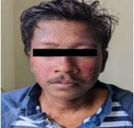
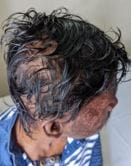
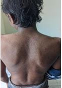
SLE is an autoimmune disease with multisystem manifestations. SLE is more common in females typically beginning in the childbearing years suggesting a role for both hormones and as yet uncharacterized sex-related factors in the disease pathogenesis. The prevalence of the disease being one male patient for every 8-15 patients, the disease manifestations in male patients are considered to be graver than in females. Here is a case reporting of two males who came with different presentations of SLE , the first patient presented with rash,fever,alopecia,and arthralgia of small joints , the second patient presented with fever, easy fatiguability ,generalised edema ,breathlessness on exertion .Both of them were evaluated and found to have strong positive for ANA and ds DNA and EULAR ACR criteria for SLE was applied and both were diagnosed as SLE .They were treated with steroids and other immunosuppressants .Clinical improvement was noticed in both patients.

Keywords: SLE, male, Anemia, Alopecia, ANA

Corresponding Author How to Cite this Article To Browse
S. Sucharitha, Post graduate, Department of General Medicine, NRI Medical College and General Hospital, Chinnakakani, Mangalagiri, Mangalagiri, Andhra Pradesh, .
Email:
A. Pavithira, S. Sucharitha, A Tale of Two Male SLES (Systemic lupus erythematosus). Int J Med Res Rev. 2022;10(6):171-175.
Available From
https://ijmrr.medresearch.in/index.php/ijmrr/article/view/1406

Manuscript Received Review Round 1 Review Round 2 Review Round 3 Accepted
2022-11-24 2022-11-26 2022-12-03 2022-12-10 2022-12-17
Conflict of Interest Funding Ethical Approval Plagiarism X-checker Note
Nil Nil Yes 19%

© 2022by A. Pavithira, S. Sucharithaand Published by Siddharth Health Research and Social Welfare Society. This is an Open Access article licensed under a Creative Commons Attribution 4.0 International License https://creativecommons.org/licenses/by/4.0/ unported [CC BY 4.0].

Introduction

SLE is an autoimmune disease which is more common in females and has a multi system involvement with varied presentations.

Here we have two male patients who had two different presentations of SLE.

One patient is young presented with fever, dermatological and haematological manifestations with skeletal system involvement and diagnosed as SLE and treated accordingly and significant improvement was seen.

The other patient is a 62 years old man severe fatiguability, with anaemia and jaundice with features of congestive cardiac failure and was diagnosed as SLE and treated accordingly.

From the above cases, though SLE is rare in males we should have in mind regarding possible clinical presentation so that early diagnosis and appropriate treatment can be provided.

Case 1

A 21 years old male patient presented with erythematous rash over the face and back. He also complained of fever as intermittent episodes over 2 months, alopecia, easy fatiguability and arthralgia involving the small joints of both hands.

On examination the rash was found to be discoid rash with non-scarring alopecia(fig).

ijmrr_1406_01.jpg

ijmrr_1406_02.JPG

ijmrr_1406_03.JPG

Investigations revealed the following:

Hemoglobin: 6.6 gm/dL with schistocytes in the peripheral smear and increased reticulocyte count.


Direct Coomb's test: Positive

LDH: 650

Platelet count: 80,000/cu.mm

Complement C3: 0.38 (low)

Complement C4: 0.049 (low)

ANA: Strong Positive with a nuclear fine granular pattern

Anti-ds-DNA: Positive

Anti SmD1: Positive ++

Anti SS-A/Ro 60: Positive ++

24 hours urinary protein: 504mg/day

The patient was diagnosed to be having SLE and was started on Tab Prednisone 40 mg OD, Tab MMF 1g BD and Tab Hydroxychloroquine 400mg BD. The rash, arthralgia and alopecia improved in 3 weeks significantly. Hemoglobin improved to 10 gm/dL in 4 weeks.

Case 2

A 62 years old male presented with pedal edema, fever, breathlessness on exertion and easy fatiguability gradually progressing over a month. Examination revealed jaundice and hepatosplenomegaly.

Investigations revealed the following:

Hemoglobin: 2.5gm/dL with schistocytes in the peripheral smear and elevated reticulocyte count.

WBC count: 10,500/cumm.

Platelet count: 30,000/cumm.

LDH: 549 IU

Direct Coomb's test: Positive

LFT: elevated direct bilirubin

ANA: Strong Positive

Anti-ds-DNA: Positive

Complement C3: 0.31 (low)

Complement C4: 0.029 (low)

24 hours urinary protein: 160mg/day

The patient was diagnosed to have SLE with AIHA in congestive cardiac failure and started on

Tab Prednisone 40 mg/day and Tab Hydroxychloroquine 400mg BD. The hemoglobin improved to 8 gm/dL in 4 weeks with complete resolution of congestive features.

Discussion

Systemic lupus erythematosus (SLE) is autoimmune, heterogeneous, multi system disease with relapsing and remitting course with the rise in the occurrence of autoimmune diseases due to better detection methods, it is important to understand and revisit the clinical profiling of SLE patients based on their gender.

SLE is more common in women than men, and its incidence is being markedly increasing in the women of child bearing years.[1]

Female to male ratio of 8–15:1 and this suggests some hormonal influence.[2]

Potential causes of the female predilection for SLE include the effects of estrogen and its hydroxylation, decreased androgen levels, hyperprolactinemia and differences in gonadotropin-releasing hormone (GnRH) signalling.[3]

The most accepted theory in explaining the female predilection is drawn from the fact that levels of estrogen metabolites, 2-hydroxy and 16-hydroxy estrones are found elevated in women with SLE when compared to women without SLE.[4]

The disease is found to be more severe in women more than 40 years of age, male gender and paediatric age group according to a review study done by Pons-estel et al. [5]

SLE shows significant sex-specific features. Though the incidence may favour the female sex more, the complications tend to contradict this favour and men are affected by a more severe disease with regard to both renal and extra-renal manifestations.

Additionally, men are at a higher risk of developing ESRD which may require an increased awareness and monitoring in clinical practice. [6]

The impairment occurs in older men have higher mortality in one year than women with SLE, suggesting that even men with lupus have a more complex clinical course than women.[7]

Haematological involvement may exist more frequently in male SLE.


Male dominance in haemolytic anaemia, lymphopenia, and thrombocytopenia were reported in several studies. [8,9]

Serositis, discoid rash and higher incidence of subcutaneous lupus erythematosis found more commonly in males. [10,11]

Specker et al in their study showed the strikingly high incidence of thrombotic complications, cardiovascular damage, and ESRD in male SLE patients compared to their female counterparts.[12]

In a study done by Molina JF et al, Males were found to have increased incidence of renal disease, anti-ds DNA, vascular thrombosis.[13]

Garcia MA et al study showed that fever and weight loss at onset are found more common in male SLE.[14]

In spite of the above conclusions having been drawn in the past from many studies SLE is still not being given its deserving place in the differential diagnoses if the patient is a male.

Conclusion

The female predilection for SLE makes the suspicion of SLE in males far-fetched and hence results in delay in the workup and diagnosis. The above two cases prove that age and gender should not limit our suspicion of SLE as both are males and the second patient is elderly.

Reference

01. Cervera R, Khamashta MA, Font J, Sebastiani GD, Gil A, Lavilla P, Mejía JC, Aydintug AO, Chwalinska-Sadowska H, de Ramón E, Fernández-Nebro A, Galeazzi M, Valen M, Mathieu A, Houssiau F, Caro N, Alba P, Ramos-Casals M, Ingelmo M, Hughes GR; European Working Party on Systemic Lupus Erythematosus. Morbidity and mortality in systemic lupus erythematosus during a 10-year period: a comparison of early and late manifestations in a cohort of 1,000 patients. Medicine (Baltimore). 2003 Sep;82(5):299-308. doi: 10.1097/01.md.0000091181.93122.55. PMID: 14530779 [Crossref][PubMed][Google Scholar]

02. Ortona E, Pierdominici M, Maselli A, Veroni C, Aloisi F, Shoenfeld Y. Sex-based differences in autoimmune diseases. Ann Ist Super Sanita. 2016 Apr-Jun;52(2):205-12.

doi: 10.4415/ANN_16_02_12. PMID: 27364395 [Crossref][PubMed][Google Scholar]

03. Yacoub Wasef SZ. Gender differences in systemic lupus erythematosus. Gend Med. 2004 Aug;1(1):12-7. doi: 10.1016/s1550-8579(04)80006-8. PMID: 16115579 [Crossref][PubMed][Google Scholar]

04. Kassi E, Moutsatsou P. Estrogen receptor signaling and its relationship to cytokines in systemic lupus erythematosus. J Biomed Biotechnol. 2010;2010:317452. doi: 10.1155/2010/317452. Epub 2010 Jun 10. PMID: 20617147; PMCID: PMC2896666 [Crossref][PubMed][Google Scholar]

05. Pons-Estel GJ, Alarcón GS, Scofield L, Reinlib L, Cooper GS. Understanding the epidemiology and progression of systemic lupus erythematosus. Semin Arthritis Rheum. 2010 Feb;39(4):257-68. doi: 10.1016/j.semarthrit.2008.10.007. Epub 2009 Jan 10. PMID: 19136143; PMCID: PMC2813992 [Crossref][PubMed][Google Scholar]

06. Ramírez Sepúlveda, J. I. , Bolin, K. , Mofors, J. et al. Sex differences in clinical presentation of systemic lupus erythematosus. Biol Sex Differ 10, 60 (2019). [Article][Crossref][PubMed][Google Scholar]

07. Prete PE, Majlessi A, Gilman S, Hamideh F. Systemic lupus erythematosus in men: a retrospective analysis in a Veterans Administration Healthcare System population. J Clin Rheumatol. 2001 Jun;7(3):142-50. doi: 10.1097/00124743-200106000-00003. PMID: 17039119 [Crossref][PubMed][Google Scholar]

08. Sthoeger ZM, Geltner D, Rider A, Bentwich Z. Systemic lupus erythematosus in 49 Israeli males: a retrospective study. Clin Exp Rheumatol. 1987 Jul-Sep;5(3):233-40. PMID: 3427838 [Crossref][PubMed][Google Scholar]

09. Kaufman LD, Gomez-Reino JJ, Heinicke MH, Gorevic PD. Male lupus: retrospective analysis of the clinical and laboratory features of 52 patients, with a review of the literature. Semin Arthritis Rheum. 1989 Feb;18(3):189-97. doi: 10.1016/0049-0172(89)90061-9. PMID: 2648578 [Crossref][PubMed][Google Scholar]

10. Font J, Cervera R, Navarro M, Pallarés L, López-Soto A, Vivancos J, Ingelmo M. Systemic lupus erythematosus in men: clinical and immunological


characteristics. Ann Rheum Dis. 1992 Sep;51(9):1050-2. doi: 10.1136/ard.51.9.1050. PMID: 1417135; PMCID: PMC1004835 [Crossref][PubMed][Google Scholar]

11. Kyttaris VC. Systemic lupus erythematosus: from genes to organ damage. Methods Mol Biol. 2010;662:265-83. doi: 10.1007/978-1-60761-800-3_13. PMID: 20824476; PMCID: PMC3153363 [Crossref][PubMed][Google Scholar]

12. Specker C, Becker A, Lakomek HJ, Bach D, Grabensee B. Systemischer Lupus Erythematodes bei Männern--Eine andere Krankheitsprognose? [Systemic lupus erythematosus in men--a different prognosis?]. Z Rheumatol. 1994 Nov-Dec;53(6):339-45. German. PMID: 7871906 [Crossref][PubMed][Google Scholar]

13. Molina JF, Drenkard C, Molina J, Cardiel MH, Uribe O, Anaya JM, Gomez LJ, Felipe O, Ramirez LA, Alarcon-Segovia D. Systemic lupus erythematosus in males. A study of 107 Latin American patients. Medicine (Baltimore). 1996 May;75(3):124-30. doi: 10.1097/00005792-199605000-00002. PMID: 8965681 [Crossref][PubMed][Google Scholar]

14. Garcia MA, Marcos JC, Marcos AI, Pons-Estel BA, Wojdyla D, Arturi A, Babini JC, Catoggio LJ, Alarcon-Segovia D. Male systemic lupus erythematosus in a Latin-American inception cohort of 1214 patients. Lupus. 2005;14(12):938-46. doi: 10.1191/0961203305lu2245oa. PMID: 16425573 [Crossref][PubMed][Google Scholar]