E-ISSN:2320-8686
P-ISSN:2321-127X

Review Article

Axial Spondyloarthritis

International Journal of Medical Research and Review

2022 Volume 10 Number 5 September-October
Publisherwww.medresearch.in

The window of opportunity in axial spondyloarthritis: A Stitch in Time

Ghosh P.1*, Kumar Sarma P.2, Shenoy P.3, Mukherjee D.4, Desai A.5
DOI: https://doi.org/10.17511/ijmrr.2022.i05.04

1* Parasar Ghosh, Professor and HOD, Department of Clinical Immunology and Rheumatology, Institute of Postgraduate Medical Education and Research and SSKM Hospital, Kolkata, West Bengal, India. https://orcid.org/0000-0001-9023-5169

2 Pradip Kumar Sarma, HOD, Department of Rheumatology and Clinical Immunology, Excel Care Hospital, Guwahati, Assam, India. https://orcid.org/0000-0003-2043-3682

3 Padmanabha Shenoy, Medical Director, Center For Arthritis and Rheumatism Excellence, Kerala, India. https://orcid.org/0000-0002-7666-1361

4 Dhrubojyoti Mukherjee, Senior Medical Scientific Liaison, Novartis Healthcare Private Limited, India. https://orcid.org/0000-0003-4104-4117

5 Akshay Desai, Franchise Medical Head, Novartis Healthcare Private Limited, India. https://orcid.org/0000-0001-7491-0558

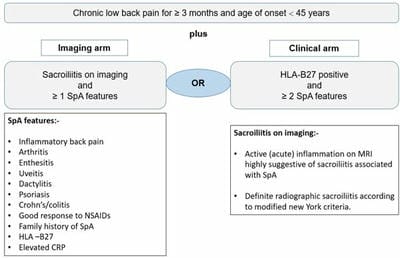
Axial spondyloarthritis comprising both non-radiographic axial spondyloarthritis and ankylosing spondylitis has a deleterious impact on the patient’s quality of life with a detrimental outcome of structural damage. Although in the current era of diagnostic advancements, axSpA can be diagnosed early within a short period after the onset of symptoms, but still there is a delay of up to several years in many parts of the world. The concept of a window of opportunity is primarily derived from rheumatoid arthritis, which is relevant in the context of axSpA based upon the early diagnosis and to commence highly effective treatment with biologics like anti-TNF and anti-IL-17 to modify the disease process for arresting structural damage or syndesmophytes formation. Still, challenges exist for early diagnosis of SpA in patients with low back pain which ultimately creates a barrier to effective treatment initiation. More robust researches along with the available evidence on both the aspects of clinical and imaging factors are the way forward for the early identification of susceptible individuals for early intervention with a better outcome.

Keywords: Ankylosing Spondylitis, Axial Spondyloarthritis, Non-Radiographic Axial Spondyloarthritis, Magnetic Resonance Imaging, Sacroiliac Joint, Sacroiliitis

Corresponding Author How to Cite this Article To Browse
Parasar Ghosh, Professor and HOD, Department of Clinical Immunology and Rheumatology, Institute of Postgraduate Medical Education and Research and SSKM Hospital, Kolkata, West Bengal, India.
Email:
Parasar Ghosh, Pradip Kumar Sarma, Padmanabha Shenoy, Dhrubojyoti Mukherjee, Akshay Desai, The window of opportunity in axial spondyloarthritis: A Stitch in Time. Int J Med Res Rev. 2022;10(5):154-165.
Available From
https://ijmrr.medresearch.in/index.php/ijmrr/article/view/1398

Manuscript Received Review Round 1 Review Round 2 Review Round 3 Accepted
2022-10-04 2022-10-06 2022-10-13 2022-10-20 2022-10-27
Conflict of Interest Funding Ethical Approval Plagiarism X-checker Note
Nil Nil Yes 17%

© 2022by Parasar Ghosh, Pradip Kumar Sarma, Padmanabha Shenoy, Dhrubojyoti Mukherjee, Akshay Desaiand Published by Siddharth Health Research and Social Welfare Society. This is an Open Access article licensed under a Creative Commons Attribution 4.0 International License https://creativecommons.org/licenses/by/4.0/ unported [CC BY 4.0].

Introduction

Axial spondyloarthritis (axSpA) is a chronic inflammatory autoimmune disease under the umbrella of spondyloarthritis (SpA) which predominantly affects the axial skeleton including sacroiliac joints and spine. axSpA is also further classified into radiographic (ankylosing spondylitis, AS) which reveals positive SI joint X-Ray for sacroiliitis as per the modified New York criteria; or non-radiographic respectively as per MRI evaluation. Apart from the inflammatory low back pain, AxSpA can also manifest as peripheral involvement (arthritis, enthesitis, or dactylitis) or extra-articular features (uveitis, psoriasis, inflammatory bowel disease); which often is also associated with a higher prevalence of the human leukocyte antigen HLA-B27. [1].

The estimated global prevalence of overall SpA is around 1%, 0.32–0.7% for axSpA and 0.01–0.2% for AS. [2]. The main characteristics of ankylosing spondylitis are inflammatory back pain, radiographic sacroiliitis, excess spinal bone formation, and a high prevalence of HLA-B27. [3]. HLA‐B27 plays a pivotal role in the pathogenesis of axial spondyloarthritis (axSpA) via four possible hypotheses; arthritogenic-peptide, homodimer, misfolding and intracellular microbial survival hypothesis where all of these mechanisms have a role in disease predisposition but no single one can entirely explain the relationship in disease predisposition. [1].

Therapeutic options for spondyloarthritis treatment (both axial and peripheral) have been expanded in recent years, by the use of targeted therapies. Apart from the T-helper type 1 pathway dominant cytokine TNF-a, the involvement of the interleukin-23/ T-helper type 17 (IL-23/Th17 axis) has determined the way for the development of drugs targeting IL-17 cytokines which are key disease-modifying targets. [4]. There are currently five anti-TNF agents and two IL-17 inhibitors approved for the treatment of axSpA.

In axial spondyloarthritis, ‘window of opportunity is a relatively new concept which can be correlated with a similar concept in rheumatoid arthritis wherein early treatment with disease-modifying anti-rheumatic drugs changes the outcome favourably in the management of rheumatoid arthritis, as it is observed that the response rates are higher and damage is substantially reduced.

A plethora of available evidence suggests that early biologic intervention in axSpA patients can effectively suppress inflammation and has the potential to improve functioning and quality of life with an obvious advantage of preventing further irreversible outcomes such as disability due to structural damage of the axial skeleton. Suggestively the concept of window of opportunity was also coined and reframed in the context of axial spondyloarthritis as well. [5].

ijmrr_1398_01.JPG

Figure 1. ASAS classification criteria of axial-SpA (Redrawn from [17])

Treatment Modalities: Based on new evidence and an expert consensus, the American College of Rheumatology (ACR), the Spondylitis Association of America, and the Spondyloarthritis Research and Treatment Network released updated recommendations for the treatment of axial spondyloarthritis (axSpA). [3].

The introduction of biological agents effectively targeting TNF and IL-17A has been game-changers in the management of axial spondyloarthritis (axSpA). AxSpA management typically requires both pharmacological and non-pharmacological interventions. Currently, pharmacological options involve licensed drug treatments for axSpA which are NSAIDs and biologic DMARDs (bDMARDs) targeting TNF or IL-17A. NSAIDs are 1st line drug therapies for axSpA for efficacious symptomatic management but there is uncertainty regarding their safety in long-term use and relation to radiographic progression. TNF inhibitors have actively established themselves in the management of active axSpA since long before the introduction of IL-17A inhibitors. Also, indirect comparison studies have shown IL-17 inhibitors to be equally efficacious to TNF inhibitors in patients with axial SpA. [6] [7]. Non-pharmacological strategies involve mainly physical rehabilitation, exercise therapies etc. [8].


American College of Rheumatology recommends the treatment with NSAIDs, although no preferred choice of NSAIDs is advocated. Despite treatment with NSAIDs, if the active axial disease persists then TNFi and IL-17i are strongly recommended treatments over no treatment with TNFI and IL-17i. Moreover, it is also recommended that treatment with sulfasalazine or methotrexate should be considered only in patients with prominent peripheral arthritis or when biologics are not available or contraindicated. Among the class of drugs, IL-17 inhibitors (secukinumab, ixekizumab) and TNFis (certolizumab, etanercept, infliximab, adalimumab, and golimumab) are also strongly recommended, although no particular TNFi is recommended as the preferred choice. [3].

Imaging: Imaging plays a very important and central role in diagnosing, managing and performing follow-ups in patients with axSpA. In recent years, there have been huge technical developments in imaging CT and MRI resulting in higher feasibility for their usage in clinical practice for detecting structural lesions. Currently, advanced techniques of CT provide high-resolution images with a lesser amount of radiation, whereas the availability of novel multiple MRI sequences for high-resolution images of axial joints has impacted the early diagnosis of SpA. Although low-dose CT of SI joints has superior diagnostic accuracy than X-Ray, EULAR has recommended plain radiographs as the primary imaging modality due to cost and wide availability. [9]. MRI of SI joints has transformed the early diagnostic evaluation of axSpA due to its unique capability to detect accurately the inflammatory lesions as well as the post-inflammatory structural lesions, which enables diagnosis at a much earlier stage than CT and X-Ray. [10]. Hence, the ASAS 2009 classification criteria consider MRI-evident sacroiliitis in the imaging arm for including the patients at an earlier stage of the disease. As per the consensus definition of the ASAS/OMERACT MRI working group, the presence of inflammation in three or more anterior or posterior vertebral corners on at least two consecutive sagittal slices determines the specificity of axSpA, which is profuse in younger patients as degenerative changes are less frequently expected. [11].

Challenges in management

Challenges in diagnosis: A delay of 5-10 years between the appearance of the

first (chronic) symptoms of the disease and diagnosis is a big obstruction to early treatment of axSpA. [12]. A major reason for this delay is a low level of awareness about axSpA and AS in general among non-rheumatologists. Patients with low back pain are primarily evaluated first by general practitioners, orthopaedics, neurologists, physiotherapists etc., which often leads to a delayed journey of probable axSpA to a specialist rheumatologist due to a lack of awareness. [13]. Interestingly, differentiating between mechanical versus inflammatory back pain is also a major challenge among primary practitioners for early diagnosis of probable axSpA patients. [14].

The considerable delay between the onset of signs & symptoms and diagnosis of axSpA is due to the indistinguishable nature of axSpA related pain from other kinds of back pain, ill-defined criteria for diagnosis, delay from the patient side and referrals to the specialists. This delayed diagnosis of axSpA eventually results in poor clinical outcomes, high disease progression, deficiency in physical function, increased structural damage, aggravated healthcare costs, increased chances of depression and negative psychological impact compared to those who had an earlier diagnosis of axSpA. [12].

As per, the Assessment of Spondyloarthritis International Society (ASAS) classification criteria the axSpA can be defined as the presence of sacroiliitis with one SpA feature (“imaging arm”), or human leukocyte antigen (HLA)-B27 presence with at least two SpA features (“clinical arm”). [15]. Clinical data has consistently shown that younger age, HLAB27-negativity and female gender are often associated with a longer delay in the diagnosis. [16]. Moreover, the longer diagnostic delay is also associated with the absence of other SpA features of peripheral and extra-articular manifestations. Longer diagnostic delays are also evident at a younger age with chronic low back pain, which might be related to the lack of seriousness by both the younger patients and physicians. [13].

The wide availability of MRI in hospitals and awareness of the importance of its usage among physicians and healthcare providers cannot prevent the restrictions on its usage. Other reasons may include the quality of healthcare systems in the region and the cost of MRI and associated procedures. [15].


The diagnosis of patients encountered in a rheumatology clinic with chronic back pain of short symptom duration cannot be relied on an X-Ray of the SI joint, since radiographic changes are often absent at the early stage of the disease and thus require an MRI to rule out the presence of inflammation of the axial skeleton. Thus MRI lesions have obvious utility in both diagnosis and ruling out of the disease. Moreover, in this context, bone marrow edema seen in the SI joints of axSpA patients, can also be seen in other states like in normal subjects especially joggers, hockey players, or even in other mechanical/degenerative conditions and postpartum women. [18]. Often interobserver differences in the interpretation exist between the readers for assessment of X-Ray and MRI scans which might affect the confirmation of axSpA, and thus in such instances often the clinical arm of ASAS 2009 criteria is relied upon by many clinicians. [19]. This disparity in X-ray reading was observed in DESIR cohort data, in which disagreement between local and central readers was seen in around 28% of the patients, which ultimately resulted in a change of classification of 7.9% of the patients. [20]. Thus, education and training of radiologists in this regard might also be a potential step in the direction of reducing the delay in diagnosis seen in this disease.

Challenges in treatment; Introduction of effective biologic agents targeting TNF and IL-17A have led to multiple available treatment options, despite that treatment, recommendations are lacking certainty for deciding an optimised treatment strategy for a patient. Guidelines recommend NSAIDs as 1st line of treatment for axSpA but uncertainty remains about their safety profile for prolonged duration and effect in reducing radiographic progression. [21] [8].

Long-term cardiovascular safety with NSAIDs is also a point of concern for many medical practitioners especially due to chronic usage of axSpA. [8].

Clinical data from randomised controlled trials and real-world evidence also demonstrate that a substantial proportion of axSpA patients can show inadequate response to treatment (primary failure), lose response over time (secondary failure), or develop treatment-emergent adverse events. [22]. Immunogenicity of anti-drug antibodies against the protein-based monoclonal antibodies is a well-documented phenomenon which is the

main contributor to treatment failure with biological therapies, especially with anti-TNFs like Infliximab and adalimumab. [23].

Moreover, incidences of reactivation of latent tuberculosis with anti-TNFs are another drawback posing a challenge to treatment adherence. [24]. In this context, the long-term safety data of newer biologics targeting IL-17 has demonstrated an effective option for latent tuberculosis-positive or high-risk individuals. [25].

Since the current treatment paradigm is to reduce inflammation and disease activity, which should ultimately lead to reduced radiographic changes, long-term head-to-head studies among biologics are warranted to determine the optimised targeted pathways for inhibiting radiographic progression. Likewise, the first-in-class study SURPASS is ongoing to evaluate the superiority of an IL-17 inhibitor over a TNF inhibitor (adalimumab biosimilar), in reducing spinal radiographic progression in axSpA patients. [26]. It remains to be determined whether an aggressive treat-to-target approach with clinical remission or low disease activity will enable more effective retardation of radiographic progression in axSpA. [8].

Nevertheless, the high cost of therapy with biologics remains a challenge affecting treatment adherence, particularly in a resource-limited healthcare setting. However, published HEOR reports are available with comparative cost-effectiveness of biologics in axSpA; [27]. but often becomes a challenge to easily extrapolate due to vast differences in healthcare funding, reimbursement policies and different cost of biologics between the countries.

Concept of the window of opportunity

Two main treatment targets in axial spondyloarthritis (axSpA) could be defined:

1. Reduction of inflammation resulting in control of signs and symptoms such as pain and stiffness

2. Prevention or retardation of structural damage progression in the spine resulting in preservation of functional status and improvement in the long-term outcome. [28].

In contrast to the effective control of axSpA symptoms, currently, the more challenging task is to handle the disease modification (radiological spinal progression in axSpA).


In context to axSpA, a ‘window of opportunity’ is a certain time period (from the first few months to years of disease onset) in which resolution of inflammation would not lead to activation of new bone formation in the spine in the future. [28] [29].

It has been well validated that SI joint and spine MRI lesions in the sequences of STIR/T1/T2 can effectively serve as a surrogate marker for the prediction of future structural damage. [30]. In the context of the window of opportunity, early diagnosis and early treatment with biologics can significantly halt the radiographic progression. Various studies have already revealed that the presence of fatty infiltration or fat metaplasia at the vertebral corners can predict the formation of syndesmophytes or new bone formation, especially at the same vertebral site of prior inflammation. [31].

Moreover, the disease activity indices also positively correlate with the inflammation detected in the SI joints and spine on MRI, which further aids in monitoring the disease prognosis. Consequently, the selection of patients for intervention with biologics early in the disease course can be derived from the presence of MRI structural lesions.

Especially in young SpA patients with active symptoms despite NSAID therapy these factors are helpful to consider intensification of therapy leading to biologic agents.

It is also noteworthy that several cohort studies concluded that in patients of non-radiographic axSpA, evident MRI inflammation of SI joints is a reliable predictor of progression from non-radiographic to radiographic stage or X-Ray evident sacroiliitis as per New York criteria. [32]. Moreover, data suggest that during a 5-year follow-up of DESIR cohort, inflammation on MRI was the sole predictor of radiographic progression i.e., shift from nrAxSpA to AS in both the subsets of HLA-B27-positive as well as HLA-B27-negative patients. [33].

It is a plausible strategy that early diagnosis of nrAxSpA and early intervention with biologics may well benefit to relieve symptoms and improve functioning, but also benefit in forestalling radiographic damage as much as possible. There is good evidence of biologics reducing MRI-detected inflammatory lesions and subsequently affecting the transition states by inhibiting structural progression [34-37].

Table 1: Predictors of radiographic progression in axial spondyloarthritis. [30] [38]

Demographics Serum Biomarkers Disease activity index MRI features
Male gender CRP ASDAS Baseline syndesmophytes
Smoking ESR BASDAI Bone marrow edema
Obesity HLA B27   Fat metaplasia
MMP-3    
VEGF    
Calprotectin    
Visfatin    
MIF    

CRP: C-reactive protein; ESR: erythrocyte sedimentation rate; MMP-3: matrix-metalloproteinase-3; VEGF: vascular endothelial growth factor (VEGF); ASDAS: ankylosing spondylitis disease activity score; BASDAI: bath ankylosing spondylitis activity index; MRI: magnetic resonance. 

Table 2: Biologics approved in axial spondyloarthritis. [19]

  European Union United States
Biologics AS Nr-axSpA AS Nr-axSpA
  Anti-TNFs Adalimumab Approved Approved Approved --
Infliximab Approved -- Approved --
Etanercept Approved Approved Approved --
Golimumab Approved Approved Approved --
Certolizumab Approved Approved Approved Approved
  Anti-IL-17 Secukinumab Approved Approved Approved Approved
Ixekizumab Approved Approved Approved Approved

Biologics in the treatment of axial spondyloarthritis

Axial spondyloarthritis can affect routine activities like schooling, work, and the social life of a patient, and therefore it should be borne in mind that the goals of treatment, in the order of priority, are to reduce signs & symptoms (disease severity), prevention of structural damage and disability, maintain a healthy quality of life and improve work productivity and community participation. While taking risks and benefits into account, NSAIDs (up to maximum dose) should be used for symptomatic treatment for patients with pain and stiffness. [21]. The TNFi have transformed the treatment path of axSpA and more recently the discovery of IL-17 inhibitors has provided effective therapeutic alternatives.


Table 3: Studies appraising the effect of bDMARDs on spinal radiographic progression of axial SpA.

Ref Type of study Number of participants Type of patients bDMARDs Follow up duration Definition of radiographic progression Radiographic outcomes    
Haroon N, et al. 2013 [48] Prospective 334 AS TNFi 2.9 years more than 1 mSASSS unit/year were considered progressors. < 50% reduction in the odds of progression* with TNFi (OR 0.52 [95% 0.3-0.88]
Baraliakos X, et al. 2014 [51] Retrospective 56 AS Infliximab 8 years NA Radiographic inhibition at year 4 and 8 (no differences in the first 4 years)
Molnar C, et al. 2018 [52] Prospective 432 AS TNFi 10 years progression was defined as an increase in ≥2 mSASSS units in 2 years 50% reduction in the odds of progression with prior use of TNFi (OR 0.50, 95% CI 0.28 to 0.88). Radiographic inhibition on TNFi for ≥ 2 years (higher in inactive disease)
Braun J, et al. 2014 [53] RCT 233 AS Golimumab 4 years Progression was defined as an mSASSS increase of ≥2 points from baseline to week 208. Less than one third of patients progressed at year 4.  
Jeong H, et al. 2018 [54] Cohort 151 AS TNFi 102.9 ± 54.9 months NA Radiographic progression was associated with delay in starting TNFi; although mean change of mSASSS/year was 1.01 units/year.
Van der Heijde D, et al. 2018 [35] RCT 325 AS and nr-axSpA Certolizumab 4 years Progression was defined as an mSASSS increase of ≥2 points) from baseline to week 96. No progression in 80.6% at year 4.
Braun J, et al. 2017 [55]   RCT 168 AS Secukinumab 2 years No progression was defined as mSASSS change from baseline to year 2 of ≤ 0. No progression in ~ 80% at year 2.
Braun J, et al. 2019 [36] RCT extension 171 AS Secukinumab 4 years No progression was defined a mSASSS change from baseline to year 4 of <2. No progression in 79% at year 4.

There are five licensed anti-TNF drugs (adalimumab, certolizumab, etanercept, golimumab and infliximab) for the indication of AS and four (adalimumab, etanercept, certolizumab and golimumab) for the indication of nr-axSpA (in the US, only certolizumab was approved for the indication of nr-axSpA). (see table 1) Recently, among IL-17 blockers, secukinumab has been approved by both the European Medicines Agency (EMA) and the US Food and Drug Administration (FDA) for the indication of AS and nr-axSpA. Ixekizumab was also recently approved in the US for nr-axSpA and by EMA for both AS and nr-axSpA. [19].

Randomized controlled trials with anti-TNF like adalimumab, etanercept and certolizumab in patients with nr-axSpA have been reported with significant improvement in the ASAS 40 responses. [39] [37] [40] [41]. Elevated CRP and active MRI inflammation have been found as good predictors for response to TNF inhibitors in nr axSpA patients. [42] Likewise, the IL-17 inhibitors have also demonstrated significant efficacy in both biologic-naïve and TNFi-experienced patients with r-axSpA. [43] [44, 45]. In PREVENT study, treatment with secukinumab in patients with active nr-axSpA revealed a significant and sustained improvement in the signs and symptoms along with the aspects of pain, mobility, and health-related quality of life. Moreover, SI joint bone marrow edema was also reported to be significantly reduced with secukinumab treatment compared to placebo. [45]. Similarly, ixekizumab was also reported to be effective for early improvement of symptoms and inflammation in patients with nr-axSpA. [46].

Extensive research has identified the key roles of IL-17 and TNF in axSpA pathogenesis leading to new bone formation in the vertebral corners. In areas of inflammation, studies have shown that TNF majorly triggers the inflammation-derived osteoclastogenesis leading to bone erosion, whereas IL-17 predominantly promotes both osteoblastic and osteoclastic activity leading to new bone formation via the Wnt/β-catenin signalling pathway [47]. In a similar context, it has been reported that disease progression is more likely to affect patients with delay (>10 years) in starting biologics therapy as compared to those started earlier. [48]. Also, new bone formation is more likely in MRI-evident advanced inflammatory lesions and proceeds through a process of fatty infiltration,


supporting a window of opportunity for disease modification. [29]. Early treatment with biologics can resolve both the spinal inflammation and fatty changes in the vertebral edges. [49]. Significant advances in the research of axSpA elucidate that beyond the efficacy of anti-TNF, anti-IL-17 offers an alternative therapeutic target with a substantial effect on the disease-modifying process with an established safety profile. [50].

Advantages of early introduction of biologics: An early diagnosis may prove to be a key for the management of axSpA, as the delayed diagnosis leads to a delay in the treatment of the disease at the appropriate time and may result in permanent damage, work impairment, poor quality of life and high healthcare cost. [12]. Recently results of a study known as SKIPPAIN, revealed that apart from the effect on disease activity measures, secukinumab treatment was also effective in reducing spinal pain in patients with axSpA as early as Week 8. [56]. Thus early management of spinal pain and disease activity in patients with axSpA through newer biologics like Secukinumab can also ameliorate the impact of pain on both physical and mental well-being. Duration of therapy and the appropriate timing of biological initiation is important for the optimal effect on the rate of structural damage in the spine of axSpA patients. On a similar note, clinical studies have shown that the delay in starting biologics of more than 10 years was associated with higher structural progression compared to those who started earlier. [48]. Another longitudinal study in patients less than 10 years of disease duration has shown that early suppression of inflammation with tight control of CRP by TNFi, significantly decreases spinal radiographic progression in early AS when compared to NSAIDs treatment. [57]. Moreover, it is also reported that patients treated with biologics early in the disease course are twice as likely to achieve clinical remission. [58]. Longitudinal studies with axSpA patients have demonstrated that younger age and shorter disease duration at the time of biologic initiation were significant predictors of ASDAS-CRP remission as an achievable goal. [59]. Treatment with newer biologics like secukinumab in AS patients has also been shown to reduce MRI-evident spinal inflammation as early as week 16 and was maintained through 4 years of treatment. [36]. Moreover in nr-axSpA patients, secukinumab treatment was also reported to reduce

SI joint edema on MRI at week 16 which also sustained up to week 52. [45]. It has also been reported that after 2 years of treatment with secukinumab, there was a resolution of both spinal vertebral corner inflammatory lesions and fatty lesions respectively. [49].

Furthermore, several studies have concluded that in axSpA patients with clinical remission or low disease activity in the early disease course, a dose tapering strategy of biologics by increasing the dosing interval or decreasing the dosage can also be achieved which also reduces the risk of infections and financial burden. [60] [61] [62]. The possibility of diagnosing axSpA early, together with the advent of new therapeutic options for the treatment of nr-axSpA and AS, have significantly improved the care of affected individuals. [63].

Conclusion

The early diagnosis of axSpA, together with the advent of new therapeutic options like biologics for the treatment of nr-axSpA and AS, have significantly improved the outcome in affected individuals. The current need of the hour is to diagnose the disease early and introduce biologics early to delay structural progression. Since the diagnostic delay is majorly due to delayed referral to a rheumatologist, thus the concept of early diagnosis and referral strategy should be implemented in our primary healthcare set-up through effective disease awareness programmes. The training of radiologists for picking up subtle signs in MRI images might also be a potential step in this direction. In the current era of modern medicine, hopefully, the ongoing extensive research would lead to validated optimal biomarkers for predicting the course of early disease and an early intervention strategy. It is well validated that, early effective treatment intervention with biologics can alter the disease outcome by halting structural progression. The introduction of IL-17A blockade increasing the choice of cytokine target in axSpA beyond TNF marks a step forward in the management of AS and nr-axSpA

Authors' contributions: All authors have contributed significantly to the conception, design, or acquisition of data or analysis and interpretation of data. All authors have participated in drafting, reviewing, and/or revising the manuscript and have approved its submission.


Abbreviation: TNF (tumour necrosis factor), IL-17 (interleukin 17), SI joint (sacroiliac joint), MRI (magnetic resonance imaging), CT (computed tomography), mSASSS (modified Stokes Ankylosing Spondylitis Spine Score), NSAIDs (non-steroidal anti-inflammatory drugs), bDMARDs (biological disease-modifying antirheumatic drugs).

Reference

01. Lim CSE, Sengupta R, Gaffney K. The clinical utility of human leucocyte antigen B27 in axial spondyloarthritis. Rheumatology (Oxford). 2018 Jun 1;57(6):959-968. doi: 10.1093/rheumatology/kex345 [Crossref][PubMed][Google Scholar]

02. Siebert, Stefan, Raj Sengupta, and Alexander Tsoukas, eds. Axial spondyloarthritis. Oxford University Press, 2016. . [Crossref][PubMed][Google Scholar]

03. Ward MM, Deodhar A, Gensler LS, Dubreuil M, Yu D, Khan MA, et al. 2019 Update of the American College of Rheumatology/Spondylitis Association of America/Spondyloarthritis Research and Treatment Network Recommendations for the Treatment of Ankylosing Spondylitis and Nonradiographic Axial Spondyloarthritis. Arthritis Care Res (Hoboken). 2019 Oct;71(10):1285-1299. doi: 10.1002/acr.24025. Epub 2019 Aug 21 [Crossref][PubMed][Google Scholar]

04. Wendling D. New targeted therapies in spondyloarthritis: what are the limits? Immunotherapy. 2019 May;11(7):557-560. doi: 10. 2217/imt-2019-0007 [Crossref][PubMed][Google Scholar]

05. Robinson PC, Brown MA. The window of opportunity: a relevant concept for axial spondyloarthritis. Arthritis Res Ther. 2014;16(3):109. doi: 10.1186/ar4561 [Crossref][PubMed][Google Scholar]

06. Maksymowych WP, Strand V, Nash P, Yazici Y, Thom H, Hunger M, et al. Comparative effectiveness of secukinumab and adalimumab in ankylosing spondylitis as assessed by matching-adjusted indirect comparison. Eur J Rheumatol. 2018 Dec;5(4):216-223. doi: 10.5152/eurjrheum.2018.18162 [Crossref][PubMed][Google Scholar]

07. Betts, K. A. , et al. OP0115 Relative Efficacy of Adalimumab versus Secukinumab in Active Ankylosing Spondylitis: A Matching-Adjusted Indirect Comparison. (2016): 98-99 [Crossref][PubMed][Google Scholar]

08. Fragoulis GE, Siebert S. Treatment strategies in axial spondyloarthritis: what, when and how? Rheumatology (Oxford). 2020 Oct 1;59(Suppl4):iv79-iv89. doi: 10. 1093/rheumatology/keaa435 [Crossref][PubMed][Google Scholar]

09. Mandl P, Navarro-Compán V, Terslev L, Aegerter P, van der Heijde D, D'Agostino MA, et al. EULAR recommendations for the use of imaging in the diagnosis and management of spondyloarthritis in clinical practice. Ann Rheum Dis. 2015 Jul;74(7):1327-39. doi: 10.1136/annrheumdis-2014-206971 [Crossref][PubMed][Google Scholar]

10. Khmelinskii N, Regel A, Baraliakos X. The Role of Imaging in Diagnosing Axial Spondyloarthritis. Front Med (Lausanne). 2018 Apr 17;5:106. doi: 10.3389/fmed.2018.00106 [Crossref][PubMed][Google Scholar]

11. Hermann KG, Baraliakos X, van der Heijde DM, Jurik AG, Landewé R, Marzo-Ortega H, et al. Descriptions of spinal MRI lesions and definition of a positive MRI of the spine in axial spondyloarthritis: a consensual approach by the ASAS/OMERACT MRI study group. Ann Rheum Dis. 2012 Aug;71(8):1278-88. doi: 10.1136/ard.2011.150680 [Crossref][PubMed][Google Scholar]

12. Yi E, Ahuja A, Rajput T, George AT, Park Y. Clinical, Economic, and Humanistic Burden Associated With Delayed Diagnosis of Axial Spondyloarthritis: A Systematic Review. Rheumatol Ther. 2020 Mar;7(1):65-87. doi: 10.1007/s40744-020-00194-8 [Crossref][PubMed][Google Scholar]

13. Danve A, Deodhar A. Axial spondyloarthritis in the USA: diagnostic challenges and missed opportunities. Clin Rheumatol. 2019 Mar;38(3):625-634. doi: 10.1007/s10067-018-4397-3 [Crossref][PubMed][Google Scholar]

14. Zhao SS, Pittam B, Harrison NL, Ahmed AE, Goodson NJ, Hughes DM. Diagnostic delay in axial spondyloarthritis: a systematic review and meta-analysis. Rheumatology (Oxford). 2021 Apr 6;60(4):1620-1628. doi: 10.1093/rheumatology/keaa807 [Crossref][PubMed][Google Scholar]


15. Hammoudeh M, Abdulaziz S, Alosaimi H, Al-Rayes H, Aldeen Sarakbi H, Baamer M, et al. Challenges of diagnosis and management of axial spondyloarthritis in North Africa and the Middle East: An expert consensus. J Int Med Res. 2016 Apr;44(2):216-30. doi: 10.1177/0300060515611536 [Crossref][PubMed][Google Scholar]

16. Rusman T, van Bentum RE, van der Horst-Bruinsma IE. Sex and gender differences in axial spondyloarthritis: myths and truths. Rheumatology (Oxford). 2020 Oct 1;59(Suppl4):iv38-iv46. doi: 10.1093/rheumatology/keaa543 [Crossref][PubMed][Google Scholar]

17. Rudwaleit M, van der Heijde D, Landewé R, Listing J, Akkoc N, Brandt J, et al. The development of Assessment of SpondyloArthritis international Society classification criteria for axial spondyloarthritis (part II): validation and final selection. Ann Rheum Dis. 2009 Jun;68(6):777-83. doi: 10.1136/ard.2009.108233 [Crossref][PubMed][Google Scholar]

18. de Winter J, de Hooge M, van de Sande M, de Jong H, van Hoeven L, de Koning A, et al. Magnetic Resonance Imaging of the Sacroiliac Joints Indicating Sacroiliitis According to the Assessment of SpondyloArthritis international Society Definition in Healthy Individuals, Runners, and Women With Postpartum Back Pain. Arthritis Rheumatol. 2018 Jul;70(7):1042-1048. doi: 10.1002/art.40475 [Crossref][PubMed][Google Scholar]

19. Poddubnyy D, Sieper J. Treatment of Axial Spondyloarthritis: What Does the Future Hold? Curr Rheumatol Rep. 2020 Jul 20;22(9):47. doi: 10. 1007/s11926-020-00924-5 [Crossref][PubMed][Google Scholar]

20. van den Berg R, Lenczner G, Thévenin F, Claudepierre P, Feydy A, Reijnierse M, et al. Classification of axial SpA based on positive imaging (radiographs and/or MRI of the sacroiliac joints) by local rheumatologists or radiologists versus central trained readers in the DESIR cohort. Ann Rheum Dis. 2015 Nov;74(11):2016-21. doi: 10.1136/annrheumdis-2014-205432 [Crossref][PubMed][Google Scholar]

21. van der Heijde D, Ramiro S, Landewé R, Baraliakos X, Van den Bosch F, Sepriano A, et al. 2016 update of the ASAS-EULAR management

recommendations for axial spondyloarthritis. Ann Rheum Dis. 2017 Jun;76(6):978-991. doi: 10.1136/annrheumdis-2016-210770 [Crossref][PubMed][Google Scholar]

22. Manica SR, Sepriano A, Pimentel-Santos F, Gouveia N, Barcelos A, Branco JC, et al. Effectiveness of switching between TNF inhibitors in patients with axial spondyloarthritis: is the reason to switch relevant? Arthritis Res Ther. 2020 Aug 21;22(1):195. doi: 10. 1186/s13075-020-02288-8 [Crossref][PubMed][Google Scholar]

23. Garcês S, Demengeot J, Benito-Garcia E. The immunogenicity of anti-TNF therapy in immune-mediated inflammatory diseases: a systematic review of the literature with a meta-analysis. Ann Rheum Dis. 2013 Dec;72(12):1947-55. doi: 10.1136/annrheumdis-2012-202220 [Crossref][PubMed][Google Scholar]

24. Zhang Z, Fan W, Yang G, Xu Z, Wang J, Cheng Q, et al. Risk of tuberculosis in patients treated with TNF-α antagonists: a systematic review and meta-analysis of randomised controlled trials. BMJ Open. 2017 Mar 22;7(3):e012567. doi: 10.1136/bmjopen-2016-012567 [Crossref][PubMed][Google Scholar]

25. Elewski BE, Baddley JW, Deodhar AA, Magrey M, Rich PA, Soriano ER, et al. Association of Secukinumab Treatment With Tuberculosis Reactivation in Patients With Psoriasis, Psoriatic Arthritis, or Ankylosing Spondylitis. JAMA Dermatol. 2021 Jan 1;157(1):43-51. doi: 10.1001/jamadermatol.2020.3257 [Crossref][PubMed][Google Scholar]

26. Baraliakos X, Østergaard M, Gensler LS, Poddubnyy D, Lee EY, Kiltz U, et al. Comparison of the Effects of Secukinumab and Adalimumab Biosimilar on Radiographic Progression in Patients with Ankylosing Spondylitis: Design of a Randomized, Phase IIIb Study (SURPASS). Clin Drug Investig. 2020 Mar;40(3):269-278. doi: 10.1007/s40261-020-00886-7 [Crossref][PubMed][Google Scholar]

27. Emery P, Van Keep M, Beard S, Graham C, Miles L, Jugl SM, et al. Cost Effectiveness of Secukinumab for the Treatment of Active Ankylosing Spondylitis in the UK. Pharmacoeconomics. 2018 Aug;36(8):1015-1027. doi: 10.1007/s40273-018-0675-9 [Crossref][PubMed][Google Scholar]

28. Rios Rodriguez V, Poddubnyy


D. Old and new treatment targets in axial spondyloarthritis. RMD Open. 2015 Aug 15;1(Suppl 1):e000054. doi: 10.1136/rmdopen-2015-000054 [Crossref][PubMed][Google Scholar]

29. Maksymowych WP, Morency N, Conner-Spady B, Lambert RG. Suppression of inflammation and effects on new bone formation in ankylosing spondylitis: evidence for a window of opportunity in disease modification. Ann Rheum Dis. 2013 Jan;72(1):23-8. doi: 10.1136/annrheumdis-2011-200859 [Crossref][PubMed][Google Scholar]

30. Maksymowych WP. Biomarkers for Diagnosis of Axial Spondyloarthritis, Disease Activity, Prognosis, and Prediction of Response to Therapy. Front Immunol. 2019 Mar 7;10:305. doi: 10.3389/fimmu.2019.00305 [Crossref][PubMed][Google Scholar]

31. Maksymowych WP, Wichuk S, Chiowchanwisawakit P, Lambert RG, Pedersen SJ. Fat metaplasia on MRI of the sacroiliac joints increases the propensity for disease progression in the spine of patients with spondyloarthritis. RMD Open. 2017 Mar 30;3(1):e000399. doi: 10.1136/rmdopen-2016-000399 [Crossref][PubMed][Google Scholar]

32. Protopopov M, Proft F, Sepriano A, Landewé R, van der Heijde D, Maksymowych WP, et al. Radiographic sacroiliitis progression in axial spondyloarthritis: central reading of 5 year follow-up data from the Assessment of SpondyloArthritis international Society cohort. Rheumatology (Oxford). 2021 May 14;60(5):2478-2480. doi: 10.1093/rheumatology/keab091 [Crossref][PubMed][Google Scholar]

33. Dougados M, Sepriano A, Molto A, van Lunteren M, Ramiro S, de Hooge M, et al. Sacroiliac radiographic progression in recent onset axial spondyloarthritis: the 5-year data of the DESIR cohort. Ann Rheum Dis. 2017 Nov;76(11):1823-1828. doi: 10.1136/annrheumdis-2017-211596 [Crossref][PubMed][Google Scholar]

34. Huang Y, Chen Y, Liu T, Lin S, Yin G, Xie Q. Impact of tumor necrosis factor α inhibitors on MRI inflammation in axial spondyloarthritis assessed by Spondyloarthritis Research Consortium Canada score: A meta-analysis. PLoS One. 2020 Dec 31;15(12):e0244788. doi: 10.1371/journal.pone.0244788 [Crossref][PubMed][Google Scholar]

35. van der Heijde D, Baraliakos X, Hermann KA, Landewé RBM, Machado PM, Maksymowych WP, et al. Limited radiographic progression and sustained reductions in MRI inflammation in patients with axial spondyloarthritis: 4-year imaging outcomes from the RAPID-axSpA phase III randomised trial. Ann Rheum Dis. 2018 May;77(5):699-705. doi: 10.1136/annrheumdis-2017-212377 [Crossref][PubMed][Google Scholar]

36. Braun J, Baraliakos X, Deodhar A, Poddubnyy D, Emery P, Delicha EM, et al. Secukinumab shows sustained efficacy and low structural progression in ankylosing spondylitis: 4-year results from the MEASURE 1 study. Rheumatology (Oxford). 2019 May 1;58(5):859-868. doi: 10.1093/rheumatology/key375 [Crossref][PubMed][Google Scholar]

37. Maksymowych WP, Dougados M, van der Heijde D, Sieper J, Braun J, Citera G, et al. Clinical and MRI responses to etanercept in early non-radiographic axial spondyloarthritis: 48-week results from the EMBARK study. Ann Rheum Dis. 2016 Jul;75(7):1328-35. doi: 10.1136/annrheumdis-2015-207596 [Crossref][PubMed][Google Scholar]

38. Lorenzin M, Ometto F, Ortolan A, Felicetti M, Favero M, Doria A, Ramonda R. An update on serum biomarkers to assess axial spondyloarthritis and to guide treatment decision. Ther Adv Musculoskelet Dis. 2020 Jun 24;12:1759720X20934277. doi: 10.1177/1759720X20934277 [Crossref][PubMed][Google Scholar]

39. Sieper J, van der Heijde D, Dougados M, Mease PJ, Maksymowych WP, Brown MA, et al. Efficacy and safety of adalimumab in patients with non-radiographic axial spondyloarthritis: results of a randomised placebo-controlled trial (ABILITY-1). Ann Rheum Dis. 2013 Jun;72(6):815-22. doi: 10.1136/annrheumdis-2012-201766 [Crossref][PubMed][Google Scholar]

40. Sieper J, van der Heijde D, Dougados M, Maksymowych WP, Scott BB, Boice JA, et al. A randomized, double-blind, placebo-controlled, sixteen-week study of subcutaneous golimumab in patients with active nonradiographic axial spondyloarthritis. Arthritis Rheumatol. 2015 Oct;67(10):2702-12. doi: 10.1002/art.39257 [Crossref][PubMed][Google Scholar]

41. Dougados M, van der Heijde D, Sieper J, Braun


J, Maksymowych WP, Citera G, et al. Symptomatic efficacy of etanercept and its effects on objective signs of inflammation in early nonradiographic axial spondyloarthritis: a multicenter, randomized, double-blind, placebo-controlled trial. Arthritis Rheumatol. 2014 Aug;66(8):2091-102. doi: 10.1002/art.38721 [Crossref][PubMed][Google Scholar]

42. Sieper J, Landewé R, Magrey M, Anderson JK, Zhong S, Wang X, et al. Predictors of remission in patients with non-radiographic axial spondyloarthritis receiving open-label adalimumab in the ABILITY-3 study. RMD Open. 2019 Jun 7;5(1):e000917. doi: 10.1136/rmdopen-2019-000917 [Crossref][PubMed][Google Scholar]

43. Dougados M, Wei JC, Landewé R, Sieper J, Baraliakos X, Van den Bosch F, et al. Efficacy and safety of ixekizumab through 52 weeks in two phase 3, randomised, controlled clinical trials in patients with active radiographic axial spondyloarthritis (COAST-V and COAST-W). Ann Rheum Dis. 2020 Feb;79(2):176-185. doi: 10.1136/annrheumdis-2019-216118 [Crossref][PubMed][Google Scholar]

44. Deodhar A, van der Heijde D, Gensler LS, Kim TH, Maksymowych WP, Østergaard M, et al. Ixekizumab for patients with non-radiographic axial spondyloarthritis (COAST-X): a randomised, placebo-controlled trial. Lancet. 2020 Jan 4;395(10217):53-64. doi: 10.1016/S0140-6736(19)32971-X [Crossref][PubMed][Google Scholar]

45. Deodhar A, Blanco R, Dokoupilová E, Hall S, Kameda H, Kivitz AJ, Poddubnyy D, van de Sande M, Wiksten AS, Porter BO, Richards HB, Haemmerle S, Braun J. Improvement of Signs and Symptoms of Nonradiographic Axial Spondyloarthritis in Patients Treated With Secukinumab: Primary Results of a Randomized, Placebo-Controlled Phase III Study. Arthritis Rheumatol. 2021 Jan;73(1):110-120. doi: 10.1002/art.41477 [Crossref][PubMed][Google Scholar]

46. San Koo B, Kim TH. The role of ixekizumab in non-radiographic axial spondyloarthritis. Ther Adv Musculoskelet Dis. 2021 Jan 12;13:1759720X20986734. doi: 10.1177/1759720X20986734 [Crossref][PubMed][Google Scholar]

47. Magrey MN, Khan MA. The Paradox of Bone Formation and Bone Loss in Ankylosing Spondylitis: Evolving New Concepts of Bone Formation and Future Trends in Management. Curr Rheumatol Rep. 2017 Apr;19(4):17. doi: 10.1007/s11926-017-0644-x [Crossref][PubMed][Google Scholar]

48. Haroon N, Inman RD, Learch TJ, Weisman MH, Lee M, Rahbar MH, et al. The impact of tumor necrosis factor α inhibitors on radiographic progression in ankylosing spondylitis. Arthritis Rheum. 2013 Oct;65(10):2645-54. doi: 10.1002/art.38070 [Crossref][PubMed][Google Scholar]

49. Baraliakos X, Borah B, Braun J, Baeten D, Laurent D, Sieper J, et al. Long-term effects of secukinumab on MRI findings in relation to clinical efficacy in subjects with active ankylosing spondylitis: an observational study. Ann Rheum Dis. 2016 Feb;75(2):408-12. doi: 10.1136/annrheumdis-2015-207544 [Crossref][PubMed][Google Scholar]

50. Jones A, Ciurtin C, Ismajli M, Leandro M, Sengupta R, Machado PM. Biologics for treating axial spondyloarthritis. Expert Opin Biol Ther. 2018 Jun;18(6):641-652. doi: 10.1080/14712598.2018.1468884 [Crossref][PubMed][Google Scholar]

51. Baraliakos X, Haibel H, Listing J, Sieper J, Braun J. Continuous long-term anti-TNF therapy does not lead to an increase in the rate of new bone formation over 8 years in patients with ankylosing spondylitis. Ann Rheum Dis. 2014 Apr;73(4):710-5. doi: 10.1136/annrheumdis-2012-202698 [Crossref][PubMed][Google Scholar]

52. Molnar C, Scherer A, Baraliakos X, de Hooge M, Micheroli R, Exer P, et al. Rheumatologists of the Swiss Clinical Quality Management Program. TNF blockers inhibit spinal radiographic progression in ankylosing spondylitis by reducing disease activity: results from the Swiss Clinical Quality Management cohort. Ann Rheum Dis. 2018 Jan;77(1):63-69. doi: 10.1136/annrheumdis-2017-211544 [Crossref][PubMed][Google Scholar]

53. Braun J, Baraliakos X, Hermann KG, Deodhar A, van der Heijde D, Inman R, et al. The effect of two golimumab doses on radiographic progression in ankylosing spondylitis: results through 4 years of the GO-RAISE trial.


Ann Rheum Dis. 2014 Jun;73(6):1107-13. doi: 10.1136/annrheumdis-2012-203075 [Crossref][PubMed][Google Scholar]

54. Jeong H, Eun YH, Kim IY, Park EJ, Kim H, Lee J, et al. Effect of tumor necrosis factor α inhibitors on spinal radiographic progression in patients with ankylosing spondylitis. Int J Rheum Dis. 2018 May;21(5):1098-1105. doi: 10.1111/1756-185X.13270 [Crossref][PubMed][Google Scholar]

55. Braun J, Baraliakos X, Deodhar A, Baeten D, Sieper J, Emery P, et al. REffect of secukinumab on clinical and radiographic outcomes in ankylosing spondylitis: 2-year results from the randomised phase III MEASURE 1 study. Ann Rheum Dis. 2017 Jun;76(6):1070-1077. doi: 10.1136/annrheumdis-2016-209730 [Crossref][PubMed][Google Scholar]

56. Poddubnyy D, Pournara E, Zielińska A, Baranauskaite A, Jiménez AM, Sadhu S, et al. Rapid improvement in spinal pain in patients with axial spondyloarthritis treated with secukinumab: primary results from a randomized controlled phase-IIIb trial. Ther Adv Musculoskelet Dis. 2021 Oct 22;13:1759720X211051471. doi: 10.1177/1759720X211051471 [Crossref][PubMed][Google Scholar]

57. Park JW, Kim MJ, Lee JS, Ha YJ, Park JK, Kang EH, et al. Impact of Tumor Necrosis Factor Inhibitor Versus Nonsteroidal Antiinflammatory Drug Treatment on Radiographic Progression in Early Ankylosing Spondylitis: Its Relationship to Inflammation Control During Treatment. Arthritis Rheumatol. 2019 Jan;71(1):82-90. doi: 10.1002/art.40661 [Crossref][PubMed][Google Scholar]

58. Sieper J, Lenaerts J, Wollenhaupt J, Rudwaleit M, Mazurov VI, Myasoutova L, et al. Efficacy and safety of infliximab plus naproxen versus naproxen alone in patients with early, active axial spondyloarthritis: results from the double-blind, placebo-controlled INFAST study, Part 1. Ann Rheum Dis. 2014 Jan;73(1):101-7. doi: 10.1136/annrheumdis-2012-203201 [Crossref][PubMed][Google Scholar]

59. Shimabuco AY, Gonçalves CR, Moraes JCB, Waisberg MG, Ribeiro ACM, Sampaio-Barros PD, et al. Factors associated with ASDAS remission in a long-term study of ankylosing spondylitis patients under tumor necrosis factor

inhibitors. Adv Rheumatol. 2018 Dec 4;58(1):40. doi: 10.1186/s42358-018-0040-x [Crossref][PubMed][Google Scholar]

60. Ziade NR, Baraliakos X. Tapering bDMARDs in axial SpA - what is the current evidence? Nat Rev Rheumatol. 2019 Jun;15(6):322-324. doi: 10. 1038/s41584-019-0201-2 [Crossref][PubMed][Google Scholar]

61. Li KP, Jin JY, Yang JS, Li Y, Zhao W, Luo G, et al. Full dose, half dose, or discontinuation of etanercept biosimilar in early axial spondyloarthritis patients: a real-world study in China. Arch Med Sci. 2019 May;15(3):700-705. doi: 10.5114/aoms.2018.76141 [Crossref][PubMed][Google Scholar]

62. Gratacós J, Pontes C, Juanola X, Sanz J, Torres F, Avendaño C, et al. Non-inferiority of dose reduction versus standard dosing of TNF-inhibitors in axial spondyloarthritis. Arthritis Res Ther. 2019 Jan 8;21(1):11. doi: 10.1186/s13075-018-1772-z [Crossref][PubMed][Google Scholar]

63. Dubash S, McGonagle D, Marzo-Ortega H. New advances in the understanding and treatment of axial spondyloarthritis: from chance to choice. Ther Adv Chronic Dis. 2018 Mar;9(3):77-87. doi: 10.1177/2040622317743486 [Crossref][PubMed][Google Scholar]