E-ISSN:2320-8686
P-ISSN:2321-127X

Research Article

Non-neoplastic skin disorders

International Journal of Medical Research and Review

2022 Volume 10 Number 1 January February
Publisherwww.medresearch.in

Clinico-Histopathological Correlation of Non-Neoplastic Dermatological Lesions

Kumar V.1, Sharan A.2, Poriya S.3, kundu S.4*
DOI: https://doi.org/10.17511/ijmrr.2022.i01.04

1 Vibhuti Kumar, Chief of lab, , Dr Lal Path Lab, Purnea, Bihar, India.

2 Anita Sharan, Professor, Department of Pathology, D.Y. Patil University School of Medicine, Nerul, Navi Mumbai, India.

3 Sunita Poriya, Assistant Professor, , Geetanjali Medical College, Udaipur, Rajasthan, India.

4* Sonia kundu, Additional Senior Medical Officer, , Civil Hospital, Panipat, Haryana, India.

Objective: Non-neoplastic skin disorders are more common than neoplastic skin disorders which have included Infectious diseases, non-infectious erythematous lesions, papular & squamous disorders, connective tissue diseases, non-infectious vesiculobullous / Vesiculopustular disorders e.tc. The pattern of skin diseases varies from one country to another and across different parts of the same country. So due to the variable spectrum, the histopathology of skin diseases is varied. Each clinical presentation is common to different histopathological pictures & thus definitely requires histopathology for confirmation. Design: This study was conducted by Dr D. Y. Patil Medical College and Hospital, Navi Mumbai from October 2018 to October 2020. Subjects: One hundred and fifty non-neoplastic specimens of skin biopsies submitted in the histopathology; Department of Pathology were considered as the subjects of the study. Material and Methods: Histopathological results were presented in tabular and graphical forms. Mean, median, standard deviation and ranges were calculated for quantitative data. The Chi-square test and t-test with p<0.001 was considered significant. Results: The mean age of our study participants was 41.9 years with a standard deviation of 36.5 years. The majority of our study participants were females: 29.4 % of patients presented with infectious lesions, 22.7 % of patients presented with papulosquamous lesions, and 12.7 % of patients presented with vesiculobullous lesions. The most common infectious non-neoplastic skin lesion was leprosy, the most common papulosquamous skin lesion was dermatitis, and the commonest vesiculobullous skin lesion was Pemphigus Vulgaris. Conclusion: An integrated approach by dermatologists and pathologists is recommended to increase the accuracy of diagnosis and for better management of the patient.

Keywords: Leprosy, Non-Neoplastic skin lesions, Psoriasis, Papulo-squamous lesions, Vesicobullous lesions

Corresponding Author How to Cite this Article To Browse
Sonia kundu, Additional Senior Medical Officer, , Civil Hospital, Panipat, Haryana, India.
Email:
Vibhuti Kumar, Anita Sharan, Sunita Poriya, Sonia kundu, Clinico-Histopathological Correlation of Non-Neoplastic Dermatological Lesions. Int J Med Res Rev. 2022;10(1):24-30.
Available From
https://ijmrr.medresearch.in/index.php/ijmrr/article/view/1351

Manuscript Received Review Round 1 Review Round 2 Review Round 3 Accepted
2022-01-06 2022-01-08 2022-01-15 2022-01-22 2022-01-29
Conflict of Interest Funding Ethical Approval Plagiarism X-checker Note
Nil Nil Nil 17%

© 2022by Vibhuti Kumar, Anita Sharan, Sunita Poriya, Sonia kunduand Published by Siddharth Health Research and Social Welfare Society. This is an Open Access article licensed under a Creative Commons Attribution 4.0 International License https://creativecommons.org/licenses/by/4.0/ unported [CC BY 4.0].

Introduction

Non-neoplastic skin disorders are much more common than neoplastic disorders.[1]. It includes genodermatoses, non-infectious erythematous and papulosquamous lesions, vascular disorders, non-infectious vesiculobullous and vesicopustular diseases, connective tissue diseases, infectious diseases etc. [1]. Many non-neoplastic skin disorders can be quickly diagnosed by clinical features; need no investigations. At the other extreme, some patients need detailed investigational work up to confirm the diagnosis. [2, 3]. Skin biopsy is the single most important diagnostic technique used for the management of patients with skin disorders. Various skin biopsy techniques are punch biopsy, shave biopsy, excision biopsy and incisional biopsy. [4-6]. The interpretation of many skin biopsies requires the identification & integration of two different morphological features: The tissue reaction pattern & the pattern of inflammation. [7]. There are different types of tissue reaction patterns:  Lichenoid reaction pattern, Psoriasiform reaction pattern, Spongiotic reaction pattern, Vesiculobullous reaction pattern, Granulomatous reaction pattern (further divided into Sarcoidal, Tuberculoid, Necrobiotic, Suppurative, Foreign body granuloma), Vasculopathic reaction pattern (i.e., Neutrophilic vasculitis Granulomatous vasculitis, Neutrophilic dermatoses). [8]. Papulosquamous diseases form the largest conglomerate group of skin diseases and are characterized by scaling papules or plaques, so clinical confusion may result in their wrong diagnosis. [9]. Certain conditions like psoriasis mimic diverse dermatological conditions as they present with numerous clinical variants leading to diagnostic dilemmas for the clinician. In such cases, the histopathological diagnosis will help the dermatologist in instituting proper therapy and can vary the prognosis significantly. [9]. Therefore, histopathological analysis is important for a more definitive differentiation. The pattern of skin diseases varies from country to country & across different parts of the same country. So due to the variable spectrum, the histopathology of skin diseases is varied. Clinical presentation is restricted to only a few changes such as hyperpigmentation, hypopigmentation, macule, papule, nodule & others. Each clinical presentation is common to different histopathological pictures. Therefore, though histopathology is considered

the gold standard in dermatological diagnosis, there exist few limitations and very often a definite ‘specific’ diagnosis is not possible. In such instances, the correlation of histopathological findings with clinical findings will aid in arriving at a possible diagnosis and thereby help in the disease treatment. This study mainly includes histopathological evaluation of various types of non-neoplastic skin lesions and their clinicopathological correlations followed by the study of age, site, sex distribution and frequency in various non-neoplastic skin lesions. In the present day, there is growing awareness towards skin diseases even in our geographical area and with improvement in medical facilities, histopathological examination of clinical diagnosis of skin lesions provides valuable input in confirmation of the diagnosis. [1].

Materials And Methods

Setting and Duration of study: This prospective study was conducted by Dr D. Y. Patil Medical College and Hospital, Navi Mumbai from October 2018 to October 2020 (two years).

Sampling size: In the present study, a total of 150 patients attending the Dermatology OPD were included.

Inclusion criteria: Patients attending the Dermatology OPD with clinically diagnosed non-neoplastic skin lesions of any duration within two years (October 2018 – October 2020) and giving written consent for biopsy were registered for the study. Cases were selected regardless of their age, sex, religion, occupation and socio-economic status. The patients were examined by the dermatologist to identify the site, size, colour and distribution of the lesions and a skin biopsy was done on the patients.

Exclusion criteria: Patients with clinically diagnosed neoplastic skin lesions and patients who did not give written consent for the study were excluded from the study.

Data collection procedure: A detailed clinical history, examination findings and provisional clinical diagnosis were collected. Skin biopsies of these patients were received in the Department of Pathology in Dr D.Y. Patil University, School of Medicine, Navi Mumbai, were microscopically analyzed and correlated clinically.

The biopsy techniques commonly


employed are Punch biopsy, Superficial and deep shave biopsy, Deep incisional biopsy, Complete excision and Curettage.

The skin biopsies were dispatched in vials containing 10% formalin solution. Following fixation for 12-24 hours, the tissues were processed and embedded in paraffin and serial sections were obtained. The tissue sections are then subjected to hematoxylin and eosin staining, followed by mounting and proper labelling of the slides.

Special stains used: Other stains used were Modified Fite’s stain for Mycobacterium leprae., ZN stain for Mycobacterium tuberculosis, Van Gieson’s stain for collagen, PAS & GMS for fungus and Gram stain for differentiation of bacteria into Gram-Positive and Gram-negative Bacteria.

Ethical consideration and permission: The study was approved by Institutional Ethics Committee.

Statistical Analysis: Histopathological results were presented in tabular and graphical forms. Mean, median, standard deviation and ranges were calculated for quantitative data. The Chi-square test and t-test with p<0.001 was considered significant.

Observation and Results

Ages at initial presentation ranged from 3 to 83 years. The mean age of our study participants was 41.9 years with a standard deviation of 36.5 years. Skin lesions were more common in the age group of 41 to 50 years (22%) and least common in ages less than 10 years. (Table 1).

Table 1: Age distribution of patients with skin lesions. 

Age in years No. of patients Percentage (% )
<10 Years 6 4 %
10-20 Years 17 11.3 %
21-30 Years 32 21.3 %
31-40 Years 30 20 %
41-50 Years 33 22 %
51-60 Years 15 10 %
>61 Years 17 11.3 %
Total 150 100 %

Seventy-seven out of 150 lesions were seen in females (51.3%) and 73 out of the total 150 lesions were seen in males (48.7%), therefore females were affected more commonly than males with a male: female ratio of 0.9:1.

Figure 1 showed the distribution of gender in various age groups. Majority of the skin lesions among males presented at younger age i.e., 21 to 30 years (26%) whereas among females, the presentation was a little later i.e. at 31 to 40 years and 41 to 50 years of age (24.7% each).

Out of 150 skin lesions studied, 44 (29.4%) patients presented with infectious lesions, 34 (22.7%) patients presented with papulosquamous lesions, and 19 (12.7%) patients presented with the vesiculobullous lesion. 25 patients, who presented with infectious lesions and 23 papulosquamous lesions were males. Fourteen vesicobullous lesions were seen in females. The difference in presentation among males and females was statistically significant when the Chi-square test was applied (p<0.05) (Table 2).

Table 2: The spectrum of clinical diagnosis with gender Correlation.

Skin Lesions Females Males Total
Infectious 19 (24.7 %) 25 (34.2 %) 44 (29.4 %)
Vesicobullous Lesions 14 (18.2 %) 5 (6.8 %) 19 (12.7 %)
Papulosquamous lesions 11 (14.3 %) 23 (31.5 %) 34 (22.7 %)
Connective tissue diseases 6 (7.8 %) 5 (6.8 %) 11 (7.3 %)
Pigmentary 2 (2.6 %) 1 (1.4 %) 3 (2 %)
Cysts 4 (5.2 %) 3 (4.1 %) 7 (4.7 %)
Vascular 2 (2.6 %) 2 (2.7 %) 4 (2.7 %)
Miscellaneous 19 (24.7 %) 9 (12.3 %) 28 (18.6 %)
Total 77 (100 %) 73 (100 %) 150 (100 %)
Chi-Square Value: 13.4                  df:7              p-value:0.05

Infectious skin lesions were further classified as bacterial, viral, fungal and parasitic. Among bacterial, leprosy and TB were the most common presentations. Out of 36 infectious skin lesions, 23 (63.9%) were leprosy cases and 5 (13.9%) were cutaneous TB. Leprosy was common in males and TB among females in our study (Figure 2)

Non-Infectious skin lesions were classified as vesicobullous lesions, papulosquamous lesions, Pigmentary skin lesions and miscellaneous lesions.

Out of 114 non-infectious skin lesions, the majority (29.8%) were papulosquamous lesions, which were more common in males and 16.7% were vesicobullous lesions and were common in females. (Table 3).

Papulosquamous lesions of skin included Pityriasis Rosea, Pityriasis rubra pilaris, Pityriasis lichenoides, Psoriasis, Lichen Planus, Lichen Striatus Dermatitis,


Atrophic Dermatitis, Spongiotic Dermatitis, Pustular Dermatitis and Photodermatitis. Figure 3 showed the distribution of various papulosquamous skin lesions in our study.

Table 3: The spectrum of non-infectious skin lesions with gender Correlation. 

Disorder Males Females Total (%)
Vesicobullous Lesions 5 (9.1 %) 14 (23.7 %) 19 (16.7 %)
Papulosquamous Lesions 23 (41.8 %) 11 (18.6 %) 34 (29.8 %)
Connective Tissue Disorders 5 (9.1 %) 6 (10.2 %) 11 (9.6 %)
Pigmentary Lesions 1 (1.8 %) 2 (3.4 %) 3 (2.6 %)
Vascular Lesions 2 (3.6 %) 2 (3.4 %) 4 (3.5 %)
Cysts 7 (12.7 %) 4 (6.8 %) 11 (9.6 %)
Miscellaneous 12 (21.8 %) 20 (33.9 %) 32 (28.1 %)
Total 55 (100 %) 59 (100 %) 114 (100 %)

Other non-infectious skin lesions are Pigmentary lesions which include Intradermal Nevus, Melasma, Tattoo granuloma, Porphyria cutanea tarda, Connective tissue disorders like SLE, DLE, morphea and keloid, cysts like epidermal cysts, keratinous cysts and trichilemmal cysts, Vascular lesions like vasculitis and others. Table 4 showed the distribution of miscellaneous non-infectious skin lesions in our study.

Table 4: Histopathological Spectrum of other non-infectious skin lesions. 

Lesion Males Females Total
Connective Tissue Disorders 5 (18.5)% 6 (17.6 %) 11(18 %)
Cysts 7 (25.9 %) 4 (11.8 %) 11 (18 %)
Pigmentary lesions 1(3.7 %) 2 (5.9 %) 3 (4.9 %)
Vascular lesions 2 (7.4 %) 2 (5.9 %) 4 (6.6 %)
Others 12 (44.4 %) 20 (58.8 %) 32 (52.5 %)
Total 27 (100 %) 34 (100 %) 61 (100 %)

Microphotographs

ijmrr_1351_01.JPG
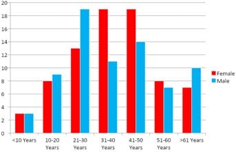
Figure 1:  Bar graph showing age and sex distribution of skin lesions.

ijmrr_1351_02.JPG
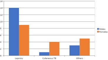
Figure 2:  Graph showing distribution of infectious skin lesions with gender.

ijmrr_1351_03.JPG
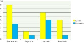
Figure 3: Bar graph showing the distribution of papulosquamous skin lesions with gender.

ijmrr_1351_04.JPG
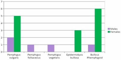
Figure 4:  Bar graph showing the distribution of vesicobullous skin lesions with gender.

ijmrr_1351_05.JPG
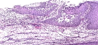
Figure 5: Pemphigus Vegetans: Suprabasal acantholysis with pseudo-epitheliomatous hyperplasia.H & E 100X.


ijmrr_1351_06.JPG
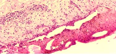
Figure 6: Bullous Pemphigoid: Lymphocytes and eosinophils are present at the dermo-epidermal junction.Eosinophilic spongiosis. H & E 100X.

ijmrr_1351_07.JPG
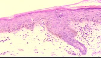
Figure 7: Guttate Psoriasis: Blunting of rete ridges, orthokeratosis, slight acanthosis, dilated blood vessels in dermal papillae.  H & E 100X.

ijmrr_1351_08.JPG
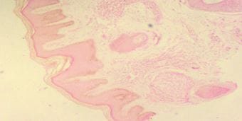
Figure 8: Indeterminate Leprosy: Orthokeratotic, hyperkeratosis, irregular acanthosis and mild spongiosis. Pandermal perineurovascular and periappendegeal chronic inflammation. H&E 40X.

ijmrr_1351_09.JPG
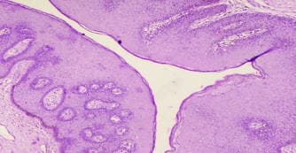
Figure 9: Condyloma Acuminata: Papillomatosis and acanthotic squamous epithelium. Vacuolated and round hyperchromatic nuclei. H&E 40X.

Discussion

The findings of this study were compared to various original studies to evaluate and analyze differences if any and the reasons for them in the distribution of non-neoplastic skin lesions according to the various clinical and pathological aspects. According to age distribution, the maximum number of cases belonged to the 41 to 50 years of age group (22%). In less than 10 years of age group, our study was lower than Megha et al. [9]. but higher than Dixit et al. [10]. and Mithila et al. [11]. In patients of 20 to 30 years of age, our study showed lower than Mithila et al. [11]. Megha et al. [9]. but higher with other studies. In 41 to 50 our study was higher than other studies. In the 51 to 60 and > 61-year age group, we had a minimum variable in studies that suggested that the maximum number of cases in their study belonged to <50 years age group (78.6%). In our studies, differences may be due to the spectrum of skin lesions which is highly variable and expected changes are quantitative rather than qualitative.

Our study showed that the majority of our cases were seen in females (51.3%). Similarly, Kusum et al. also reported female preponderance. However, studies by Dixit et al. [10]. Mithila et al. [11]. and Megha et al. [9]. showed that the incidence of non-neoplastic skin lesions was higher in males. Our hospital is a tertiary referral hospital in a predominantly urban setting. The greater awareness and social pressures of maintaining cosmetics, especially in the female gender is probably the reason for this finding in our study. An analysis of the broad spectrum of the dermatological lesions revealed that the maximum number of lesions were of infectious nature constituting 44 cases (29.4%) followed by non-infectious papulosquamous disorders constituting 34 cases (22.7%). Studies were carried out by Jyoti et al. [3].  Megha et al. [9]. Dixit et al. [10].  and Mithila et al.[11]. also corroborated our findings.

Out of 36 infectious cases, the overwhelming majority of cases constituted of leprosy comprising 23 cases (63.9%) and 5 cases of cutaneous tuberculosis were observed. Dixit et al., Mithila et al,Kusum et al. and Jyoti et al. also observed leprosy to be the single largest category of all infectious skin lesions. In contrast to our study, Veldhurthy et al. [12]. concluded


lichenoid lesions as the commonest histopathological findings (25%) followed by Hansen’s disease (23.9%). These findings probably reflect the resurgence and re-emergence of leprosy as an important skin lesion in our country. This is probably due to the neglect in the control of this disease following the stated eradication of leprosy in our country. This re-emergence of this socially relevant disease is a wake-up call to all government health authorities to tackle this problem on a war footing. The second-largest group of disorders in the present study was papulosquamous disorders constituting 22.7 % of all the cases. Similarly, Dixit et al. also found these to be commonest after infectious disorders. Out of the broad category of non-infectious, papular and squamous disorders, the majority of cases were dermatitis consisting of 13 out of 34 cases (38.2%), next was a lichenoid pattern (35.3%) and psoriasis was in 17.6 % cases. The study by Dixit et al. also showed similar findings.  However, another study by Jyoti et al. found psoriasis to be the most common group among papulosquamous disorders followed by lichenoid pattern and Kusum et al. found lichenoid lesions are most common followed by psoriasis. In the study by Grace D’Costa et al. [13]. Lichenoid lesions were the commonest (46.57%) followed by psoriasis (19.88%). The etiopathogenesis of these lesions is heavily dependent on the environmental conditions as well as exposure to different agents. These different findings reflect this variability in the conditions. The most common vesicobullous skin lesion was Pemphigus Vulgaris (42.1%) followed by Bullous Pemphigoid (36.8%). This is consistent with studies conducted by Dixit et al. Mithila et al. and Megha et al. also reported Pemphigus Vulgaris is the commonest vesicobullous skin lesion followed by Bullous Pemphigoid.

Conclusion

Based on our study, Middle-aged adults constituted the major group with a preponderance of females. The analysis of cases revealed that the maximum number of cases were from the broad group of infectious lesions (leprosy being the most common infectious disease) followed by a group of noninfectious papulosquamous lesions, followed by vesicobullous lesions. Out of the broad category of non-infectious papular and squamous disorders, the majority of cases showed dermatitis. The most common vesicobullous skin lesion was Pemphigus

Vulgaris. The spectrum of skin lesions is highly variable. Therefore, for confirmation of diagnosis and initiation of treatment, histopathological examination of the skin biopsy remains the gold standard which can be supported with other techniques to confirm the diagnosis. Lastly, an exact diagnosis of skin lesions is at times difficult without proper clinical differential diagnoses from the dermatologist. Without these inputs, the work of the pathologist becomes much more complicated and strenuous. Therefore, an integrated approach by both dermatologists and pathologists with continuous communication between the two is recommended to increase the accuracy of diagnosis and for better management of the patient.

Reference

01. Johnson Jr, B. L. Congenital diseases (genodermatosis). Levers Histopathology of the Skin (2005). [Crossref][PubMed][Google Scholar]

02. Singh, Akhil Kumar, et al. A Study on Clinico-histopathological Correlation of Skin Diseases in a Dermatological Setting. " Indian Journal of Public Health Research & Development 7. 1 (2016). [Crossref][PubMed][Google Scholar]

03. Narang, Sanjeev, and Ravi Jain. An evaluation of histopathological findings of skin biopsies in various skin disorders. Annals of Pathology and Laboratory Medicine 2. 01 (2015). [Crossref][PubMed][Google Scholar]

04. Lever WF. Histology of the skin. Histopathology of the Skin. Philadelphia. 1990:9-43 [Crossref][PubMed][Google Scholar]

05. Alguire PC, Mathes BM. Skin biopsy techniques for the internist. J Gen Intern Med. 1998 Jan;13(1):46-54. doi: 10.1046/j.1525-1497.1998.00009.x [Crossref][PubMed][Google Scholar]

06. Sleiman R, Kurban M, Abbas O. Maximizing diagnostic outcomes of skin biopsy specimens. Int J Dermatol. 2013 Jan;52(1):72-8. doi: 10.1111/j.1365-4632.2012.05731.x [Crossref][PubMed][Google Scholar]

07. Weedon, David. Weedon's Skin Pathology E-Book: Expert Consult-Online and Print. Elsevier Health Sciences, 2009. [Crossref][PubMed][Google Scholar]


08. Fox BJ, Odom RB. Papulosquamous diseases: a review. J Am Acad Dermatol. 1985 Apr;12(4):597-624. doi: 10.1016/s0190-9622(85)70084-9 [Crossref][PubMed][Google Scholar]

09. Megha Sharma. Clinicopathological study of non-Neoplastic skin lesions, JEBMH. 2020; 7(11). [Crossref][PubMed][Google Scholar]

10. Prajapati, Dixit H. , Purvi Patel, and Hansa Goswami. Non neoplastic skin lesions: A histopathological study based on punch biopsy. [Crossref][PubMed][Google Scholar]

11. Bisht, Mithila, Anjana Arya, and B. C. Choudhry. Histomorphological analysis and clinical correlation of neoplastic and non neoplastic skin lesions: a study in a tertiary care centre of Western Uttar Pradesh, India. International Journal of Research in Medical Sciences 8.8 (2020): 2820 [Crossref][PubMed][Google Scholar]

12. Veldurthy, Vijay Sreedhar, et al. Pathological study of non-neoplastic skin lesions by punch biopsy. (2015). [Crossref][PubMed][Google Scholar]

13. D' Costa G, Bharambe BM. Spectrum of non-infectious erythematous, papular and squamous lesions of the skin. Indian J Dermatol. 2010 Jul-Sep;55(3):225-8. doi: 10.4103/0019-5154.70666 [Crossref][PubMed][Google Scholar]