E-ISSN:2320-8686
P-ISSN:2321-127X

Research Article

Bronchiectasis

International Journal of Medical Research and Review

2021 Volume 9 Number 6 November December
Publisherwww.medresearch.in

Risk Factors Affecting Exacerbation of Bronchiectasis Leading to Hospitalisation in Patients Attending a Tertiary Care Setting

Ettuveetil M.1, Anandan P.2*, Akkara P.3
DOI: https://doi.org/10.17511/ijmrr.2021.i06.05

1 Mahroofa Ettuveetil, Senior Resident, Department of Pulmonary medicine, Govt. Medical College, Manjeri, Malappuram, Kerala, India.

2* Poriyanganattu Thankappan Anandan, Associate Professor, Department of Pulmonary medicine, Govt. Medical College, Kozhikode, Kerala, India.

3 Paulo Varghese Akkara, Assistant Professor, Department of Pulmonary medicine, Govt. Medical College, Kozhikode, Kerala, India.

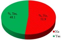
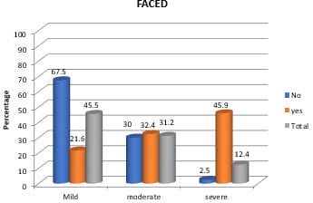
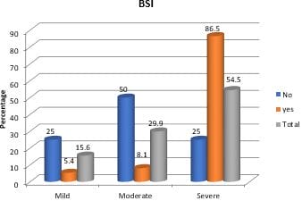
Introduction: Bronchiectasis is a chronic respiratory disease characterized by structural changes in airways, leading to recurrent episodes of cough with expectoration. Exacerbations are significant events associated with increased morbidity and mortality. Aim: To evaluate factors associated with exacerbations requiring hospitalization in patients with bronchiectasis Materials and methods: A prospective observational study was done at a tertiary care teaching hospital from December 2017 to May 2019. Demographic data, comorbidities, investigations including HRCT, sputum culture and spirometry were collected. Patients were grouped into exacerbations requiring hospitalizations and those managed as outpatients and followed up for one year. Factors associated with exacerbations were assessed and compared between the two groups. Results: 89 patients were recruited for the study, out of which 12 patients without exacerbation during the study period were excluded. The remaining 77 patients were divided into those who needed hospitalization (37) and those managed as outpatients (40). About 48.1%required hospital admission. Factors associated with exacerbations were advanced age, isolation of organisms from sputum, FEV1 less than 50%, PPI use during the study period, concomitant COPD, vaccination with pneumococcal and influenza vaccines and high severity scores. Use of LTO2, LABA/ICS, chest physiotherapy, mucolytic agents, radiological extent and previous history of exacerbations were not associated with exacerbations leading to hospitalization. Conclusion: Factors associated with bronchiectasis exacerbations leading to hospitalizations were advanced age, high bronchiectasis severity score, FEV1 less than 50%, PPI use and concomitant COPD. Vaccination for influenza and pneumococcus had a favourable impact in reducing hospitalization.

Keywords: Bronchiectasis, Exacerbation, Hospitalization

Corresponding Author How to Cite this Article To Browse
Poriyanganattu Thankappan Anandan, Associate Professor, Department of Pulmonary medicine, Govt. Medical College, Kozhikode, Kerala, India.
Email:
Mahroofa Ettuveetil, Poriyanganattu Thankappan Anandan, Paulo Varghese Akkara, Risk Factors Affecting Exacerbation of Bronchiectasis Leading to Hospitalisation in Patients Attending a Tertiary Care Setting. Int J Med Res Rev. 2021;9(6):379-387.
Available From
https://ijmrr.medresearch.in/index.php/ijmrr/article/view/1349

Manuscript Received Review Round 1 Review Round 2 Review Round 3 Accepted
2021-11-20 2021-11-22 2021-11-29 2021-12-06 2021-12-13
Conflict of Interest Funding Ethical Approval Plagiarism X-checker Note
Nil Nil Yes 18%

© 2021by Mahroofa Ettuveetil, Poriyanganattu Thankappan Anandan, Paulo Varghese Akkaraand Published by Siddharth Health Research and Social Welfare Society. This is an Open Access article licensed under a Creative Commons Attribution 4.0 International License https://creativecommons.org/licenses/by/4.0/ unported [CC BY 4.0].

Introduction

Bronchiectasis is a chronic respiratory disease presenting with cough and sputum production. Underlying structural airway changes lead to recurrent respiratory infections and impaired quality of life. Bronchiectasis has been a neglected disease. However, the past several years has generated a lot of new evidences that has refined our understanding of the disease. Exacerbations are important events that affect prognosis and quality of life in patients with bronchiectasis. In bronchiectasis, exacerbations have been associated with increased mortality and more significant impairment of lung function, as well as more severe forms of the disease with substantial concomitant health-related costs [1-3]. Exacerbations account for a large proportion of the clinical workload and the economic impact of bronchiectasis on health care systems internationally.

There is limited data on mortality after an acute exacerbation of bronchiectasis. Recent studies have reported survival in patients with acute exacerbation of bronchiectasis. In these studies, the mortality rate was 40% at one year and 60% at four years [4,5,6]. At the same time, mortality in non-hospitalized patients with bronchiectasis varies from 13% to 25%, according to some other studies [7]. Mortality related to bronchiectasis exacerbations can be decreased to a certain extent if factors affecting exacerbations that require hospitalizations are identified and managed appropriately. Patients with risk factors for severe exacerbations are likely to be hospitalized. Few data are available on risk factors and patient characteristics in bronchiectasis that might provoke exacerbations requiring hospital admission apart from severity scales. Age, Frequency of exacerbations, the severity of the disease, radiological extent, colonization with microorganisms, smoking status, previous hospitalization history and vaccinations are likely to influence exacerbations and hospitalizations. Identification of these factors may help in formulating preventive strategies and patient monitoring. A European study has identified certain factors related to host characteristics, comorbidities, severity scores, previous exacerbations and treatment-related factors etc. [8]. There is a dearth of studies from our country on how such risk factors influence exacerbations and hospitalizations.

So, we planned to evaluate whether these factors are relevant in our patient populations with bronchiectasis. 

Detailed Study Plan         

Objective: To evaluate factors associated with exacerbations requiring hospitalization in patients with bronchiectasis, about host characteristics, usual treatments, severity and history of exacerbations.

Materials & Methods

Study design: Prospective observational study

Study settings: Patients attending a tertiary care teaching hospital at Government medical college in Kerala

Study period: One and half years from December 2017

Sample size: Prevalence of hospitalization in bronchiectasis exacerbation is taken as 23.55% Hence sample size= 4pq/d2.

p=23.55 q=77.45
d is taken as 10
Sample size =72

Sampling method: Simple random sampling method of selecting patients from Out Patient Department or ward.

Inclusion criteria: Adults with a compatible clinical history consistent with chronic sputum production and frequent respiratory infections with findings suggestive of bronchiectasis in HRCT of lungs performed before study recruitment.

Exclusion criteria:

  • Patients with immunosuppression-Human immunodeficiency virus infection/acquired immune deficiency syndrome (HIV/AIDS), or taking chemotherapy or other immunosuppressive drugs.
  • Active Pulmonary Tuberculosis.
  • Cystic fibrosis.
  • Interstitial lung disease.

Patients were enrolled after getting informed consent. They were examined, and various investigations were done as per the protocol. They were followed up for one year with the


periodic review every two months. The cohort consisted of 77 patients followed up for one year and separated into two groups: patients treated as outpatients and those hospitalized at least once during the follow-up period due to exacerbation. The treating physician decided for hospital admission as per the BTS 2010 guideline. Exacerbations were defined as an acute deterioration with increased sputum volume and purulence and/systemic upset, according to BTS guidelines [9].

Demographic data, diagnosis of bronchiectasis, comorbidities, smoking, and vaccine status (influenza and pneumococcal vaccines) were recorded. Comorbid conditions recorded were Diabetes mellitus, chronic obstructive pulmonary disease (COPD), Asthma, chronic heart failure, Myocardial infarction and prior Tuberculosis. We recorded COPD as comorbidity similar to other studies. We defined bronchiectasis associated with COPD in the presence of a smoking history of at least ten pack-years with airflow obstruction (FEV1/FVC ratio < 0.7) according to the Global Initiative for Chronic Obstructive Lung Disease recommendations [10].

Data related to previous chronic infections, the number of exacerbations during the last year, and bronchiectasis severity scores (BSI, FACED) were also recorded for all patients. Usual chronic and concomitant medications included bronchodilators, inhaled/nebulized antibiotics, proton pump inhibitors, long-term oxygen therapy, and mucolytic drugs in the last six months. Microbiological diagnosis of exacerbation is performed with sputum culture. In outpatient exacerbations, microbiological tests include sputum culture and any other additional test according to physician decision. The microbial aetiology of exacerbation was defined as any positive result from the microbiological investigation, as per previous publications.

Statistical analysis: Data were analyzed using SPSS software version 22. Descriptive and inferential statistical analysis has been carried out in the present study. Results on continuous measurements are presented on Mean  SD (Min-Max), and results on categorical measures are presented in number (%). Significance is assessed at a 5 % level of significance. The following assumptions on data are made Assumptions: 1. Dependent variables should be normally distributed,

2. Samples drawn from the population should be random. Cases of the samples should be independent.

Chi-square/ Fisher Exact test has been used to find the significance of study parameters on a categorical scale between two or more groups, the non-parametric setting for Qualitative data analysis. Fisher Exact test is used when samples are very small.

Results

The study group consisted of 77 patients followed up for one year and separated into two subsets: patients treated as outpatients and those admitted to the hospital at least once during the follow-up period.37(48.1%) had been hospitalized due to exacerbation among the 77 during the study period.

ijmrr_1349_01.JPG
Figure 1. Hospitalization during follow up.

Most of the hospitalizations due to exacerbations were in the 61-70 years group [56.8%]. All patients above 50 years of age required hospitalization due to exacerbation. The distribution of males and females is almost similar in the two groups (Table1).

Table 1: Association of age and Gender in relation to hospitalization during follow up. 

variables Hospitalization during follow-up Total (n=77) P value
No (n=40) Yes (n=37)
Age in years        
<40 4(10%) 0(0%) 4(5.2%) 0.001**
40-50 16(40%) 0(0%) 16(20.8%)
51-60 16(40%) 16(43.2%) 32(41.6%)
61-70 4(10%) 21(56.8%) 25(32.5%)
Gender        
Female 22(55%) 20(54.1%) 42(54.5%) 0.934
Male 18(45%) 17(45.9%) 35(45.5%)

Chi-Square/Fisher Exact Test (p-value less than 0.05 significant) Vaccinated patients were more in the OP patients' group; 35 % patients had taken


influenza vaccine and 47.5% had the pneumococcal vaccine, 8.1% and 21.6% for IP patient respectively [significant p value].8 out of 32 patients were smokers in IP cohort whereas 3 out of 40 patients were smokers in OP group (Table2).

Table 2: Association of Smoking status, Influenza vaccine and pneumococcal vaccine in relation to hospitalization during follow up. 

variables Hospitalization during follow-up Total (n=77) P value
No (n=40) Yes (n=37)
Smoking Status 3(7.5%) 8(21.6%) 11(14.3%) 0.077+
Influenza vaccine 14(35%) 3(8.1%) 17(22.1%) 0.004**
Pneumococcal vaccine 19(47.5%) 8(21.6%) 27(35.1%) 0.017*

Chi-Square/Fisher Exact Test (p-value less than 0.05 significant)

On sputum culture, 29 out of 37 IP patients yielded any culture isolate. Out of these, 32.4% were pseudomonas, 18.9 % were MDR pathogens, and 27% were other organisms, whereas, in 31 out of 40 OP patients, no organisms were isolated in culture (Table3).

Table 3: Isolated Organism distribution with hospitalization during follow up.

Organism Hospitalization during follow-up Total (n=77)
No (n=40) Yes (n=37)
No organism 31(77.5%) 8(21.6%) 39(50.6%)
Organism 9(22.5%) 29(78.4%) 38(49.4%)
·         Pseudomonas 4(10%) 12(32.4%) 15(20.8%)
·         MDR pathogen 4(10%) 7(18.9%) 11(14.3%)
·         Other organisms 1(2.5%) 10(27%) 11(14.3%)

P 0.001**, Significant, Fisher Exact Test.

In the IP Cohort, more than half of the patients had FEV1 less than 50% of the predicted value, whereas, in the OP cohort, the entire patient population had FEV1 above 50% (P value 0.001) (Table 4). 

Table 4: FEV1 in relation to hospitalization during follow up. 

FEV1 Hospitalization during follow-up Total
No Yes
<50 0(0%) 22(59.5%) 22(28.6%)
50-70 32(80%) 15(40.5%) 47(61%)
>70 8(20%) 0(0%) 8(10.4%)
Total 40(100%) 37(100%) 77(100%)

P 0.001**, Significant, Fisher Exact Test (FEV1- Forced Expiratory Volume in 1 second).

COPD was found to be associated with exacerbations, which was more in IP patients:12 out of 37 patients [32.4%] with COPD had exacerbations, which was statistically significant [P value 0.002]. In the OP cohort, only 2 (5%) had COPD and exacerbations.

Table 5: Comorbidities in relation to hospitalization during follow up. 

  Hospitalization during follow-up Total (n=77) P value
No (n=40) Yes (n=37)
DM 10(25%) 16(43.2%) 26(33.8%) 0.091+
COPD 2(5%) 12(32.4%) 14(18.2%) 0.002**
Heart Failure 3(7.5%) 8(21.6%) 11(14.3%) 0.077+

Chi-Square/Fisher Exact Test (p-value less than 0.05 significant).

[DM- Diabetes Mellitus; COPD – Chronic Obstructive Pulmonary Disease].

54% of the patient from the IP cohort shows the involvement of more than three lobes, whereas 70 % of the patient from the OP cohort shows less than three lobes, but this difference is not statistically significant (p value0.087) (Table 6). 

Table 6: Radiological extent in relation to hospitalization during follow up. 

Radiology [HRCT, No. of lobe] Hospitalization during follow-up Total
No Yes
1 14(35%) 6(16.2%) 20(26%)
2 14(35%) 11(29.7%) 25(32.5%)
3 11(27.5%) 15(40.5%) 26(33.8%)
4 1(2.5%) 5(13.5%) 6(7.8%)
Total 40(100%) 37(100%) 77(100%)

P=0.087+, Not Significant, Fisher Exact Test.

An underlying aetiology of bronchiectasis was identified in 48.06% of the patients. The most common aetiology of bronchiectasis was post-infective (83%) following Tuberculosis (25.97%), childhood infections (12.96%) and pneumonia (3.89%). There were three patients with immunoglobulin deficiency and one case of ABPA. Evaluation of bronchiectasis severity scores with risk for hospitalizations has shown that those with high severity scores had more hospitalizations than those treated as outpatients. (Table 7).


Table 7: Association of FACED and BSI to Hospitalization during follow-up. 

variables Hospitalization during follow-up Total (n=77) P value
No (n=40) Yes (n=37)
FACED        
Mild 27(67.5%) 8(21.6%) 35(45.5%) 0.001**
Moderate 12(30%) 12(32.4%) 24(31.2%)
Severe 1(2.5%) 17(45.9%) 18(23.4%)
BSI        
Mild 10(25%) 2(5.4%) 12(15.6%) 0.001**
Moderate 20(50%) 3(8.1%) 23(29.9%)
Severe 10(25%) 32(86.5%) 42(54.5%)

Chi-Square/Fisher Exact Test (p-value less than 0.05 significant).

[BSI- Bronchiectasis severity Index, F-FEV1, A-Age, C-Chronic colonization,E radiological extent,D - Dyspnoea]ijmrr_1349_02.JPG

Figure 2: Severity scoring in bronchiectasis – FACED.

ijmrr_1349_03.JPG

Figure 3: Severity scoring in bronchiectasis – BSI.

Most of the patients from both groups (83%) used LABA and ICS. There was not much difference between the groups regarding the use of LABA/ICS. Only 6 (16.2%) patients were

using LTOT among IP patients and 3(7.5%) patients using in OP cohort. 73% of patients from the IP cohort were using mucolytics, whereas, from the OP cohort, 87.5% used mucolytics. Out of 40 OP patients, 25 were using Proton Pump Inhibitors, and 32 patients from the IP cohort were using PPI. There was a statistically significant difference among the cohorts in PPI use (p-0.035). Almost 70% of patients from the IP cohort and 82.5 % patients from the OP cohort regularly used chest physiotherapy. Hospitalization due to exacerbation in the past year was more in the IP cohort [48.6%] compared to the OP cohort [30%]; however, there was no statistical significance for this finding. From the IP cohort, 64.9 % of patients had a history of the previous hospitalization due to bronchiectasis at any time, whereas in the OP cohort, it was 50% (Table 8).

Table 8: Treatment-related factors and exacerbations during follow up. 

Previous hospitalization due to BE at any time Hospitalization during follow-up Total  
No Yes
No 20(50%) 13(35.1%) 33(42.9%) P=0.188, Chi-Square Test
Yes 20(50%) 24(64.9%) 44(57.1%)
Total 40(100%) 37(100%) 77(100%)
Chest Physiotherapy  
No 7(17.5%) 11(29.7%) 18(23.4%) P=0.205, Chi-Square Test
Yes 33(82.5%) 26(70.3%) 59(76.6%)
Total 40(100%) 37(100%) 77(100%)
MUCOLYTIC    
No 5(12.5%) 10(27%) 15(19.5%) P=0.108, Chi-Square Test  
Yes 35(87.5%) 27(73%) 62(80.5%)
Total 40(100%) 37(100%) 77(100%)
LABA/ICS  
No 7(17.5%) 6(16.2%) 13(16.9%) P=0.881, Chi-Square Test  
Yes 33(82.5%) 31(83.8%) 64(83.1%)
Total 40(100%) 37(100%) 77(100%)
PPI  
No 15(37.4%) 5(13.5%) 20(22.1%) P=0.035*, Chi-Square Test  
Yes 25(62.6%) 32(86.5%) 57(77.9%)
Total 40(100%) 37(100%) 77(100%)
Long Term O2  
No 37(92.5%) 31(83.8%) 68(88.3%) P=0.299, Fisher Exact Test  
Yes 3(7.5%) 6(16.2%) 9(11.7%)
Total 40(100%) 37(100%) 77(100%)
Hospitalization in last year due to BE
No 28(70%) 19(51.4%) 47(61%) P=0.094 Chi-Square Test  
Yes 12(30%) 18(48.6%) 30(39%)
Total 40(100%) 37(100%) 77(100%)

(p-value less than 0.05 significant) [BE- Bronchiectasis exacerbation, PPI – Proton Pump Inhibitor, LABA – Long-Acting Beta Agonist, ICS – Inhaled Corticosteroids].

Discussion

We recruited a total of 89 patients with bronchiectasis in this study. About 12 patients without exacerbations during the follow-up period were excluded from the analysis. Among the 77 study subjects, males constituted 45.6% of the study population. The mean age of the population was 52.6. The relationship between demographic characteristics and outcomes for the population shows a strong relationship between age and hospitalization due to exacerbation. Most patients requiring hospitalization due to exacerbation were above 51 years of age (P value0.001). In the OP cohort, 40% of the patients were above 50 years of age. Menendez et al. found that the mean age for severe exacerbation of bronchiectasis is73.[8]. Chalmers et al. also found a strong relationship between age and mortality [3].73 % of patients with exacerbation were above 70 years with a mean age of 74 years. Whereas in our study mean age is 62 yrs. (51 -70) in hospitalized patients.

Gender difference does not seem to be responsible for the difference in exacerbations and hospital admissions among the two groups. Pasteur et al. has found a predilection for female sex to bronchiectasis, but lung damage developed at the same rate in both genders [11]. Menendez et al. found that the male gender was associated with more severe disease, more comorbidities and higher Pseudomonas colonization. Ringshausen et al. have also reported an increase in hospitalization among older men. [12].

The data on FEV1 of our study patients have shown that 59.5% of the patients from the inpatient group had FEV1 less than 50 percent of predicted, whereas, in the OP patient group, all of them had FEV1 more than 50% (P-value 0.001). Several studies have attempted to correlate disease severity with other associated factors in the decline in FEV1 % predicted. Chalmers et al. found that FEV1 < 50% had significant relation with mortality and exacerbation of bronchiectasis; based on the Youden index, FEV1 % predicted was most discriminatory for mortality and hospital admissions and was used for subsequent

analyses of lung function. Similarly, patients with lower lung function were more frequently hospitalized and had an increased annual exacerbation frequency and worse quality of life. Menendez et al. also found similar findings. Roberts et al. found that airflow obstruction correlated with the degree of radiographic findings on CT, specifically the severity of bronchial wall thickening and disease of the small airways [13].

Evaluation of the isolation of the organism from the culture in patients with exacerbations showed that there was significantly higher culture isolation in patients with any one of the bacterial isolates, compared with non-culture isolated patients. 29 out of 37 inpatients yielded any one of culture isolate, in which 32.4% were pseudomonas. Chalmers et al. found that mortality was significantly higher in patients with chronic colonization than non-colonized patients. With the highest mortality rates associated with the isolation of pseudomonas and methicillin-resistant staphylococcus. Chalmers JD et al. also found that exacerbations were more with pseudomonas infection (40.7%) [14,15]. Pseudomonas aeruginosa infection in the respiratory tract of bronchiectasis patients is associated with worsening airway clearance and airway obstruction, worsening lung function and impaired health-related quality of life [16]. This may be due to the ability of this organism to release virulent exotoxins, form bio-films on tissue surfaces, and quickly develop hypermutable P. aeruginosa strains resistant to antibiotics, and all these factors increase bronchial damage.

Analysis of radiological severity data showed that involvement of more than three lobes in HRCT had an increased chance for hospitalizations due to exacerbations compared to less involvement in HRCT; however, this finding was not statistically significant (p-value = 0.087). The involvement of 3 or more lobes was associated with hospitalization due to exacerbation in bronchiectasis. The study by Charmers et al. shows no significant relationship between radiological severity and mortality but a significant relationship between the Reiff score and hospital admissions. Thus, involvement with more than three lobes had more hospital admissions due to exacerbations, but Menendez et al. has found no significant relationship between radiological severity and exacerbation. [8].

Analysis of the use of Proton Pump Inhibitors


(PPI) showed that, out of 37 in the inpatient's group, 32(86.5%) patients were using PPI, and from the 40 OP cohort, 25 (62.6%) were using PPI. There was a statistically significant association between the use of PPI and exacerbations. The pathophysiological mechanism behind this association is not clearly defined. Some studies suggest that PPIs modify the composition of the gut microbiome, and increase levels of oral and upper gastro-intestinal tract commensals due to changes in pH [17]. This alteration of the microbiome could contribute to more severe exacerbations requiring hospital admission. Another study in a mouse model found that an increase follows depletion of the gut microbiota in bacterial dissemination, inflammation, and even resulting in organ damage [18].             

At the study enrolment, 48.6% of the patients gave a history of hospitalization with exacerbation or respiratory tract infection in the preceding year. This shows that a prior history of hospital admissions may be relevant to future mortality, hospital admissions, exacerbations, and quality of life. Previous studies have identified prior hospitalization as the most decisive risk factor for severe exacerbation in other chronic respiratory diseases [8,19]. This might be due to the greater bronchial and systemic inflammation, as has been observed during exacerbations. This contributes to perpetuating the infection-inflammation cycle and has a negative effect on prognosis [20]. The mortality at one year in our study is significantly less compared to the mortality reported by Dupont et al. (40%) and by Alzeer et al. (34%) in patients admitted to the MICU with an acute exacerbation of bronchiectasis [3,4]. In our study, 6 out of 37(16.2%) IP patients died during follow up period and 1 out of 40(2.5%) OP patients died during follow up period, and all six patients from the IP cohort had a history of ICU admissions.

As expected, patients with bronchiectasis exacerbations (BE) with higher BSI and FACED scores had more hospital admissions because of exacerbation. BSI classified patients into low, intermediate and high scores. There was a clear difference in hospitalization due to exacerbation between patients classified as low, intermediate, and high BSI scores [P value 0.001]. Other studies also show BE patients with higher BSI and FACED scores required more hospital admissions. [19,21,22,23]. Vaccination against influenza and pneumococci are associated with a protective effect

in bronchiectasis. Our study also showed that vaccinated patients were more in the non-hospitalized group; 35 % of patients had taken influenza vaccine, and 47.5% had taken the pneumococcal vaccine, whereas 8.1% and 21.6% in hospitalized patients, respectively [ p value=0.004]. The pneumococcal vaccine has been recommended for bronchiectasis patients in the literature [24]. In this study, several factors were not associated with hospital admissions after adjustment for other included variables, such as aetiology of bronchiectasis, comorbidities like DM, heart failure, smoking status, chest physiotherapy, use of LTOT, inhaled beta2 agonist and corticosteroid. Almost all patients in our study group were using ICS/LABA. Smoking has been known to be associated with lung disease. In this study, it was found that smoking was not associated with increased risk for exacerbations; it may be because of the small study population and less proportion of smoking patients, whereas in other studies, smoking was found to be a risk factor. Possible causes are defects in the ability to clear secretions predisposing them to an increased risk of infections and increased inflammation of the lung parenchyma caused by the toxic chemicals in tobacco [25]. In our study, only 11(14.3%) out of 77 were smokers. The low number of smokers in the study group might not have sufficient power to impact the exacerbation and hospitalization.

What does this study add to existing knowledge?

This study found the use of PPI as a risk factor for exacerbations leading to hospitalization in bronchiectasis patients; this warrants further studies on this subject and necessitates judicious use of proton pump inhibitors in such patients.

Limitations of the study:  Being a study done in a tertiary care centre, most of the cases would have been in the advanced stage. Therefore our findings may not be generalizable to the population with milder bronchiectasis. Long-term follow-up was needed for assessing exacerbations; only one year of follow up was done in this study.

Conclusions

Our study found that the risk factors associated with bronchiectasis patients likely to develop exacerbations requiring hospitalization


during a one-year follow-up period are Age, Forced Expiratory Volume in 1 second (FEV1), Coexistent COPD, Culture isolation of organism, Use of Proton Pump Inhibitors (PPI) and Bronchiectasis Severity score. Pneumococcal and Influenza vaccination is found to be a protective factors.

Author contributions: Mahroofa EV- Literature review, data collection, draft manuscript preparation, statistical workup. Anandan PT – Research concept, study design, Literature review, and review of the manuscript, editing and final approval. Paulo Varghese Akkara – Study design, manuscript review, statistical review and supervision of data collection.

Reference

01. Martínez-García MA, Soler-Cataluña JJ, Perpiñá-Tordera M, Román-Sánchez P, Soriano J. Factors associated with lung function decline in adult patients with stable non-cystic fibrosis bronchiectasis. Chest. 2007 Nov;132(5):1565-72. doi: 10.1378/chest.07-0490 [Crossref][PubMed][Google Scholar]

02. Quint JK, Millett ER, Joshi M, Navaratnam V, Thomas SL, Hurst JR, et al. Changes in the incidence, prevalence and mortality of bronchiectasis in the UK from 2004 to 2013: a population-based cohort study. Eur Respir J. 2016 Jan;47(1):186-93. doi: 10.1183/13993003.01033-2015 [Crossref][PubMed][Google Scholar]

03. Chalmers JD, Smith MP, McHugh BJ, Doherty C, Govan JR, Hill AT. Short- and long-term antibiotic treatment reduces airway and systemic inflammation in non-cystic fibrosis bronchiectasis. Am J Respir Crit Care Med. 2012 Oct 1;186(7):657-65. doi: 10.1164/rccm.201203-0487OC [Crossref][PubMed][Google Scholar]

04. Onen ZP, Gulbay BE, Sen E, Yildiz OA, Saryal S, Acican T, Karabiyikoglu G. Analysis of the factors related to mortality in patients with bronchiectasis. Respir Med. 2007 Jul;101(7):1390-7. doi: 10.1016/j.rmed.2007.02.002 [Crossref][PubMed][Google Scholar]

05. Dupont M, Gacouin A, Lena H, Lavoué S, Brinchault G, Delaval P, et al. Survival of patients with bronchiectasis after the first ICU stay for respiratory failure. Chest. 2004 May;125(5):1815-20. doi: 10.1378/chest.125.5.1815 [Crossref][PubMed][Google Scholar]

06. Finklea JD, Khan G, Thomas S, Song J, Myers D, Arroliga AC. Predictors of mortality in hospitalized patients with acute exacerbation of bronchiectasis. Respir Med. 2010 Jun;104(6):816-21. doi: 10.1016/j.rmed.2009.11.021 [Crossref][PubMed][Google Scholar]

07. Goeminne PC, Nawrot TS, Ruttens D, Seys S, Dupont LJ. Mortality in non-cystic fibrosis bronchiectasis: a prospective cohort analysis. Respir Med. 2014 Feb;108(2):287-96. doi: 10.1016/j.rmed.2013.12.015 [Crossref][PubMed][Google Scholar]

08. Menéndez R, Méndez R, Polverino E, Rosales-Mayor E, Amara-Elori I, Reyes S, et al. Factors associated with hospitalization in bronchiectasis exacerbations: a one-year follow-up study. Respir Res. 2017 Sep 30;18(1):176. doi: 10.1186/s12931-017-0659-x [Crossref][PubMed][Google Scholar]

09. Pasteur MC, Bilton D, Hill AT; British Thoracic Society Bronchiectasis non-CF Guideline Group. British Thoracic Society guideline for non-CF bronchiectasis. Thorax. 2010 Jul;65 Suppl 1:i1-58. doi: 10.1136/thx.2010.136119 [Crossref][PubMed][Google Scholar]

10. Global strategyfor diagnosis, management and prevention of COPD 2017 UPDATE; e GOLD website www. goldcopd. org. [Crossref][PubMed][Google Scholar]

11. Pasteur MC, Helliwell SM, Houghton SJ, Webb SC, Foweraker JE, Coulden RA, et al. An investigation into causative factors in patients with bronchiectasis. Am J Respir Crit Care Med. 2000 Oct;162(4 Pt 1):1277-84. doi: 10.1164/ajrccm.162.4.9906120 [Crossref][PubMed][Google Scholar]

12. Ringshausen FC, de Roux A, Diel R, Hohmann D, Welte T, Rademacher J. Bronchiectasis in Germany: a population-based estimation of disease prevalence. Eur Respir J. 2015 Dec;46(6):1805-7. doi: 10.1183/13993003.00954-2015 [Crossref][PubMed][Google Scholar]

13. Roberts HR, Wells AU, Milne DG, Rubens MB, Kolbe J, Cole PJ, et al. Airflow obstruction in bronchiectasis: correlation between computed tomography features and pulmonary function tests. Thorax. 2000 Mar;55(3):198-204. doi: 10.1136/thorax.55.3.198 [Crossref][PubMed][Google Scholar]


14. Davies G, Wells AU, Doffman S, Watanabe S, Wilson R. The effect of Pseudomonas aeruginosa on pulmonary function in patients with bronchiectasis. Eur Respir J. 2006 Nov;28(5):974-9. doi: 10.1183/09031936.06.00074605 [Crossref][PubMed][Google Scholar]

15. Chalmers JD, Aliberti S, Blasi F. Management of bronchiectasis in adults. Eur Respir J. 2015 May;45(5):1446-62. doi: 10.1183/09031936.00119114 [Crossref][PubMed][Google Scholar]

16. Finch S, McDonnell MJ, Abo-Leyah H, Aliberti S, Chalmers JD. A Comprehensive Analysis of the Impact of Pseudomonas aeruginosa Colonization on Prognosis in Adult Bronchiectasis. Ann Am Thorac Soc. 2015 Nov;12(11):1602-11. doi: 10.1513/AnnalsATS.201506-333OC [Crossref][PubMed][Google Scholar]

17. Jackson MA, Goodrich JK, Maxan ME, Freedberg DE, Abrams JA, et al. Proton pump inhibitors alter the composition of the gut microbiota. Gut. 2016 may;65(5):749-56. doi: 10.1136/gutjnl-2015-310861 [Crossref][PubMed][Google Scholar]

18. Schuijt TJ, Lankelma JM, Scicluna BP, de Sousa e Melo F, Roelofs JJ, de Boer JD, et al. The gut microbiota plays a protective role in the host defence against pneumococcal pneumonia. Gut. 2016 Apr;65(4):575-83. doi: 10.1136/gutjnl-2015-309728 [Crossref][PubMed][Google Scholar]

19. Chalmers JD, Goeminne P, Aliberti S, McDonnell MJ, Lonni S, Davidson J, et al. The bronchiectasis severity index. An international derivation and validation study. Am J Respir Crit Care Med. 2014 Mar 1;189(5):576-85. doi: 10.1164/rccm.201309-1575OC [Crossref][PubMed][Google Scholar]

20. Alzeer AH, Masood M, Basha SJ, Shaik SA. Survival of bronchiectatic patients with respiratory failure in ICU. BMC Pulm Med. 2007 Dec 10;7:17. doi: 10.1186/1471-2466-7-17 [Crossref][PubMed][Google Scholar]

21. Martinez-Garcia MA, Athanazio RA, Girón R, Máiz-Carro L, de la Rosa D, Olveira C, et al. Predicting high risk of exacerbations in bronchiectasis: the E-FACED score. Int J Chron Obstruct Pulmon Dis. 2017 Jan 18;12:275-284. doi: 10.2147/COPD.S121943 [Crossref][PubMed][Google Scholar]

22. Rosales-Mayor E, Polverino E, Raguer L, Alcaraz V, Gabarrus A, Ranzani O, et al. Comparison of two prognostic scores (BSI and FACED) in a Spanish cohort of adult patients with bronchiectasis and improvement of the FACED predictive capacity for exacerbations. PLoS One. 2017 Apr 6;12(4):e0175171. doi: 10.1371/journal.pone.0175171 [Crossref][PubMed][Google Scholar]

23. . . PLoS One. 2017 Apr 6;12(4):e0175171. doi: 10.1371/journal.pone.0175171 [Crossref][PubMed][Google Scholar] [Crossref][PubMed][Google Scholar]

24. McDonnell MJ, Aliberti S, Goeminne PC, Restrepo MI, Finch S, Pesci A, et al. Comorbidities and the risk of mortality in patients with bronchiectasis: an international multicentre cohort study. Lancet Respir Med. 2016 Dec;4(12):969-979. doi: 10.1016/S2213-2600(16)30320-4 [Crossref][PubMed][Google Scholar]

25. Aliberti S, Mantero M, Mirsaeidi M, Blasi F. The role of vaccination in preventing pneumococcal disease in adults. Clin Microbiol Infect. 2014 May;20 Suppl 5(0 5):52-8. doi: 10.1111/1469-0691.12518 [Crossref][PubMed][Google Scholar]

26. Peinado VI, Barberá JA, Abate P, Ramírez J, Roca J, Santos S, Rodriguez-Roisin R. Inflammatory reaction in pulmonary muscular arteries of patients with mild chronic obstructive pulmonary disease. Am J Respir Crit Care Med. 1999 May;159(5 Pt 1):1605-11. doi: 10.1164/ajrccm.159.5.9807059 [Crossref][PubMed][Google Scholar]