E-ISSN:2320-8686
P-ISSN:2321-127X

Research Article

Spectrum

International Journal of Medical Research and Review

2021 Volume 9 Number 5 September-October
Publisherwww.medresearch.in

Clinical Spectrum and Management of hydatidosis – A Prospective Study

Pandey S.1, Kumar Shukla R.2, Chawla K.3, Mishra A.4*
DOI: https://doi.org/10.17511/ijmrr.2021.i05.04

1 Sanjay Pandey, Senior Consultant, Department of Surgery, District Female Hospital, Mirzapur, Uttar Pradesh, India.

2 Rakesh Kumar Shukla, Assistant Professor, Department of Anatomy, Autonomous State Medical College and Hospital, Mirzapur, Uttar Pradesh, India.

3 Kavita Chawla, Professor, Department of Physiology, Moti Lal Nehru Medical College, Prayagraj, Uttar Pradesh, India.

4* Archana Mishra, Assistant Professor, Department of Biochemistry, Moti Lal Nehru Medical College, Prayagraj, Uttar Pradesh, India.

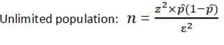
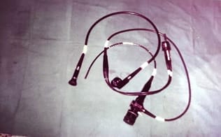
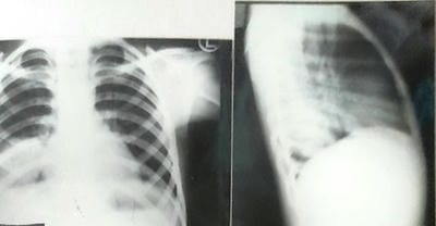
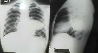
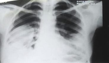
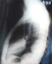
Aim: The objective of this original research article is a Clinical Spectrum and Management of hydatidosis – A Prospective Study. Material & method: -The present study includes 150 patients with intrathoracic space-occupying lesions, of which 24 patients with Hydatid cyst were seen at SS hospital Banaras Hindu University, Varanasi, UP. Most of the patients studied were from the thoracic surgery section, while few patients were taken from other departments of SS hospital. The age of those patients varied widely, starting from 10 years to 60 years. During this study, patients were investigated in systemic order with the progression of symptoms from the onset, development of latest symptoms and treatment taken before if any, history of tuberculosis, chronic cough, smoking, and contact with were recorded. The patient's vitals were recorded, and routine pathological investigation including blood count, hemoglobin and specific procedure like radiological method, Casoni diagnostic test and bronchoscopy were performed. Result: Maximum patients were in the age group of 41 to 50 years. The maximum age group was 60 years, and the minimum age group was ten years. Maximum age group 21-30 years, among which this has been observed. No cases were seen above 41 years of age. Hemoglobin was found adequate among the cases. Total Leucocytes count was found higher among the patients. In 63% cases, found above 10000 per microliter. ESR found raised among the patients and in 50% of cases observed more than 40 mm in 1st hour. Cough with/without expectoration, Chest pain, and Dyspnoea were observed in almost all cases. The right side of the Lung had 50% involvement; the Left side of the Lung had 37.5% involvement, whereas 12.5% bilateral involvement. Conclusion: Mini-thoracotomy is an efficient and safe option for managing intact or ruptured solitary pulmonary hydatid cysts.

Keywords: Multiple hydatid cysts, Hydatid Cyst, Hydatidosis, Mini – Thoracotomy

Corresponding Author How to Cite this Article To Browse
Archana Mishra, Assistant Professor, Department of Biochemistry, Moti Lal Nehru Medical College, Prayagraj, Uttar Pradesh, India.
Email:
Sanjay Pandey, Rakesh Kumar Shukla, Kavita Chawla, Archana Mishra, Clinical Spectrum and Management of hydatidosis – A Prospective Study. Int J Med Res Rev. 2021;9(5):298-305.
Available From
https://ijmrr.medresearch.in/index.php/ijmrr/article/view/1334

Manuscript Received Review Round 1 Review Round 2 Review Round 3 Accepted
2021-08-20 2021-08-30 2021-09-10 2021-09-16 2021-09-22
Conflict of Interest Funding Ethical Approval Plagiarism X-checker Note
No Nil Yes 9%

© 2021 by Sanjay Pandey, Rakesh Kumar Shukla, Kavita Chawla, Archana Mishra and Published by Siddharth Health Research and Social Welfare Society. This is an Open Access article licensed under a Creative Commons Attribution 4.0 International License https://creativecommons.org/licenses/by/4.0/ unported [CC BY 4.0].

Introduction

Hydatidosis (HD) is an ancient disease and even was known to Hippocrates. It can virtually affect any organ within the body from head to toe, and also the most typically affected organs are found to be the liver and lungs [1]. The occurrence of Echinococcosis at unusual sites is approximately 8-10%. Hydatidosis is a significant pathological state within the infested areas of the globe, mainly in sheep-raising communities. But attributable to increased travel and tourism worldwide, it is often found anywhere, even in developed countries [2]. It can involve any organ and mimic almost any pathological condition. The clinical picture depends upon the involved organs, their effects on adjacent structures, complications because of secondary infection, rupture, and anaphylaxis caused by hydatid cysts [3].

The first asymptomatic phase and delayed presentation make early diagnosis difficult. The varied complications of the disease, instead of the disease itself, are tougher to treat. The infection might undertake a 'malignant' course within the sense of a high rate of recurrence, which further enforces the importance of meticulous evaluation and proper planning of treatment and better preventive methods aiming at control and eradication of this age-old disease [4].

Further studies are therefore needed to enhance our understanding of the varied clinical problems caused by this parasite generally and more specifically to reassess the surgical nature, complications, and fatality of this disease to use a more appropriate surgical procedure to human Echinococcosis [5]. Surgery for infestation has always been considered because of the gold standard in terms of therapy despite significant advances in medical treatment and interventional radiology [6]. With time, the treatment for abdominal infestation is undergoing revolutionary changes. The era of open surgery with its associated large incision and prolonged stay is now being challenged by lesser invasive procedures [7]. With the evolution of laparoscopic surgeries, laparoscopic treatment of infestation has also evolved [8]. Hydatid cyst may be a parasitic, infectious disease, which is endemic in many places worldwide, like the Mediterranean countries, Iran, India, Australia, and South America. In step with World Health Organization (WHO), the annual incidence of Cystic Echinococcus is up to 220per 100,000 inhabitants in these countries [9].

The causal parasite of the disease is Echinococcus granulosus. Humans can function as intermediate hosts for this organism. It always infects human organs separately or in groups, especially the liver and, therefore, the lungs. The hydatid cyst grows slowly and asymptomatically in most cases to the extent that some cysts may exceed 20 cm in diameter. This expansive growth can seriously damage the tissue of the hosting organ and makes spontaneous, traumatic, or intra-operative rupture of the cyst easier. The optimal treatment targets are complete elimination of the parasite, preservation of the utmost of the healthy tissue, and prevention of recurrence by avoiding the spillage of the cystic fluid and dissemination of the cyst contents [9, 10]. The incidence of hydatidosis at unusual sites in India (as also reported by other authors from India) isbeyond that in other parts of the planet [11]. The liver is that the commonest site for hydatidosis (60–80% of cases), followed by lungs (10–30%), spleen (5%), and other organs (5%) [12].

Pulmonary Echinococcosis is most typical within the right Lung and lower lobes especially. Seventy-five to ninety percent of those cysts are solitary [12]. Both lung involvements are seen in 2–30% of cases. Multiple cysts are seen in 30% of the patients [12]. Synchronous pulmonary and hepatic infestation may occur in 4–25% of cases [13]. Pulmonary and hepatic hydatid, if on the identical side, will be removed via thoracotomy and phrenotomy with similar morbidity rates and similar postoperative outcomes [14]. Bilateral lung cysts usually require a two-stage procedure [14]. However, bilateral thoracotomy within the same setting is another surgical approach for excision with an identical postoperative outcome [15, 16]. The transdiaphragmatic method is effective and safe and prevents patients from requiring a second surgery [15]. The median sternotomy is a choice to access bilateral pulmonary hydatids. The single-stage bilateral minimally invasive approach in some selected groups of patients has also been reported to be a secure option [17].

Material and Methods

Patients presenting with clinical or radiological shreds of evidence of intrathoracic masses were subjects of this study. Most of the patients came to the thoracic surgery section directly. At the same time, other was referred from the department of ENT, radiotherapy, and medicine of SS hospital Banaras Hindu University, Varanasi, UP.


Inclusion Criteria:-

  1. Age group 10-60 yrs, all sex, occupation, religion
  2. All the patients were admitted to the department of surgery with the diagnosis of Lung echinococcosis.
  3. Patients giving consent to be a part of the study.
  4. The patient who followed up a minimum of two years.

Exclusion Criteria:-

  1. Patients having hydatidosis who have already undergone surgery previously.
  2. Recurrent echinococcosis.
  3. Patients who refused consent to be a part of the study.
  4. Patients who didn't come for follow-up a minimum of one year.

The sample size was calculated using the formula through the department of statistics at Banaras Hindu University.

ijmrr_1334_06.jpg

Where

-Z is that the z score at Confidence Interval 75% is 1.15

-ε is that the margin of error, assumed 5%

-p̂ is that the population proportion assumed 50%

This implies 133 or more measurements/surveys are needed to have& a confidence level of 75% that the 000 value is within ±5% of the measured/surveyed value. Ethical clearance was obtained from the moral committee of the Institute of Medical Sciences, Banaras Hindu University. Total 150 cases of intrathoracic masses were included, and this study was carried for two years after obtaining consent from each patient. This study was a prospective study. During this study, patients were investigated in systemic order include name, age, sex, position, occupation, address, and chief complaint. Their complaints with duration were noted. Progression of symptoms from the onset, development of latest symptoms and treatment taken before if any, history of tuberculosis, chronic cough, smoking, and contact were recorded.

The patient's vitals were recorded, and routine pathological investigation, including blood count, hemoglobin work, was tired all cases.

The specialized procedure followed:-

  • Casoni diagnostic test
  • Radiological method
  • Bronchoscopy

ijmrr_1334_01.jpg

Fig 1: Fibre optic flexible bronchoscope

Casoni diagnostic test- This is often a hypersensitivity-based diagnostic test accustomed detects hydatidosis. Although once a severe difficulty in diagnosing infestation, it's largely been superseded by newer, more sensitive, specific, and safer serological tests.

In this technique, sterile fluid (0.25 mL) of hydatid cyst origin is injected into one arm whilst 0.25 mL of normal saline is injected into the opposite arm to act as an effect. Care must be taken as there's a risk of an anaphylactic reaction. Observation of a wheel at the positioning of injection of the hydatid cyst origin fluid at half-hour post-injection is thought to be a positive test. Observation of a delayed reaction is additionally possible.

Bronchogram/bronchography: The radiographic examination of the bronchial tree employing a positive contrast agent, which shows the bronchial branches as white shadows on an X-ray film. It required the introduction of dye into the bronchial tree followed by the radiograph, which outlines the cast of the bronchial tree on the chest radiograph. Bronchography gives valuable information about the control of bronchi and their abnormalities. Two methods of Bronchography - Trans bronchoscopic and Direct bronchoscopic. Direct Bronchoscopic:- Performed by Percutaneous trans tracheal technique, nasotracheal fine tube intubation, and injecting in laryngo pharynx using the curved cannula


Computed tomography: Performed for extent and precise location of mediastinal masses, small pulmonary lesions and extent of mediastinal lymphadenopathy, pathology of major vessels, and compression or displacement of esophagus.

Bronchoscopy: Bronchoscopy is a crucial method of investigation and in conjunction with radiology. It's the foremost helpful procedure concerning broncho pulmonary neoplasm. This can be meted out using either a rigid or flexible fiberoptic bronchoscope.

Instruments used are rigid bronchoscope which is actually a lightweight carrying tube with a connecting cable to a lightweight source. The instrument permits direct viewing of the inside of the larynx trachea and main segmental bronchi. An entire range of rigid bronchoscopes includes man and feminine, adolescent, child infant, and suckling. There are direct and angle telescopes that allow magnification and visualization of these bronchial openings, which don't seem to be in direct line of the bronchoscopic field of vision. Accessory includes straight and angle bronchial biopsy punches and foreign body extractors of various sizes.

ijmrr_1334_02.jpg

Fig 2: Chest PA of 10 years old child showing hydatid cyst in the right lower lobe.

ijmrr_1334_03.jpg

Fig 3: Solitary hydatid cyst in the left lower lobe with no reaction in adjacent lung tissue.

ijmrr_1334_04.jpg ijmrr_1334_05.jpg

Fig 4: Radiograph of a 25 years old female patients showing multiple infected bilateral hydatid cysts

Preoperative bronchoscopy was done, and all patients received Albendazole 15 mg/kg/day orally in divided doses for at least four weeks before surgery. A double-lumen endotracheal tube was incorporated for all the patients. Right-sided lung and liver hydatid cysts were selected for simultaneous excision by thoracophrenotomy approach. Bilateral lung hydatid cysts underwent bilateral lateral thoracotomy, with the side of impending rupture being operated upon first. Barrett's manoeuvre was achieved by low tidal high positive pressure ventilation. Scolicidal agent—3% hypertonic saline—was used. Bronchial leaks were closed with 3–0 prolene intermittent sutures. A radial phrenotomy incision was made safeguarding the phrenic nerve.


Observations

The present study includes 150 patients with intrathoracic space-occupying lesions, of which 24 patients with Hydatid cyst were seen at SS hospital Banaras Hindu University, Varanasi, UP. Most of the patients studied were from the thoracic surgery section, while few patients were taken from other departments of SS hospital. The age of those patients varied widely, starting from 10 years to 60 years. 

Table 1: - Age Distribution of Intrathoracic Lesions Cases

Age Group (Years) No of Cases Percentage
10-20
21-30
31-40
41-50
51-60
27
21
27
54
21
18
14
18
36
14
Total = 150

Maximum patients were in the age group of 41 to 50 years. The leading age group was 60 years, and the minimum age group was ten years.

Table 2:- Age Distribution of Hydatid Cyst Cases among Intrathoracic Lesions

Age Group (Years) No of Cases Percentage
10-20
21-30
31-40
41-50
51-60
03
12
09
-
-
12.5
50
37.5
-
-
Total = 24

Maximum age group 21-30 years, among which this has been observed. No cases were seen above 41 years of age.

Table 3: - Hemoglobin Pattern in Hydatid cyst cases

Hb gm% No of Cases Percentage
8-10
10-12
12-14
06
06
12
25
25
50
Total 24 100.0

Hemoglobin was found adequate among the cases.

Table 4 :- Total Leucocytes Pattern in Hydatid cyst cases

Total Leucocytes Count No of Cases Percentage
5000-10000
10000-15000
09
09
37.5
37.5
>15000 06 25
Total 24 100.0

Total Leucocytes count was found higher among the cases. In 63% cases, found above 10000 per microliter.

Table 5: - ESR Pattern in Hydatid cyst cases

ESR No of Cases Percentage
< 20
20-40
>40
03
09
12
12.5
37.5
50
Total 24 100.0

ESR found raised among the cases and in 50% cases observed more than 40 mm in 1st hour.

Table 6:- Presenting Symptoms in Hydatid cyst cases

S.No Symptoms No of Cases (%)
1
2
3
4
5
Cough with/without expectoration
Chest pain
Dyspnoea
Fever
Other symptoms (Headache, Nausea, Vomiting)
18
15
15
05
09
75
62.5
62.5
21
37.5

Cough with/without expectoration, Chest pain, and Dyspnoea were observed in almost all cases.

Table 6:- Relative frequency of mass in Lung

Mass Lesion No of Cases Percentage
Left side
Right side
Bilateral
09
12
03
37.5
50.0
12.5
Total 24 100.0

The right side of the Lung had 50% involvement; the Left side of the Lung had 37.5% involvement, whereas 12.5% bilateral involvement.

Other Important Observations

  • Twenty-one (87.5%) cases found positive with Casoni skin test
  • Nine patients had a history of association with the dog.
  • Six patients had salty yellow sputum
  • Twenty-one patients operated, and successful enucleation of the cyst was done in eighteen cases.

Discussion

Hydatid disease may be an animal disease, which is decreasing nowadays, with the modified lifestyle of the people and improved sanitation.


Published studies during this field are only a few and limited to a minimal number of cases. This is often thanks to the chance of cystic rupture and, therefore, the spillage of its content during operation, resulting in recurrence within the future, and to the problem in controlling the accompanied bronchial fistulas and, consequently, the residual cavity, which increases complication rates and lengthen hospital stay. Such complications are also why some surgeons suggest preserving this system for dead cysts only [18]. Our study comprised 24 patients over 24 months. The maximum cohort among were 21-30 years, observed 50% of total cases. No cases were seen above 41 years old. In our study youngest was ten years, and also the oldest was 40 years. In a study, the mean age was 20.7 years [14].

In a study, the mean age was 24.3 years with nine years because the youngest and 72 years oldest patient among patients [15, 17]. Cough with/without expectoration (75%), pain (62.5%), and Dyspnoea (62.5%) were observed in the majority of cases. The same as the study by Lone, who reported pain (75%) [14]. The mean hemoglobin was 11.5 g/dl. Fifteen patients (62.5%) had raised total leukocyte count against 4% in Lone's study in Kashmir [14]. All patients received albendazole therapy 15 mg/kg/day for a minimum of 4–6 weeks before surgery [12]. In an exceedingly study on 21 patients with Echinococcosis who underwent albendazole treatment, some researchers found a 77.3% incidence of cyst rupture [19]. In our study, both lung involvements were seen in three patients (12.5%).

Biswas et al., in their study on 216 cases of pulmonary hydatids, found 42 patients with concomitant hepatic hydatid cysts (19.44%) and 18 patients with bilateral lung hydatids (8.33%) [15, 17]. Erdogan et al. studied 142 patients between 1990 and 2004 in Turkey and located 27 cases (19%) of lung and liver hydatid cysts and 12 cases (8.45%) of bilateral lung hydatids [20]. This shows that the share of both lung involvements in various series remains unaffected. All the cases underwent contrast-enhanced tomography of the thorax and, therefore, the upper abdomen in relevant cases. Twenty-one patients operated, and successful enucleation of the cyst was wiped out of eighteen cases. Three patients refused the surgery. Out of 21 cases that were operated on, 3 of them had infected cyst of left lower lobe treated as a case of chronic empyema after removal of ecto and endocyst.

Moreover, the routine application of prophylactic pharmacological therapy with Albendazole after surgery helped prevent a recurrence, which reached zero in some studies [21, 22]. However, the follow-up period in a number of these studies was short (about six months) and not sufficient to detect all cases of recurrence [21, 23, 24]. The operative time in our study was shorter than that reported in some similar studies [24, 25] under the operative easiness through mini-thoracotomy. Those studies were limited to small cysts that are but 7 cm in diameter, which isn't usually amid bronchial fistulas, while our study included large cysts that reached 15 cm in diameter.

The duration of hospital stay in our study was relatively short, thanks to the easiness of control over bronchial fistulas and closure of the residual cavity after cyst removal via mini-thoracotomy, and thus avoiding prolonged postoperative air leakage (a single case out of 130 operations). In some studies [21, 24, 26], where the mini-thoracotomy approach wasn't used, prolonged postoperative air leakage was the only common complication that had led to an extended hospital stay and to an increased complications rate that reached 13.3% within the Levent Alpay study [25]. The rate of infectious complications was high in our series, within which we reported three cases: empyema and abscess formation within the place of the removed cyst. This can be in step with the results of the Milind study [27] and requires reconsideration of the role of thoracoscopic surgery in such cases and searching for a more robust alternative.

Conclusion

In conclusion, the case series revealed that mini-thoracotomy is an efficient and safe option for managing intact or ruptured solitary pulmonary hydatid cysts. Further studies in future controlled design are needed to match this approach to other modalities of management.

Disclosure

The authors declare that there is no conflict of interest.

Authors Contribution

Conception and design: Dr Sanjay Pandey

Collection and assembly of data: Dr Sanjay Pandey and Dr Rakesh Kumar Shukla


Data analysis and interpretation: Dr Archana Mishra

Manuscript writing: All authors

Final approval of manuscript: All authors

Accountable for all aspects of the work: All authors

Reference

01. PUMP KK. Echinococcosis (hydatid disease): a review and report of a case of secondary Echinococcosis. Can Med Assoc J. 1963 Jul 13;89(2):73-8. [Crossref][PubMed][Google Scholar]

02. Barnes TS, Deplazes P, Gottstein B, Jenkins DJ, Mathis A, Siles-Lucas M, et al. Challenges for diagnosis and control of cystic hydatid disease. Acta Trop. 2012 Jul;123(1):1-7. doi: 10.1016/j.actatropica.2012.02.066 [Crossref][PubMed][Google Scholar]

03. Vuitton DA. Echinococcosis and allergy. Clin Rev Allergy Immunol. 2004 Apr;26(2):93-104. doi: 10.1007/s12016-004-0004-2 [Crossref][PubMed][Google Scholar]

04. Irabedra P, Ferreira C, Sayes J, Elola S, Rodríguez M, Morel N, et al. Control programme for cystic echinococcosis in Uruguay. Mem Inst Oswaldo Cruz. 2016 May 24;111(6):372-7. doi: 10.1590/0074-02760160070 [Crossref][PubMed][Google Scholar]

05. Romero-Torres R, Campbell JR. An interpretive review of the surgical treatment of hydatid disease. Surg Gynecol Obstet. 1965 Oct;121(4):851-64. [Crossref][PubMed][Google Scholar]

06. Palanivelu C, Jani K, Malladi V, Senthilkumar R, Rajan PS, Sendhilkumar K, et al. Laparoscopic management of hepatic hydatid disease. JSLS. 2006 Jan-Mar;10(1):56-62. [Crossref][PubMed][Google Scholar]

07. Buia A, Stockhausen F, Hanisch E. Laparoscopic surgery: A qualified systematic review. World J Methodol. 2015 Dec 26;5(4):238-54. doi: 10.5662/wjm.v5.i4.238 [Crossref][PubMed][Google Scholar]

08. Zaharie, Florin, et al. "Open or laparoscopic treatment for hydatid disease of the liver? A 10-year single-institution experience. " Surgical endoscopy. 27;6(2013): 2110-2116. [Crossref][PubMed][Google Scholar]

09. Guidelines for treatment of cystic and alveolar Echinococcosis in humans. WHO Informal Working Group on Echinococcosis. Bull World Health Organ. 1996;74(3):231-42. [Crossref][PubMed][Google Scholar]

10. Bagheri R, Haghi SZ, Amini M, Fattahi AS, Noorshafiee S. Pulmonary hydatid cyst: analysis of 1024 cases. Gen Thorac Cardiovasc Surg. 2011 Feb;59(2):105-9. doi: 10.1007/s11748-010-0690-z [Crossref][PubMed][Google Scholar]

11. Kayal A, Hussain A. A comprehensive prospective clinical study of hydatid disease. ISRN Gastroenterol. 2014 Mar 9;2014:514757. doi: 10.1155/2014/514757 [Crossref][PubMed][Google Scholar]

12. Shields T W, Reed C, LoCicero III J, Feins R. General Thoracic surgery 7th edition. Philadelphia, LWW. (2009). [Crossref][PubMed][Google Scholar]

13. Kurul IC, Topcu S, Altinok T, Yazici U, Tastepe I, Kaya S, Cetin G. One-stage operation for hydatid disease of Lung and liver: principles of treatment. J Thorac Cardiovasc Surg. 2002 Dec;124(6):1212-5. doi: 10.1067/mtc.2002.127314 [Crossref][PubMed][Google Scholar]

14. Lone GN, Bhat MA, Ali N, Bashir A, Garcoo SA. Single-stage bilateral minimally invasive approach for pulmonary hydatid disease: an alternative technique. J Thorac Cardiovasc Surg. 2002 Nov;124(5):1021-4. doi: 10.1067/mtc.2002.122315 [Crossref][PubMed][Google Scholar]

15. Biswas B, Ghosh D, Bhattacharjee R, Patra A, Basuthakur S, Basu R. One stage surgical management of hydatid cyst of Lung & liver—by right thoracotomy & phrenotomy. Indian Journal of Thoracic and Cardiovascular Surgery. 20;2(2004)88-90. [Crossref][PubMed][Google Scholar]

16. Aydin Y, Çelik M, ULAŞ A B, EROĞLU A. Transdiaphragmatic approach to liver and lung hydatid cysts. Turkish Journal of Medical Sciences. 42;Sup 2(2012):1388-1393. [Crossref][PubMed][Google Scholar]

17. Biswas B, Ghosh D, Bhattacharjee R, Patra A, Basuthakur S, Basu R. One stage bilateral thoracotomy for hydatid cysts of both lungs. Indian Journal of Thoracic and Cardiovascular Surgery. 20;3(2004):126-128. [Crossref][PubMed][Google Scholar]


18. Auldist, Alex W, Russell Blakelock. "Pulmonary hydatid disease". Pediatric Thoracic Surgery, Springer, London. 2009;161-167. [Crossref][PubMed][Google Scholar]

19. Usluer O, Kaya SO, Samancilar O, Ceylan KC, Gursoy S. The effect of preoperative albendazole treatment on the cuticular membranes of pulmonary hydatid cysts: should it be administered preoperatively?. Kardiochir Torakochirurgia Pol. 2014 Mar;11(1):26-9. doi: 10.5114/kitp.2014.41926 [Crossref][PubMed][Google Scholar]

20. Erdogan A, Ayten A, Kabukcu H, Demircan A. One-stage transthoracic operation for the treatment of right lung and liver hydatid cysts. World J Surg. 2005 Dec;29(12):1680-6. doi: 10.1007/s00268-005-0130-x [Crossref][PubMed][Google Scholar]

21. Uchikov AP, Shipkov CD, Prisadov G. Treatment of lung hydatidosis by VATS: a preliminary report. Can J Surg. 2004 Oct;47(5):380-1. [Crossref][PubMed][Google Scholar]

22. Creţu CM, Codreanu RR, Mastalier B, Popa LG, Cordoş I, Beuran M, et al. Albendazole associated to surgery or minimally invasive procedures for hydatid disease--how much and how long. Chirurgia (Bucur). 2012 Jan-Feb;107(1):15-21. [Crossref][PubMed][Google Scholar]

23. Ettayebi F, M Benhannou. "Echinococcus granulosus cyst of the lung: treatment by thoracoscopy". Pediatric Endosurgery and Innovative Techniques. 7;1(2003):67-70. [Crossref][PubMed][Google Scholar]

24. Parelkar SV, Gupta RK, Shah H, Sanghvi B, Gupta A, Jadhav V, et al. Experience with video-assisted thoracoscopic removal of pulmonary hydatid cysts in children. J Pediatr Surg. 2009 Apr;44(4):836-41. doi: 10.1016/j.jpedsurg.2008.11.029 [Crossref][PubMed][Google Scholar]

25. Alpay L, Lacin T, Ocakcioglu I, Evman S, Dogruyol T, Vayvada M, Baysungur V, Yalcinkaya I. Is Video-Assisted Thoracoscopic Surgery Adequate in Treatment of Pulmonary Hydatidosis?. Ann Thorac Surg. 2015 Jul;100(1):258-62. doi: 10.1016/j.athoracsur.2015.03.011 [Crossref][PubMed][Google Scholar]

26. Bagheri R, Haghi SZ, Amini M, Fattahi AS, Noorshafiee S. Pulmonary hydatid cyst: analysis of 1024 cases. Gen Thorac Cardiovasc Surg. 2011 Feb;59(2):105-9. doi: 10.1007/s11748-010-0690-z [Crossref][PubMed][Google Scholar]

27. Tullu MS, Lahiri KR, Kumar S, Oak SN. Minimal access therapy in pediatric pulmonary hydatid cysts. Pediatr Pulmonol. 2005 Jul;40(1):92-5. doi: 10.1002/ppul.20231 [Crossref][PubMed][Google Scholar]