E-ISSN:2320-8686
P-ISSN:2321-127X

Research Article

Population

International Journal of Medical Research and Review

2021 Volume 9 Number 5 September-October
Publisherwww.medresearch.in

Prevalence of Bronchogenic carcinoma in the North Indian Population

Pandey S.1, Mishra A.2, Chawla K.3, Kumar Shukla R.4*
DOI: https://doi.org/10.17511/ijmrr.2021.i05.05

1 Sanjay Pandey, Senior Consultant, Department of Surgery, District Female Hospital, Mirzapur, Uttar Pradesh, India.

2 Archana Mishra, Assistant Professor, Department of Biochemistry, MLN Medical College, Prayagraj, Uttar Pradesh, India.

3 Kavita Chawla, Professor, Department of Physiology, Moti Lal Nehru Medical College, Prayagraj, Uttar Pradesh, India.

4* Rakesh Kumar Shukla, Assistant Professor, Department of Anatomy, Autonomous State Medical College and Hospital, Mirzapur, Uttar Pradesh, India.

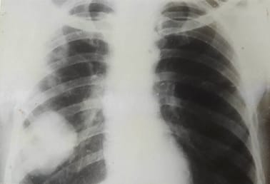
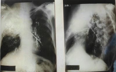
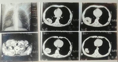
Aim: This original research article aims to study the Prevalence of Bronchogenic carcinoma in the North Indian Population. Material & Method: The present study includes 150 patients with intrathoracic space-occupying lesions, of which 42 patients with bronchogenic carcinoma were seen at SS hospital Banaras Hindu University, Varanasi, UP. Most of the patients studied were from the thoracic surgery section, while few patients were taken from other departments of SS hospital. The age of those patients varied widely, starting from 10 years to 60 years. During this study, patients were investigated in systemic order with the progression of symptoms from the onset, development of latest symptoms and treatment taken before if any, history of tuberculosis, chronic cough, smoking, and contact with were recorded. Result: Maximum patients were in the age group of 41 to 50 years. Forty-two patients were diagnosed with Bronchogenic carcinoma, a definitive diagnosis was confirmed by Clinical & Radiological (chest X-ray & CT Scan) in 24 (16%), by Bronchoscopic examination & Biopsy in 15 (10%) and by thoracotomy in 03(2%). The maximum age group among the cases were 41-60. The most common symptom was Cough with expectoration in 33 patients (78.5%), followed by Haemoptysis and Dyspnea in 30 cases (71.4%). Fever was least common with 6 cases. It was observed that 18 patients (43%) had the habit of smoking for more than 18 years. Conclusion: This study provides a framework for assessing the prevalence of bronchogenic carcinoma in the North Indian population and proves that smoking is a potential risk factor in bronchogenic carcinoma. Prevalence of Bronchogenic Carcinoma among all cases of Intrathoracic Lesions Cases 28 % and among all subjects who undergone for screening in one year – 2.84%.

Keywords: Epidemiology, India, Lung Cancer, Nonsmokers, Smokers, Lung Cancer Diagnosis, Bronchogenic Carcinoma

Corresponding Author How to Cite this Article To Browse
Rakesh Kumar Shukla, Assistant Professor, Department of Anatomy, Autonomous State Medical College and Hospital, Mirzapur, Uttar Pradesh, India.
Email:
Sanjay Pandey, Archana Mishra, Kavita Chawla, Rakesh Kumar Shukla, Prevalence of Bronchogenic carcinoma in the North Indian Population. Int J Med Res Rev. 2021;9(5):306-314.
Available From
https://ijmrr.medresearch.in/index.php/ijmrr/article/view/1333

Manuscript Received Review Round 1 Review Round 2 Review Round 3 Accepted
2021-08-10 2021-08-20 2021-08-30 2021-09-17 2021-09-22
Conflict of Interest Funding Ethical Approval Plagiarism X-checker Note
No Nil Yes 7%

© 2021 by Sanjay Pandey, Archana Mishra, Kavita Chawla, Rakesh Kumar Shukla and Published by Siddharth Health Research and Social Welfare Society. This is an Open Access article licensed under a Creative Commons Attribution 4.0 International License https://creativecommons.org/licenses/by/4.0/ unported [CC BY 4.0].

Introduction

Cancer is that the second leading reason of death globally after cardiovascular diseases. Patients with cancer generally have a poorer prognosis in low-income and middle-income countries, including India, thanks to relatively low cancer awareness, late diagnosis, and the lack of or inequitable access to affordable curative services than patients in high-income countries [1, 2]. Bronchogenic carcinoma was once wont to describe a selected form of cancer that arises within the bronchi (the large airways of the lungs) and bronchioles (the small branches into which the airways divide).

Today, it's considered another name for carcinoma generally. We check with tracheal, bronchus, and carcinoma as carcinoma during this report for simplicity. The number of incident carcinoma cases in India in 2016 was 67 000 (95% UI 63 000–72 000), 72•2% of which were in males, and therefore the prevalent cases were 74 000 (70 000–80 000). This cancer was the second most familiar incident cancer among males in 2016. The age-standardized incidence rate of carcinoma varied eight times in both sexes combined across the states of India in 2016. The crude carcinoma incidence rate in males was highest in Kerala and Mizoram, and in females was highest in Mizoram and Manipur.

There was a 6•3 times difference between the very best and lowest state-specific crude disability-adjusted life-years DALY rates for carcinoma in 2016. The crude DALY rate for carcinoma in 2016 was highest in Mizoram, followed by Kerala, Manipur, Jammu, and Kashmir. Carcinoma was the primary or second leading reason behind cancer deaths in 19 states for males and four for females in 2016. Tobacco use and pollution were the top risk factors within the Global Burden of Diseases (GBD) for carcinoma in India in 2016, to which 43•2% and 43•0% of the carcinoma DALYs might be attributed, respectively [3].

There is a lack in our current understanding of the changing epidemiological trends of carcinoma among Indian patients. While the worldwide trend of an increase in adenocarcinoma appears to be paralleled in India, we don't completely understand the alarming rise in carcinoma incidence among nonsmokers. We have, specifically, a limited understanding of the impact of the factors that are unique to our regions, like the presence of indoor pollution, the utilization of domestic or biomass fuel exposure, the presence or lack of micronutrients in

our diet, occupational exposure, and also the possible contribution of infectious pathogens like mycobacteria. Smoking tobacco, both cigarettes, and beedis are the principal risk factors for the causation of carcinoma in Indian men; however, among Indian women, the association with smoking isn't strong, suggesting that there may be other risk factors besides smoking [4]. This study was planned within the Department of surgery Banaras Hindu University Varanasi, UP, because this can be one of all the foremost prestigious institutes in the province with adequate infrastructures, faculties, and patient outflow likewise as no study has been found during this region under such parameters with substantial evidence.

Materials and Methods

This study was performed in the Department of Surgery, Institute of Medical Sciences, Banaras Hindu University, Varanasi, Uttar Pradesh. The total duration of this study was two years, and this study was a prospective and retrospective case study—a total of 1478 patients undergone for screening process in one year. Most of the patients came to the thoracic surgery section directly. At the same time, other was referred from the Department of ENT, radiotherapy, and medicine of SS hospital Banaras Hindu University, Varanasi, UP.

ijmrr_1333_01.jpg

Bronchoscope

Inclusion criteria:

  1. age bracket 10-60 yrs, all sex, occupation, religion
  2. Histologically or cytologically confirmed advanced.
  3. The first tumour and regional lymph gland metastases had to be measurable or a minimum of ready to be evaluated by imaging studies.
  4. Had histological or cytological documentation of

NSCLC, including epithelial cell carcinoma, adenocarcinoma (bronchoalveolar cell carcinoma), and enormous cell and anaplastic carcinoma (including giant- and clear-cell carcinomas). Patients included unresectable or inoperable stage III disease, including N2–N3 disease and any T stage, or those with T4 and nodal stages. Patients with N3 disease were eligible if all gross diseases could be encompassed within the radiation boost field. All patients had the measurable or assessable disease measured by chest radiography, CT, or MRI performed within 28 days of registration. Assessable lesions included ill-defined masses related to post-obstructive changes or mediastinal or hilar lymphadenopathy measurable only in one dimension. A radiation oncologist saw all patients before enrollment in the study. Additional eligibility criteria included a CALGB PS of 0–1, weight loss of two months.

  1. Histologically or cytologically confirmed diagnosis of inoperable IIIA or IIIB NSCLC suitable for radical RT, WHO/ECOG PS 0–2, a measurable or evaluable neoplastic lesion in keeping with WHO criteria, adequate bone marrow.
  2. Previously untreated initially unresectable (or inoperable for medical reasons) non-metastatic NSCLC (histologically or cytologically confirmed) without homolateral malignant pleural effusion and homolateral (except for upper lobe lesion) or hetero-lateral supraclavicular lymph gland involvement; no functional or anatomical contraindication to chest irradiation; an assessable or measurable lesion had to be present. Patients shouldn't have a previous history of malignancy except for non-melanoma skin carcinoma or in place carcinoma of the cervix and 'cured' neoplasm.

Exclusion criteria:

  1. Postoperative thoracic recurrence or a history of any previous or concurrent cancer (except that of the skin) unless the patient had shown no evidence of disease for >5 years. Patients with malignant pleural effusion were also excluded.
  2. Patients with pleural effusion or distant metastases weren't eligible. Patients were excluded if they'd had previous invasive malignant tumours aside from squamous or basal cell carcinoma of the skin within five years of randomization or previous RT or CTX.
  1. Myocardial infarct within the past three months, uncontrolled congestive cardiopathy, uncontrolled arrhythmia, over a minimal pleural effusion, previous CTX or RT for this malignancy, weight loss >5% within the past three months, pregnant or lactating women.
  2. 4. Patients with stage T3N0 or N1 weren't eligible. Patients with scalene, supraclavicular or contralateral hilar node involvement or direct invasion of the vertebral body or pleural effusion exudative, bloody, or cytologically proven to contain malignant cells were ineligible. Patients with completely resected tumours, who were pregnant or who had previously received CTX or RT were also excluded
  3. Previous CTX or RT, history of other malignancy (except for in place cervical carcinoma or non-melanoma skin carcinoma), pregnancy Significant pleural effusions, previous systemic CTX, previous RT to the thorax or total surgical resection, brain metastases, active concurrent malignancy, severe medical or psychiatric illness, history of significantcardiac disease.
  4. Active uncontrolled infection or a fever >38.3ºC, unstable disorder, previous malignancy (except for in place carcinoma of the cervix or adequately treated cutaneous basal or epithelial cell carcinoma). The sample size was calculated using the formula through the Department of statistics at Banaras Hindu University.

ijmrr_1333_02.jpg

Where
Z is that the z score at Confidence Interval 75% is 1.15
ε is that the margin of error, assumed 5%
p̂ is that the ;population proportion assumed 50%

This means 133 or more measurements/surveys are needed to own a confidence level of 75% that the 000 value is within ±5% of the measured/surveyed value.

Ethical clearance was obtained from the moral committee of the Institute of Medical Sciences, Banaras Hindu University.

Total 150 cases of intrathoracic masses were included, and this study was carried for two years after obtaining consent from each patient.


This study may be a prospective and retrospective study. During this study, patients were investigated in systemic order include name, age, sex, legal status, occupation, address, and chief complaint.

Their complaints with duration were noted. Progression of symptoms from the onset, development of latest symptoms and treatment taken before if any, history of tuberculosis, chronic cough, smoking, and contact were recorded. The patient's vitals were recorded, and routine pathological investigation, including blood count, hemoglobin work, was tired all cases.

The specific procedure followed:-

  • Radiological method
  • Sputum
  • Bronchoscopy

Biopsies: lymphoid tissue biopsy, accurate cut biopsy, and excisional Biopsy done.

Bronchogram/bronchography: The radiographic examination of the bronchial tree employing a positive contrast agent, which shows the bronchial branches as white shadows on an X-ray film. It required the introduction of dye into the bronchial tree followed by the radiograph, which outlines the cast of the bronchial tree on the chest radiograph. Bronchography gives valuable information about the control of bronchi and their abnormalities.

Two methods of Bronchography: Trans bronchoscopic and Direct bronchoscopic.
Direct Bronchoscopic:- Performed by Percutaneous trans tracheal technique, nasotracheal fine tube intubation, and injecting in laryngo pharynx using the curved cannula

Computed tomography: Performed for extent and precise location of mediastinal masses, small pulmonary lesions and extent of mediastinal lymphadenopathy, pathology of major vessels, and compression or displacement of esophagus.

Cytology: the target is to spot abnormal (malignant) cells, sheds within the tract and bronchial secretions.

Bronchoscopy: Bronchoscopy is a required method of investigation and in conjunction with radiology. It's the foremost helpful procedure concerning broncho pulmonary neoplasm. This can be meted out using either a rigid or flexible fiberoptic bronchoscope.

Instruments used are rigid bronchoscope which is a lightweight carrying tube with a connecting cable to a light source. The instrument permits direct viewing of the inside of the larynx trachea and main segmental bronchi. An entire range of rigid bronchoscopes includes man and feminine, adolescent, child infant, and suckling. There are direct and angle telescopes that allow magnification and visualization of these bronchial openings, which don't seem to be in direct line of the bronchoscopic field of vision. Accessory includes straight and angle bronchial biopsy punches and foreign body extractors of various sizes.

Observation

The present study includes 150 patients with intrathoracic space-occupying lesions, of which 42 patients with bronchogenic carcinoma were seen at SS hospital Banaras Hindu University, Varanasi, UP. Most of the patients studied were from the thoracic surgery section, while few patients were taken from other departments of SS hospital. The age of those patients varied widely, starting from 10 years to 60 years. The bulk of the patients were young adults and middle-aged persons, with 35 males and seven females.

Table 1: Age Distribution of Intrathoracic Lesions Cases

Age Group (Years) No of Cases Percentage
10-20
21-30
31-40
41-50
51-60
27
21
27
54
21
18
14
18
36
14
Total = 150

Maximum patients were in the age group of 41 to 50 years. The leading age group was 60 years, and the minimum age group was ten years.

Table 2: Definitive Diagnostic Procedure

S.No. Diagnostic Procedure Cases Diagnosed
1 Clinical & Radiological 24
2 Bronchoscopic examination & Biopsy 15
3 Thoracotomy 03
Total 42

Out of 150 patients with intrathoracic lesions, 42 patients diagnosed for Bronchogenic carcinoma, a definitive diagnosis was confirmed by Clinical & Radiological (chest X-ray & CT Scan) in 24 (16%), by Bronchoscopic examination & Biopsy in 15 (10%) and by thoracotomy in 03(2%).


The maximum age group among the cases were 41-60, and male completely outnumbered the female (35 male and seven female).

ijmrr_1333_03.jpg

Figure 1:Chest Radiograph showing sharply defined Ovoid mass in the dorsal segment of the right lower lobe.

Table 3: Presenting Symptoms in Bronchogenic Carcinoma

S.No Symptoms No of Cases Percentage
1
2
3
4
5
6
7
Cough with expectoration
Haemoptysis
Chest pain
Dyspnoea
Fever
Loss of weight
Other symptoms (Headache, Nausea, Vomiting)
33
30
24
30
6
12
15
78.5
71.4
57.1
71.4
14.3
28.6
35.7

The most common symptom was Cough with expectoration in 33 cases (78.5%), followed by Haemoptysis and Dyspnea in 30 patients (71.4%). Fever was least common with 6 cases.

ijmrr_1333_04.jpg

Figure 2
Bronchogenic carcinoma of the right upper lobe with intact inter lobar fissure bulge download by the growth.
Figure 3
Bronchogram shows complete cutoff of upper lobe bronchus on right side.

ijmrr_1333_05.jpg

Figure 4: CT Scan of thorax reveals central necrosis in lesion

Table 4: Hemoglobin Pattern in Bronchogenic Carcinoma

Hb gm% No of Cases Percentage
<8
8-10
>10
6
36
-
14.3
85.7
-
Total 42 100.0

All the patients had hemoglobin less than ten gm%, and most of the patients had leucocytosis ranging between 10000-15000 per mm3 suggestive of secondary infection with ESR between 20-40 mm in 1st hour.

Table 5: Habit of Smoking in Bronchogenic Carcinoma

Group No of Cases Percentage (%)
Smokers 18 43
Non Smokers 24 57

It was observed that 18 patients (43%) had the habit of smoking for more than 18 years.

Prevalence

Prevalence of Bronchogenic Carcinoma among all cases of Intrathoracic Lesions Cases (150) – 28 %

Prevalence of Bronchogenic Carcinoma among all cases 1478 cases who undergone for screening in one year – 2.84%

Discussion

India exhibits heterogeneity in cancer. Population-based cancer registries (PBCRs ) provide statistics on the occurrence and outcome of cancer during a geographically defined population. They also provide the framework for assessing the control of cancer within the community. Hospital-based cancer registries (HBCRs) under the National Cancer Registry Programme–National Centre for Disease Informatics and Research of Indian Council of


Medical Research are concerned with the recording of data on patients with cancer seen during a particular hospital. They are mainly used for reviewing clinical performance and, therefore, the hospital cancer program [5].

The incidence rates of Aizawl district were observed to be seven times and four times that of Osmanabad and Beed PBCRs in males and females, respectively. The best cancer incidence rate was observed within the Northeast region (6 PBCRs for males and 4 PBCRs for females) than other areas within the country. The leading sites of cancer within the NE region were nasopharynx, hypopharynx, oesophagus, stomach, liver, gallbladder, larynx, lung, breast, and porta. The NE region lacks the desired infrastructure for specialized treatment facilities, human resources, as seen by the low 5-year survival of breast, cervix, and head and neck cancer compared with the remainder of India. Many patients with cancer from the NE region are travelling outside the NE for treatment and cancer care [6, 7]. Local cultural factors and lifestyle choices may have contributed to the heterogeneity in cancer incidence patterns and differences in India, as was seen in Thailand [8].

Lung (9 PBCRs), mouth (9 PBCRs), oesophagus (5 PBCRs), stomach (4 PBCRs), and nasopharynx (1 PBCR) cancers were the foremost common cancers in men. Carcinoma was the leading site in metropolitan cities and the southern region, whereas mouth cancer was the top site within the West and Central regions. Carcinoma and oral/mouth cancer were the foremost common cancers among males within the Indian subcontinent [9]. There are cancers of several anatomic sites known to be related to tobacco utilization [10]. Supported PBCR data, almost one-third of the cancers were associated with the utilization of tobacco in India. India state-level disease burden initiative cancer collaborators estimated that tobacco use was the best contributing risk factor for cancer in India. In India,carcinoma is attributed to tobacco use and pollution, the leading risk factors [11].

As per the World Cancer Observatory, GLOBOCAN 2008 report, males predominate with a male: female ratio of 4.5:1, and this ratio varies with age and smoking status. The percentage increased progressively till 51–60 years so remained steady. Various reports have noted that the smoker: nonsmoker ratio is high at 20:1. After 40 years, the epithelial cell type was the foremost common in

smokers, and adenocarcinoma was expected within nonsmokers. The present demographic pattern of carcinoma in India appears to be almost like that of Western countries approximately 40 years ago. There seems to be a marginal increase within the mean age of diagnosis of lung cancers in India over the years from 52.16 years during 1958–1985 to 54.6 years during 1985–2001. The above findings also support this study. During this study, 35 males and seven females were diagnosed with carcinoma. The utmost people was 41 to 50, and over one-third were smokers (43%).

Approximately 70% of cancers in India were potentially preventable through modifiable risk factors [12]. The earliest stage of carcinoma is commonly not related to any symptoms. The foremost common symptoms related to carcinoma are common in benign presentations within the community and particularly amongst smokers. Therefore the discriminative utility of most of those symptoms in isolation is low. Positive predictive values (PPVs) for various carcinoma symptoms, both alone and together, are determined from a case-control study.

Importantly PPVs for every sign are higher in smokers and people over the age of 70 [13]. With the very best PPV of two., 4–7.5% [14], unexplained hemoptysis always warrants further investigation. Haemoptysis, however, could be a feature of only a few fifths of lung cancers [15, 16] therefore, the absence of this symptom shouldn't provide reassurance. In this study, we found the foremost common symptom was Cough with expectoration in 33 cases (78.5%), followed by Haemoptysis and Dyspnea in 30 patients (71.4%).

While guidelines have streamlined access to diagnosis for a few, concern has been raised that this approach might prioritize patients with classical presentations, like hemoptysis, at the expense of those with symptoms that reflect less advanced disease and would therefore have the foremost to achieve from early diagnosis [13]. In fact, in 2013, only 28% of England's carcinoma cases were diagnosed through the country's '2-week-wait' urgent referral pathway. In many cases, appropriately urgent action may have occurred outside the 2-week-wait path, for example, through automatic referral following a suspicious CXR or through routine surveillance for pulmonary nodules. Although declining proportionally, diagnoses following emergency presentations remained the most common route of diagnosis at 35% [17].


Such diagnoses are related to the poorest outcomes. However, ;the explanations for this are likely to be complex. Doubtless, they include the more inferior performance status, more advanced disease, and greater levels of socio-economic deprivation of patients who present during this way [18]. The first-line investigation of suspected ;carcinoma remains the CXR. CXR has the advantage of being cheap and accessible [19], with an occasional radiation dose of 0.02 mSv like three days of natural background [20].

Unfortunately, CXR includes a significant false-negative rate, with roughly 75–80% [21-24]. One study has reported that 10% of the CXRs of carcinoma patients were initially reported as normal, with an extra 13% reported as abnormal but with no suspicion of carcinoma [21]. Despite its limitations, evidence suggests that strategies to extend CXR uptake can yield improvements in referral rates and possibly improve the early detection of lung cancer [25, 26].

Our study also proves the above statements because Out of 150 patients with intrathoracic lesions, 42 patients diagnosed for Bronchogenic carcinoma, a definitive diagnosis was confirmed by Clinical & Radiological (chest X-ray & CT Scan) in 24 (16%), by Bronchoscopic examination & Biopsy in 15 (10%) and by thoracotomy in 03(2%).

Conclusion

This study provides a framework for assessing the prevalence of bronchogenic carcinoma in the North Indian population. Evidence of the study concludes the occurrence of bronchogenic carcinoma among males with a habit of smoking for more than 18 years. Cough with expectoration followed by Haemoptysis and Dyspnea are primary clinical features among the diagnosed cases. Depending on the level of risk, strategies including safety netting, planning a repeat Chest X-Ray after an appropriate interval or an urgent referral for further investigation such as CT or secondary care assessment may be reasonable.

Outcome Of This Study

  • Prevalence of Bronchogenic Carcinoma among all cases of Intrathoracic Lesions Cases (150) – 28 %
  • Prevalence of Bronchogenic Carcinoma among all cases 1478 cases who undergone for screening in one year – 2.84%

Disclosure

The authors declare that there is no conflict of interest.

Authors Contribution

Conception and design: Dr Sanjay Pandey

Collection and assembly of data: Dr Sanjay Pandey and Dr Rakesh Kumar Shukla

Data analysis and interpretation: Dr Archana Mishra

Manuscript writing: All authors

Final approval of manuscript: All authors

Accountable for all aspects of the work: All authors

Reference

01. Chalkidou K, Marquez P, Dhillon PK, Teerawattananon Y, Anothaisintawee T, Gadelha CA, Sullivan R. Evidence-informed frameworks for cost-effective cancer care and prevention in low, middle, and high-income countries. Lancet Oncol. 2014 Mar;15(3):e119-31. doi: 10.1016/S1470-2045(13)70547-3 [Crossref][PubMed][Google Scholar]

02. Sivaram S, Majumdar G, Perin D, Nessa A, Broeders M, Lynge E, et al. Population-based cancer screening programmes in low-income and middle-income countries: regional consultation of the International Cancer Screening Network in India. Lancet Oncol. 2018 Feb;19(2):e113-e122. doi: 10.1016/S1470-2045(18)30003-2 [Crossref][PubMed][Google Scholar]

03. India State-Level Disease Burden Initiative Cancer Collaborators. The burden of cancers and their variations across the states of India: the Global Burden of Disease Study 1990-2016. Lancet Oncol. 2018 Oct;19(10):1289-1306. doi: 10.1016/S1470-2045(18)30447-9 [Crossref][PubMed][Google Scholar]

04. Noronha V, Pinninti R, Patil VM, Joshi A, Prabhash K. Lung cancer in the Indian subcontinent. South Asian J Cancer. 2016 Jul-Sep;5(3):95-103. doi: 10.4103/2278-330X.187571 [Crossref][PubMed][Google Scholar]


05. Muir Calum S, R G Skeet. Cancer registration: principles and methods, Eds, Ole Møller Jensen, D Maxwell Parkin, and Robert MacLennan, Vol 95. Lyon: IARC. 1991. [Crossref][PubMed][Google Scholar]

06. National Centre for Disease Informatics and Research. Report on cancer burden in north eastern states of India. Bengaluru, India, national cancer registry programme (NCRP-ICMR). [Crossref][PubMed][Google Scholar]

07. Ngaihte P, Zomawia E, Kaushik I. Cancer in the NorthEast India: Where we are and what needs to be done?. Indian J Public Health. 2019 Jul-Sep;63(3):251-253. doi: 10.4103/ijph.IJPH_323_18 [Crossref][PubMed][Google Scholar]

08. Pongnikorn D, Daoprasert K, Waisri N, Laversanne M, Bray F. Cancer incidence in northern Thailand: Results from six population-based cancer registries 1993-2012. Int J Cancer. 2018 May 1;142(9):1767-1775. doi: 10.1002/ijc.31203 [Crossref][PubMed][Google Scholar]

09. Bray F, Ferlay J, Soerjomataram I, Siegel RL, Torre LA, Jemal A. Global cancer statistics 2018: GLOBOCAN estimates of incidence and mortality worldwide for 36 cancers in 185 countries. CA Cancer J Clin. 2018 Nov;68(6):394-424. doi: 10.3322/caac.21492 [Crossref][PubMed][Google Scholar]

10. Overall evaluations of carcinogenicity: an updating of IARC Monographs volumes 1 to 42. IARC Monogr Eval Carcinog Risks Hum Suppl. 1987;7:1-440. [Crossref][PubMed][Google Scholar]

11. India State-Level Disease Burden Initiative Cancer Collaborators. The burden of cancers and their variations across the states of India: the Global Burden of Disease Study 1990-2016. Lancet Oncol. 2018 Oct;19(10):1289-1306. doi: 10.1016/S1470-2045(18)30447-9 [Crossref][PubMed][Google Scholar]

12. Gandhi AK, Kumar P, Bhandari M, Devnani B, Rath GK. Burden of preventable cancers in India: Time to strike the cancer epidemic. J Egypt Natl Canc Inst. 201729(1):11-18. doi: 10.1016/j.jnci. 2016.08.002 [Crossref][PubMed][Google Scholar]

13. Hamilton, William Trevor. Towards earlier diagnosis of cancer in primary care: a population-based case-control study of colorectal, lung and prostate cancer. Diss. University of Bristol, 2005. [Crossref][PubMed][Google Scholar]

14. Shim J, Brindle L, Simon M, George S. A systematic review of symptomatic diagnosis of lung cancer. Fam Pract. 2014 Apr;31(2):137-48. doi: 10.1093/fampra/cmt076 [Crossref][PubMed][Google Scholar]

15. Hamilton W, Peters TJ, Round A, Sharp D. What are the clinical features of lung cancer before the diagnosis is made? A population based case-control study. Thorax. 2005 Dec;60(12):1059-65. doi: 10.1136/thx.2005.045880 [Crossref][PubMed][Google Scholar]

16. Walter FM, Rubin G, Bankhead C, Morris HC, Hall N, Mills K, Dobson C, Rintoul RC, Hamilton W, Emery J. Symptoms and other factors associated with time to diagnosis and stage of lung cancer: a prospective cohort study. Br J Cancer. 2015 Mar 31;112(Suppl 1):S6-13. doi: 10.1038/bjc. 2015.30 [Crossref][PubMed][Google Scholar]

17. National Cancer Intelligence Network. Routes to diagnosis. London: NCIN. 2015; 2016-2013. workbook [cited Aug 14 2018]. Available from: [Article][Crossref][PubMed][Google Scholar]

18. Beckett P, Tata LJ, Hubbard RB. Risk factors and survival outcome for non-elective referral in non-small cell lung cancer patients--analysis based on the National Lung Cancer Audit. Lung Cancer. 2014 Mar;83(3):396-400. doi: 10.1016/j.lungcan. 2013.10.010 [Crossref][PubMed][Google Scholar]

19. Hamilton W. Cancer diagnosis in primary care. Br J Gen Pract. 2010 Feb;60(571):121-8. doi: 10.3399/bjgp10X483175 [Crossref][PubMed][Google Scholar]

20. Public Health England. Patient dose information: guidance. . 2008 [cited Nov 27 2018]. Available from: [Article][Crossref][PubMed][Google Scholar]

21. Stapley S, Sharp D, Hamilton W. Negative chest X-rays in primary care patients with lung cancer. Br J Gen Pract. 2006 Aug;56(529):570-3. [Crossref][PubMed][Google Scholar]

22. Fernández V, Alonso JL, Munuera L, Moya JL, Lasa B, Suárez A, Gutiérrez J. Análisis de los casos de cáncer de pulmón diagnosticados en el Servicio de Medicina Interna del Hospital de Navarra: enero de 2001 a septiembre de 200 [Analysis of lung cancer cases diagnosed in an internal medicine department: from January 2001 to September 2006]. An Sist Sanit Navar. 2007;30(3):353-62. doi: 10.4321/s1137-66272007000500004 [Crossref][PubMed][Google Scholar]


23. Bjerager M, Palshof T, Dahl R, Vedsted P, Olesen F. Delay in diagnosis of lung cancer in general practice. Br J Gen Pract. 2006 Nov;56(532):863-8. [Crossref][PubMed][Google Scholar]

24. Mitchell E, U Macleod, and G Rubin. "Cancer in primary care: an analysis of significant event audits (sea) for diagnosis of lung cancer and cancers in teenagers and young adults 2008-2009". (2009). [Crossref][PubMed][Google Scholar]

25. Kennedy MPT, Cheyne L, Darby M, Plant P, Milton R, Robson JM, et al. Lung cancer stage-shift following a symptom awareness campaign. Thorax. 2018 Dec;73(12):1128-1136. doi: 10.1136/thoraxjnl-2018-211842 [Crossref][PubMed][Google Scholar]

26. Athey VL, Suckling RJ, Tod AM, Walters SJ, Rogers TK. Early diagnosis of lung cancer: evaluation of a community-based social marketing intervention. Thorax. 2012 May;67(5):412-7. doi: 10.1136/thoraxjnl-2011-200714 [Crossref][PubMed][Google Scholar]