E-ISSN:2320-8686
P-ISSN:2321-127X

Research Article

Etiological

International Journal of Medical Research and Review

2021 Volume 9 Number 4 July-August
Publisherwww.medresearch.in

Etiological classification of seizures in pediatric age group (6-12years) – MRI study

Gopalakrishnan N.1, Gaffoor M.2*
DOI: https://doi.org/10.17511/ijmrr.2021.i04.08

1 Nirmalkumar Gopalakrishnan, Associate Professor, Sri Venkateshwaraa Medical College Hospital and Research Centre, Puducherry, India.

2* Mohammed Ansari Gaffoor, Assistant Professor, Sri Venkateshwaraa Medical College Hospital and Research Centre, Puducherry, India.

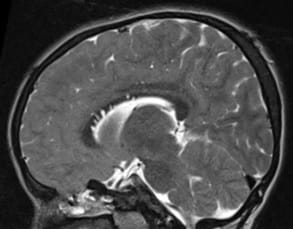
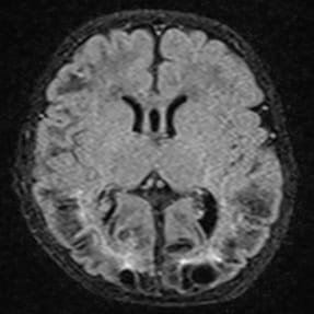
Background: A seizure is an occurrence of signs or symptoms due to abnormal excessive or synchronous neuronal activity in the brain. The present study aims to study the etiological factors and clinical profile for new-onset seizures in children aged 6-12 years and to determine the frequency of Magnetic resonance imaging (MRI) abnormalities in the pediatrics age group with new-onset unprovoked seizure and those with inadequately investigated longstanding epilepsy and classify the etiology based on the MRI findings. Methods: A prospective study involving a total of 50 patients was recruited aged between 6 to 12 years. All of them underwent neuro-imaging with MRI. Uncooperative patients were imaged following sedation and monitoring by the anesthetist. All children aged 6-12 years who presented with new-onset seizures were included. All MR images were obtained at a 3-mm section thickness except magnetization-prepared rapid gradient-echo images, which are obtained at a 1.8-mm section thickness. Results: Of the 50 patients 28 presented with generalized tonic-clonic seizures, 12 with simple partial seizures, 10 with complex partial seizures. Generalized seizures were a more common presentation than partial seizures in children 6-12 years of age. Conclusion: With the positivity of the MRI in the new-onset seizure in children between 6-12 years in our study gives an important aspect of the essential factor of imaging in pediatric new-onset seizures.

Keywords: Seizures, Children, MRI

Corresponding Author How to Cite this Article To Browse
Mohammed Ansari Gaffoor, Assistant Professor, Sri Venkateshwaraa Medical College Hospital and Research Centre, Puducherry, India.
Email:
Gopalakrishnan N, Gaffoor A. Etiological classification of seizures in pediatric age group (6-12years) – MRI study. Int J Med Res Rev. 2021;9(4):257-262.
Available From
https://ijmrr.medresearch.in/index.php/ijmrr/article/view/1316

Manuscript Received Review Round 1 Review Round 2 Review Round 3 Accepted
2021-06--30 2021-07-10 2021-07-20 2021-07-26 2021-08-19
Conflict of Interest Funding Ethical Approval Plagiarism X-checker Note
No Nil Yes 6%

© 2021 by Nirmalkumar Gopalakrishnan, Mohammed Ansari Gaffoor and Published by Siddharth Health Research and Social Welfare Society. This is an Open Access article licensed under a Creative Commons Attribution 4.0 International License https://creativecommons.org/licenses/by/4.0/ unported [CC BY 4.0].

Introduction

A seizure is an occurrence of signs or symptoms due to abnormal excessive or synchronous neuronal activity in the brain. When the above is associated with motor components then they are known as convulsions. Epilepsy is a condition characterized by recurrent (two or more) unprovoked seizures occurring 24 hours apart. [1]. Seizures are common in the pediatric age group and occur in 10% of children. Less than 1/3rd of seizures in children are caused by epilepsy. [2]. The etiological spectrum of acute symptomatic seizures in developing countries is different from developed countries. In developing countries like India, intracranial infections still constitute the single largest cause of childhood seizures. [3-5]. The tremendous advances in neuro-radiological investigations and electroencephalogram (EEG) have resulted in a series of noninvasive diagnostic procedures. However, it is not always that the information derived from these investigations changes the line of management and there is always a concern regarding cost and radiation exposure. [6,7].

Hence it is essential to evolve a method to identify children with symptomatic epilepsy by clinical criteria so that seizures are better managed and expensive investigations can be judiciously used. The present study aims to study the etiological factors and clinical profile for new-onset seizures in children aged 6-12 years and to determine the frequency of MRI abnormalities in the pediatrics age group with new-onset unprovoked seizure and those with inadequately investigated longstanding epilepsy and classify the etiology based on the MRI findings.

Material and Methods

A prospective study involving pediatric patients presenting with new-onset, recurrent and refractory seizures to the pediatric department was done. A total of 50 patients were recruited aged between 6 to 12 years. Following evaluation by a pediatrician and a neurologist, all these patients underwent appropriate investigations. All of them underwent neuro-imaging with MRI. Uncooperative patients were imaged following sedation and monitoring by the anesthetist.

Inclusion criteria: All children aged 6-12 years who presented with new-onset seizures were included.

Exclusion criteria: Children with a previous history of seizures, developmental retardation, and prior neurological abnormality, seizure mimics, and trauma were excluded. After getting informed written consent from the parents, the data were collected and entered in a Pro forma.

Magnetic Resonance Imaging was performed on a Siemens 1.5 Tesla Magnetom scanner. The protocol at our institution includes the following sequences,

  • Axial T1-weighted spin-echo (SE), T2-weighted fast spin-echo (FSE), and fluid-attenuated inversion recovery (FLAIR)
  • Coronal magnetization prepared rapid gradient-echo (MP-RAGE), T2-weighted spin-echo (SE)
  • Sagittal T1-weighted spin-echo (SE) and
  • Axial diffusion-weighted single-shot spin-echo-planar imaging

All MR images were obtained at a 3-mm section thickness except magnetization-prepared rapid

gradient-echo images, which are obtained at a 1.8-mm section thickness.

Results

Of the 50 patients, 28 presented with generalized tonic-clonic seizures, 12 with simple partial seizures, 10 with complex partial seizures (Refer Table.1). Table 2 shows the correlation of Generalized tonic-clonic seizures (GTCS) with the positive finding was the most common (15 patients), followed by simple partial seizures (CPS) in 10 patients, complex partial seizures (SPS) in 8 patients. In this study, the most prevalent cause of epilepsy was Cryptogenic (26%), followed by Hypoxic Ischemic Encephalopathy (20%) (Refer Table. 3). Of the 50 patients who underwent MRI study, 46% had normal imaging and 54% had abnormal imaging as shown in Table 4.

Table 1: Age distribution in this study

Age -group Generalized Partial seizure N(22) Total
N(28)
6-7 3 2 5
7-8 6 7 13
8-9 4 6 10
9-10 7 2 9
10-11 4 3 7
11-12 4 2 6
Total 28 22 50

Table 2: Correlation of type of seizure with Positive findings

S. No Type of seizure Positive finding Normal Total
1 Generalized Tonic-Clonic Seizures (GTCS) 15 13 28
2 complex partial seizures (CPS) 8 2 10
3 simple partial seizures (SPS) 10 2 12

Table 3: Cause of epilepsy in the order of prevalence

S. No Cause of epilepsy No. of cases Percentage (%)
1 Cryptogenic 23 46
2 Hypoxic Ischemic Encephalopathy 10 20
3 Focal cortical dysplasia 6 12
4 Mucopolysaccharidosis 3 6
5 Infection tuberculoma 1 2
6 Infection Acute disseminated encephalomyelitis (ADEM) 1 2
7 Metachromatic Leukodystrophy 1 2
8 Corpus Callosum Dysgenesis 1 2
9 Dandy Walker variant 1 2
10 Malformed Adenohypophysis 1 2
11 Macrocephaly 1 2
12 Microcephaly 1 2
  Total 50 100%

Table 4: MRI study

MRI study No of subjects Percentage (%) p-Value
Normal imaging 23 46 0.04972
Abnormal Imaging 27 54 0.0214

ijmrr_1316_01.jpg

Figure 1: T2W sagittal image showing prominent perivascular spaces in the corpus callosum.

ijmrr_1316_02.jpg

Figure 2: T2 FLAIR Axial image showing bilateral subcortical white matter hyperintensities and porencephalic cysts consistent with hypoxic-ischemic injury.

Discussion

Epilepsy is a brain disease characterized by abnormal electrical activity causing seizures or unusual behavior, sensations, and occasionally loss of consciousness. The pathophysiological basis is the loss of normal regulation of neurons resulting in a state of relative hyperexcitability. The new 2017 ILAE seizure classification has three levels, seizure type, epilepsy type, and epilepsy syndrome. Seizures are classified into focal onset, generalized onset, and unknown onset. Epilepsy type-level includes focal epilepsy, generalized epilepsy, combined generalized and focal epilepsy, and unknown epilepsy. Epilepsy syndrome represents a cluster of features that tend to occur in conjunction.

Focal epilepsy includes unifocal and multifocal disorders and seizures involving one hemisphere. The seizure types include focal aware seizures, focal impaired awareness seizures, focal motor seizures, focal non-motor seizures, and focal to bilateral tonic-clonic seizures. Absence, tonic-clonic, myoclonic, atonic, and tonic seizures are subtypes of generalized seizures. In combined generalized and focal epilepsy the patients present with both focal and generalized seizures. In the unknown class, the epilepsy type is indeterminate. Origin, pathophysiology, and clinical manifestations are beyond the scope of this manuscript.


ILAE etiological classification has six categories namely structural, genetic, infectious, metabolic, immunological, and unknown. In our study etiological classification of epilepsy in the pediatric age group (6-12years) based on MRI findings in 50 patients was evaluated. Epilepsy has a significant psychosocial and financial burden on society in developing countries. The advent of MRI has revolutionized the evaluation and management of seizures. In our research, we studied the role of MRI in identifying subtle and gross anatomical /structural pathologies that are instrumental in causing epilepsy. MRI was preferred over CT scan in our study due to lack of ionizing radiation and excellent soft-tissue contrast allowing the detailed depiction of anatomy with precision.

Our study population consisted of 56% of patients who had presented with a history of generalized tonic-clonic seizures. Thus a majority of the patients presenting with generalized seizure is in tune with other studies conducted in developing countries. In the present study, generalized tonic-clonic seizures (GTCS) were the most common type seen in 56% followed by complex partial seizures (CPS) in 20%, simple partial seizures (SPS) in 24%. Another study by Saha SP et al concluded that simple partial seizures constituted 10.5% of total seizures, complex partial constituted 21.1% of total seizures. [8]

In a study made by Singhi S et al, seizures were partial complex in 65% and simple partial in 35%. [9] Present results are comparable with these studies. We found positive MRI findings in 56% of the patients presenting with seizures. Our study had a relatively higher percentage of positive findings because of inclusion criteria of patients presenting with a minimum of 2 episodes of seizure. The patients visiting our hospital are from the rural background, who visits the hospital only when they are seriously ill. In our study population, 60% of the patients had a previous history of multiple episodes of seizures.

The other reason could also be due to the high resolution 1.5 tesla MRI scanner used. In our study structural abnormality could not be detected in 45.6% of patients. Among these 55% had presented with generalized tonic-clonic seizures. Hence cases with generalized tonic-clonic seizures constituted maximum to the cryptogenic cause. The most common cause of seizure based on MRI findings in our study was found to be cryptogenic.

In cases of drug-resistant focal epilepsy localization of the epileptogenic zone is essential for surgical planning before a focal cortical resection. MRI is an important tool in defining the etiology of epilepsy and helps in prognostic counseling and formulating treatment strategies. MRI negative epilepsy is encountered in 20-30% of patients with temporal epilepsy and 20-40% of those with extra-temporal epilepsy. In these cases, SPECT may prove to be useful in the localization of the epileptogenic zone. Ictal SPECT assesses the cerebral perfusion during seizures. However, Hypoxic Ischemic Encephalopathy (HIE) was found to be the largest group to have positive MRI findings in our study constituting 20% of the cases. This finding does not correlate with older studies conducted in developing tropical countries. This could be attributed to the poor percentage of the institutional delivery rate and neglected peri-natal care in our region. Infectious etiology was the commonest cause in the earlier studies conducted in similar rural areas. However, the widespread availability of low-cost antimicrobials due to the flourishing world-class pharmaceutical companies has reduced infectious load.

HIE is higher in our region because of the poor availability of perinatal care. Lack of trained personnel, hospitals, lack of timely availability of transportation facilities, etc. have been the main contributing factors. HIE is assessed on three basic parameters: the level of brain maturation at the time of insult, severity, and the duration of hypoperfusion event. HIE injury to the grey matter (deep grey matter and cortex) demonstrate characteristic T1 weighted hyperintensities and variable T2 weighted intensities in the grey matter depending on the time of imaging and pathological condition such as hemorrhage, gliosis, encephalomalacia, periventricular leukomalacia, and porencephaly. Focal cortical dysplasia (FCD) accounted for the second-largest group of known etiology constituting 12% of the total cases. It is described as a distinct type of malformation of cortical development where there is the arrest of the neuronal migrations from the periventricular region to the cortex. Epilepsy due to FCD is refractile to medical management and hence accurate anatomical delineation of the epileptogenic foci using MRI plays a pivotal role in surgical management. They consist of subtle focal changes in the form of subcortical T2 hyperintensities extending from the periventricular region to the cortex.


Other characteristics include focal cortical thickening and blurring of grey-white matter junction. Two cases of infectious origin were reported. One was a case of tuberculoma. We reported 2% of metabolic storage disorders. One of them was metachromatic leukodystrophy demonstrating bilateral confluent areas of high signal intensities in per ventricular white matter and sparing of U-fibres in T2 weighted imaging.

The three other cases were mucopolysaccharidosis which had J-shaped sella, prominent perivascular spaces, and white matter intensities on T2 weighted imaging. In our present study (n=50) we had 54% positive findings in MRI as our study population was opted between 6- 12 years mostly to exclude febrile seizures. Hence with this, our study implies the importance of imaging in seizures even in the pediatric age group.

As we do not have many Indian studies done with MRI imaging we have compared with CT scan studies to impact the importance of imaging in the seizure in children. A study done by Kanitkar M et al concluded that CT scan abnormality was detected in 66% of cases. [10]. Single or multiple tuberculomas were noted in 24% cases, cortical atrophy in 14% cases, focal calcification in 10%, hemorrhage in 3%, and subdural effusions in 2%.

In a study done by Baheti R et al, 50% of children with partial seizure and 35% of children with generalized seizure had abnormal CT findings. [11]. Jagar H et al observed abnormal CT findings in 68% of children with partial seizures. [12]. Shinnar S et al observed 21% of children had abnormalities; focal encephalomalacia and cerebral dysgenesis were the most common abnormality. [13].

Berg AT et al observed abnormality in 12.7% of those imaged. [14]. Misra S et al observed CT Brain was abnormal in 75% of children with the seizure disorder, the ring-enhancing lesion was the commonest abnormality (54%) followed by brain atrophy. [15].

Conclusion

Generalized seizures were a more common presentation than partial seizures in children 6-12 years of age. Positivity of the MRI in the new-onset seizure in children between 6-12 years in our study emphasizes the importance of imaging in pediatric new-onset seizures.

Reference

01. Verma A, Kunju PAM, Kanhare S, Maheshwari N. IAP Textbook of Pediatric Neurology. New Delhi- Jaypee Brothers Medical Publishers (p) Ltd. 2014;112. [Crossref][PubMed][Google Scholar]

02. Behrman, Kleigman, Jenson. Nervous system In- Nelson text book of paediatrics. 18th ed, 2nd vol. p 2457-2478. [Crossref][PubMed][Google Scholar]

03. Rayamajhi A, Singh R, Prasad R, Khanal B, Singhi S. Study of Japanese encephalitis and other viral encephalitis in Nepali children. Pediatr Int. 2007 Dec;49(6)978-84. doi: 10.1111/j.1442-200X.2007.02495.x [Crossref][PubMed][Google Scholar]

04. Hauser WA, Beghi E. First seizure definitions and worldwide incidence and mortality. Epilepsia. 2008;49 Suppl 1;8-12. doi: 10.1111/j.1528-1167.2008.01443.x [Crossref][PubMed][Google Scholar]

05. Chen CY, Chang YJ, Wu HP. New-onset seizures in pediatric emergency. Pediatr Neonatol. 2010 Apr;51(2)103-11. doi: 10.1016/S1875-9572(10)60019-8 [Crossref][PubMed][Google Scholar]

06. Sharma S, Riviello JJ, Harper MB, Baskin MN. The role of emergent neuroimaging in children with new-onset afebrile seizures. Pediatrics. 2003 Jan;111(1)1-5. doi: 10.1542/peds.111.1.1 [Crossref][PubMed][Google Scholar]

07. Pearce MS, Salotti JA, Little MP, McHugh K, Lee C, Kim KP, et al. Radiation exposure from CT scans in childhood and subsequent risk of leukaemia and brain tumours- a retrospective cohort study. Lancet. 2012 Aug 4;380(9840)499-505. doi: 10.1016/S0140-6736(12)60815-0 [Crossref][PubMed][Google Scholar]

08. Saha S P, Bhattachrya S, Roy B K, Basu A, Maity A, Das S K. A prospective incidence study of epilepsy in a rural community of West-Bengal, India". Neurology Asia. (2008). [Crossref][PubMed][Google Scholar]

09. Singhi, Sunit, and Pratibha Singhi. "Clinical profile and etiology of partial seizures in North Indian infants and children". Journal of Epilepsy. 10;1(1997)32-36. [Crossref][PubMed][Google Scholar]


10. Kanitkar M, Purkayastha SB, Deshpande Nd. Role of ct scan in partial seizures in children. Med J Armed Forces India. 1994 Jan;50(1)23-26. doi: 10.1016/S0377-1237(17)31032-8 [Crossref][PubMed][Google Scholar]

11. Baheti, Ramesh, B D Gupta. "A study of CT and EEG findings in patients with generalised ar partial seizures is western Rajasthan". Journal, Indian Academy of Clinical Medicine. 4;1(2003)25-9. [Crossref][PubMed][Google Scholar]

12. Hussain J, Srinivasan S, Serane VT, Mahadevan S, Elangovan S, Bhuvaneswari V. Cranial computed tomography in partial motor seizures. Indian J Pediatr. 2004 Jul;71(7)641-4. doi: 10.1007/BF02724126 [Crossref][PubMed][Google Scholar]

13. Shinnar S, Berg AT, Moshé SL, Petix M, Maytal J, Kang H, et al. Risk of seizure recurrence following a first unprovoked seizure in childhood- a prospective study. Pediatrics. 1990 Jun;85(6)1076-85. [Crossref][PubMed][Google Scholar]

14. Berg AT, Testa FM, Levy SR, Shinnar S. Neuroimaging in children with newly diagnosed epilepsy- A community-based study. Pediatrics. 2000 Sep;106(3)527-32. doi: 10.1542/peds.106.3.527 [Crossref][PubMed][Google Scholar]

15. Misra S, Das BK, Srivastava AK. Neuroimaging study in children with seizures. JNPS. 2007;27(1)13-6. [Crossref][PubMed][Google Scholar]