E-ISSN:2320-8686
P-ISSN:2321-127X

Case Report

Diagnosis

International Journal of Medical Research and Review

2021 Volume 9 Number 1 January-February
Publisherwww.medresearch.in

The Potential of Prenatal Diagnosis in the Early Detection of Congenital Malformations

Albu C.1*, Albu D.2, Albu S.3
DOI: https://doi.org/10.17511/ijmrr.2021.i01.08

1* Cristina-Crenguta Albu, Associate Professor, Department of Genetics, University of Medicine and Pharmacy Carol Davila, Bucharest, Romania.

2 Dinu-Florin Albu, Associate Professor, Department of Obstetrics Gynecology, Clinical Hospital of Obstetrics and Gynecology Prof. Dr. Panait Sirbu, Bucharest, Romania.

3 Stefan-Dimitrie Albu, PhD Student, Doctoral School, University of Medicine and Pharmacy Carol Davila, Bucharest, Romania.

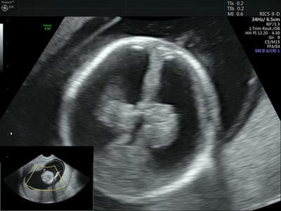
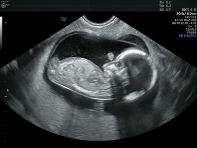
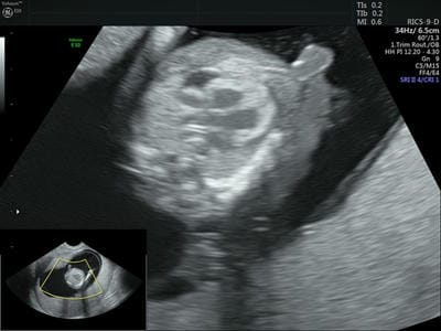
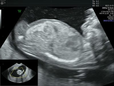
We present a unique association of fetal malformations very early diagnosed by ultrasound examination, at 14 weeks of gestation. A 28-year-old pregnant female, was addressed in a private medical center from Bucharest, Romania, for a routine ultrasound screening. A detailed ultrasound evaluation of the fetus showed numerous and significant cephalic and heart malformations. The ultrasound examination of the fetal head suggest the diagnosis of fetal lobar hydrocephalus, and the ultrasound examination of the fetal heart suggest the transposition of the great vessels. The parents were informed about the severity of the fetal malformations and decided to terminate the pregnancy due to medical reasons. Anatomopathological examination confirmed the prenatal diagnosis. First trimester ultrasonography was crucial in the early prenatal diagnosis and management of the malformed fetus with a unique association of fetal malformations.

Keywords: Ultrasound investigation, Prenatal diagnosis, Lobar hydrocephalus, Transposition of the great vessels

Corresponding Author How to Cite this Article To Browse
Cristina-Crenguta Albu, Associate Professor, Department of Genetics, University of Medicine and Pharmacy Carol Davila, Bucharest, Romania.
Email:
Albu CC, Albu DF, Albu SD. The Potential of Prenatal Diagnosis in the Early Detection of Congenital Malformations. Int J Med Res Rev. 2021;9(1):54-57.
Available From
https://ijmrr.medresearch.in/index.php/ijmrr/article/view/1233

Manuscript Received Review Round 1 Review Round 2 Review Round 3 Accepted
2020-12-11 2021-01-04 2021-01-14 2021-01-22
Conflict of Interest Funding Ethical Approval Plagiarism X-checker Note
No Nil Yes 9%

© 2021 by Cristina-Crenguta Albu, Dinu-Florin Albu, Stefan-Dimitrie Albu and Published by Siddharth Health Research and Social Welfare Society. This is an Open Access article licensed under a Creative Commons Attribution 4.0 International License https://creativecommons.org/licenses/by/4.0/ unported [CC BY 4.0].

Introduction

Congenital hydrocephalus is a rare isolated or associated multifactorial condition of genetic or epigenetic cause, characterized by an abnormal excess of cerebrospinal fluid within the cerebral ventricle with ventricular dilatation [1].

The most common causes of congenital hydrocephalus include aqueductal stenosis, neural tube defect (spina bifida), arachnoid cysts, Chiari malformations, and Dandy-Walker syndrome [2].

Genetic research in laboratory animals illustrate that genetic factors play a considerable role in the pathogenesis of hydrocephalus, but it is difficult to evaluate whether data gained from laboratory animals can be extrapolated to humans [3, 4].

The transposition of the great vessels, one of the most severe cyanotic heart defects, with an incidence of 1 in 3,500-5,000 live births, is a congenital heart malformation with unknown etiology, very rarely associated with genetic syndromes [5,6]. Both isolated congenital hydrocephalus and isolated congenital heart malformation are very serious malformations [7-9]. The association of both malformations in the same fetus has not been described so far.

Case Report

We present a rare case report of a 28-year-old pregnant female, at 14 weeks of gestation, who was addressed in a private medical center from Bucharest, Romania, in February 2019, for a routine antenatal ultrasonographic screening.

The sonography was performed with a General Electric Voluson E10 Ultrasound device. After the patient was informed about the examination, with her informed consent for ultrasonographic investigation, the transabdominal ultrasound was done by an expert ultrasonographer. The ultrasound scanning highlight a unique fetus 14.4 weeks old, in evolution, with an estimated fetal weight of 83g.

A detailed ultrasound evaluation of the fetus showed numerous and significant malformations. At the level of the cephalic extremity: the biparietal diameter was 33.1mm, the occipitofrontal diameter was 32.7 mm and head circumference was 103.3 mm. The cerebral ventricles occupy almost the entire cerebral hemisphere and the cerebral cortical mantle thickness was about 1 mm.

Furthermore, the choroid plexuses are visible with a "bell tongue" appearance, ultrasound features that suggest the diagnosis of fetal lobar hydrocephalus (Figure 1). The nasal bone was visible: 1.8 mm, and the nuchal fold, registered a value of 2.1 mm.

At the level of the thorax, the anteroposterior diameter was 24 mm and the transverse diameter was 20.5 mm. Also, the spine has a normal conformation, without visible abnormalities over 0.5 cm (Figure 2).

ijmrr_1233_01.jpg

Fig. 1: First trimester ultrasonography, 2D examination - Fetal lobar hydrocephalus

ijmrr_1233_02.jpg

Fig. 2: First trimester ultrasonography, 2D examination - Mid-sagittal section of the fetus at 14 weeks' gestation.

At the examination of the fetal heart, we highlighted the tetracameral heart, with atrioventricular valves with incomplete appearance, important tricuspid, and mitral regurgitation, with the appearance of the atrioventricular heart, as well as the parallelism of the large vessels at the base of the heart, in the initial portion.


The crossing is not visualized, which suggest probably the transposition of the great vessels (Figure 3). At the same time, the presence of a blade of pericardial fluid, with a hydropericardium appearance, was highlighted.

ijmrr_1233_03.jpg

Fig. 3: First trimester ultrasonography, 2D examination - Transposition of the great vessels.

At the examination of the fetal abdomen, we highlighted: abdomen with normal conformation, anteroposterior diameter: 23 mm, transverse diameter: 25.4 mm, abdominal circumference: 76.1 mm, and ileal hyperechogenicity (Figure 4). Also, ultrasound examination of the fetus showed the presence of a double contour of the trunk and the fetal skull, probably due to a moderate and generalized subtegumentary edema.

ijmrr_1233_04.jpg

Fig. 4: First trimester ultrasonography, 2D examination - Mid-sagittal section of the fetus, at 14 weeks' gestation, anatomic details.

The parents were informed about the severity of the fetal malformations and decided to terminate the pregnancy due to medical reasons.

Anatopathological examination confirmed the prenatal diagnosis.

Discussion

A detailed ultrasonographic examination of the fetal anatomy during the first trimester of pregnancy can detect about half of the major structural defects in both low-risk and high-risk pregnancies. The benefits of ultrasound examination are early prenatal diagnosis of severe congenital malformations reassurance, and relatively easy decision to terminate the pregnancy for medical reasons if required [10, 11]. Genetic counseling after prenatal diagnosis of severe fetal malformations is very difficult because of the negative prognosis, but the early prenatal detection of extremely severe birth defects and proper case management is crucial for the prevention of newborns with congenital disabilities [12]. In conclusion, the first trimester ultrasonography was crucial, for the early prenatal diagnosis and management of a severely affected fetus, with a unique association of lobar hydrocephalus and transposition of the great vessels.

Authors' contributions

All authors, ACC, ADF and ASD, contributed equally to preparing, review and editing of the article. All authors read and approved the final version of the manuscript.

Reference

  1. Partington MD. Congenital hydrocephalus. Neurosurg Clin N Am. 2001;12(4)737-742.
  2. Tully HM, Dobyns WB. Infantile hydrocephalus- a review of epidemiology, classification and causes. Eur J Med Genet. 2014;57(8)359-368.
  3. Zhang J, Williams MA, Rigamonti D. Genetics of human hydrocephalus. J Neurol. 2006;253(10)1255-1266.
  4. Venkataramana NK, Mukundan CR. Evaluation of functional outcomes in congenital hydrocephalus. J Pediatr Neurosci. 2011;6(1)4-12.

  1. Martins P, Castela E. Transposition of the great arteries. Orphanet J Rare Dis. 2008;3;27.
  2. Albu CC, Albu DF, Albu SD, Patrascu A, Musat AR, Goganau A. Early Prenatal Diagnosis of an Extremely Rare Association of Down Syndrome and Transposition of the Great Vessels. Rev Chim. 2019;70:2574-2578.
  3. Rao PS. Management of Congenital Heart Disease- State of the Art-Part II-Cyanotic Heart Defects. Children (Basel). 2019;6(4)54.
  4. Unolt M, Putotto C, Silvestri LM, Marino D, Scarabotti A, Massaccesi V, et al. Transposition of great arteries: new insights into the pathogenesis. Front Pediatr. 2013;1:11.
  5. Forcelini CM, Mallmann AB, Crusius PS, Seibert CA, Crusius MU, Zandoná DI, et al. Down syndrome with congenital hydrocephalus- case report. Arq Neuropsiquiatr. 2006;64(3B)869-871.
  1. Kashyap N, Pradhan M, Singh N, Yadav S. Early Detection of Fetal Malformation, a Long Distance Yet to Cover! Present Status and Potential of First Trimester Ultrasonography in Detection of Fetal Congenital Malformation in a Developing Country- Experience at a Tertiary Care Centre in India. J Pregnancy. 2015;623059.
  2. Karim JN, Roberts NW, Salomon LJ, Papageorghiou AT. Systematic review of first-trimester ultrasound screening for detection of fetal structural anomalies and factors that affect screening performance. Ultrasound Obstet Gynecol. 2017;50(4)429-441.
  3. Montaguti E, Balducci A, Perolo A, Emanuela Angeli E, Gargiulo G, Pilu G, et al. Prenatal diagnosis of congenital heart defects and voluntary termination of pregnancy. Am J Obstet Gynecol MFM. 2020;2;100207.