E-ISSN:2320-8686
P-ISSN:2321-127X

Research Article

Undiagnosed

International Journal of Medical Research and Review

2020 Volume 8 Number 3 May-June
Publisherwww.medresearch.in

Role of pleural biopsy in patients of undiagnosed exudative pleural effusion

Sanwalka N.1, Kumar R.2, Sanwalka M.3*
DOI: https://doi.org/10.17511/ijmrr.2020.i03.08

1 Nidhi Sanwalka, Post graduate resident, Rukmani Devi Beni Prasad Jaipuria Hospital, Jaipur, Rajasthan, India.

2 Ramesh Kumar, Post Graduate Resident, Rukmani Devi Beni Prasad Jaipuria Hospital, Jaipur, Rajasthan, India.

3* Madhvi Sanwalka, Post Graduate Resident, National Institute of Medical Sciences and Research, Jaipur, Rajasthan, India.

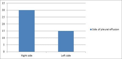
Introduction: Sometimes etiological diagnosis of pleural fluid is not possible by cytology, biochemical and microbiological examinations and labeled as undiagnosed exudative pleural effusion. Our aim of this study to make an etiological diagnosis in such undiagnosed exudative cases with pleural biopsy. Material and method: In this study patients with undiagnosed exudative pleural effusion, where the diagnosis was not made by laboratory investigations were included. Pleural tissue was obtained by Abram’s Needle and sent for histopathology and culture to find mycobacterium tuberculosis. Result: Out of 45 patients 34 (75.5%) were males and 11 (24.5%) were females. The side of pleural effusion was right-sided in 30 (66.6%) and left-sided in 15 (33.4%). The mean value of polymorphs and lymphocytes count was 7.24% and 92.76% respectively. Pleural fluid was hemorrhagic in 10 (22.22%) patients, straw-colored in 30 (71.11%) patients, and clear in 5 (11.11%) patients. The mean level of glucose was 65.66 mg/dl, the lowest being nil and highest being 110 mg/dl. The mean level of protein was 5.54 gm/dl (range 3.7-7.21 gm/dl). The mean value of the pH of pleural fluid was 65.44. Histopathology showed granulomatous inflammation compatible with tuberculosis in 24 (53.3%) cases, metastatic malignancy in 7 (15.5%) cases, chronic inflammation in 10 (22.3%) cases. In 4 (8.9%) cases pleural tissue was inadequate to give any opinion. Among 7 cases of malignancy, 5 (71.42%) cases showed adenocarcinomas and 2 (28.58%) cases showed squamous cell carcinoma. Conclusion: This study suggests that tuberculosis and malignancy are the two common etiologies for exudative pleural effusion. The role of pleural biopsy is pivotal as it helps in making the diagnosis in the majority of cases where other laboratory investigations fail to provide a diagnosis.

Keywords: Etiology, Exudative, Undiagnosed, Pleural effusion, Pleural biopsy, Tuberculosis, Malignancy

Corresponding Author How to Cite this Article To Browse
Madhvi Sanwalka, Post Graduate Resident, National Institute of Medical Sciences and Research, Jaipur, Rajasthan, India.
Email:
Sanwalka N, Kumar R, Sanwalka M. Role of pleural biopsy in patients of undiagnosed exudative pleural effusion. Int J Med Res Rev. 2020;8(3):265-268.
Available From
https://ijmrr.medresearch.in/index.php/ijmrr/article/view/1194

Manuscript Received Review Round 1 Review Round 2 Review Round 3 Accepted
2020-05-22 2020-05-28 2020-06-04 2020-06-08
Conflict of Interest Funding Ethical Approval Plagiarism X-checker Note
No Nil Yes 7%

© 2020 by Nidhi Sanwalka, Ramesh Kumar, Madhvi Sanwalka and Published by Siddharth Health Research and Social Welfare Society. This is an Open Access article licensed under a Creative Commons Attribution 4.0 International License https://creativecommons.org/licenses/by/4.0/ unported [CC BY 4.0].

Introduction

Pleural effusion is a common clinical condition that is encountered by clinicians in their practice. Pleural effusion is known as “the accumulation of fluid in the pleural space”. It is of two types i.e. transudative or exudative [1]. Transudative effusion has low protein levels and mostly due to systemic pathology like cardiac, hepatic, or renal disorders. On the other hand, exudative effusions have high protein content and are mostly due to pleural pathologies like tuberculosis, malignancy, or any pleural infection [2]. Despite the good history, clinical and radiological investigation, laboratory investigation of pleural fluid, as many as in 15-20% of the cases, it is not possible to make the diagnosis [3]. In these cases many times, the patient receives empirical treatment, without any confirmed diagnosis. Therefore in such cases, it is essential to make the etiological diagnosis in exudative pleural effusion to give proper treatment to patients. One of the modality is percutaneous needle pleural biopsy of parietal pleura, which can diagnose up to 50% of cases [4]. The first pleural biopsy was done by using the Vim Silverman needle in 1955 [5]. But from 1958 Abram’s pleural biopsy needle is used as it is safe, easy to perform, and inexpensive [6]. Subsequently, a different type of introduced by Cope and Radja, and occasionally Tru-cut biopsy needle is also used [7,8]. The aim of this study is to make an etiological diagnosis of undiagnosed exudative pleural effusion when laboratory examination of pleural fluid is failed to make a diagnosis and to find the role of percutaneous needle pleural biopsy in cases of undiagnosed exudative pleural effusion.

Material and Methods

This study was done in tertiary care hospital in Rajasthan, for one year, from August 2018 to July 2019. This was a cross-sectional study. Eighty-nine patients who had pleural effusion were evaluated thoroughly. Clinical, radiological, and laboratory investigations were done. Thoracocentesis was done and pleural fluid was sent for cytological (cell type, cell type, and malignant cells), biochemical (protein, LDH, and ADA) and microbiological (Gram and Z-N staining and culture) examinations to determine the cause of the effusion. If the diagnosis was done by these examinations then those cases were excluded by the present study. But if not then those cases were labeled as undiagnosed cases and pleural biopsy was done after taking informed consent.

All the patients who gave consent, age more than 18 years, were able to understand the procedure, normal coagulation profile was included in this study. All the patients with bleeding diathesis, not cooperative, transudative effusion, taking anticoagulants, empyema, local skin infection, hemodynamic instabilities were excluded from this study. Forty-five patients were eligible for this study.

Pleural Biopsy: The procedure of pleural biopsy was done on the patient in the sitting position and after determining the effusion side, biopsy site was selected. The area was cleaned thoroughly with antiseptic and then 1% lignocaine (local anesthetic) was infiltrated at that site. Pleural fluid aspiration in the needle confirmed the presence of free fluid. Now 0.5cm size incision was made just above the upper border of the rib of that site and Abram’s needle was introduced through it. Multiple biopsies were taken by multiple passes and then the incision site was sutured with a single stitch. Post biopsy X-ray was taken to rule out any complication (pneumothorax). Pleural tissue was placed in two vials, one with formalin and sent for histopathological examination, second in normal saline, and sent for culture (mycobacterium tuberculosis). Ethical clearance was taken from the editorial committee of the hospital. All the data collected was saved in an excel sheet and statistical analysis was done by using SPSS software.

Result

This study included forty-five patients of exudative pleural effusion in whom the diagnosis was not made by cytological, biochemical, and microbiological examinations. Out of forty-five, 34 (75.5%) were males and 11 (24.4%) were females. The side of pleural effusion was right-sided in 30 (66.6%) and left-sided in 15(33.33%).

Table-1: Distribution of cases in gender.

No of Patients Male Percentage Female Percentage
45 34 75.5 11 24.5

ijmrr_1194_01.jpg

Fig-1: Side of pleural effusion.


The majority of pleural effusion were lymphocytic. The mean value of polymorphs and lymphocytes count was 7.24% and 92.76% respectively. Pleural fluid was hemorrhagic in 10 (22.22%) patients, straw-colored in 30 (71.11%) patients, and clear in 5 (11.11%) patients.

The mean level of glucose was 65.66 mg/dl, the lowest being nil and the highest being 110 mg/dl. The mean level of protein was 5.54 gm/dl (range 3.7-7.21 gm/dl). The mean value of pH of pleural fluid was 65.44, lowest being 7.077 and highest being 7.486. ADA was measured and the average value in tuberculosis, malignancy, and chronic inflammation was 83.83, 33.33, and 84.00 U/L respectively.

Tuberculin test was positive in 4 patients, who were later diagnosed as tubercular pleural effusion by histopathology. More than one pass was taken to obtain at least three samples of pleural biopsy. The average number of passes was 5.10 (range 4-7) and the sample obtained was 3.76 (range 4-8). Histopathology showed granulomatous inflammation compatible with tuberculosis in 24 (53.3%) cases, metastatic malignancy in 7 (15.5%) cases, chronic inflammation in 10 (22.3%) cases. In 4 (8.9%) cases pleural tissue was inadequate to give any opinion.

Among 7 cases of malignancy, 5 (71.42%) cases showed adenocarcinomas and 2 (28.58%) cases showed squamous cell carcinoma. In 8 cases of chronic inflammation and 2 cases of inconclusive biopsy, antitubercular therapy was started based on clinic-radiological finding and all were improved. In two cases of inconclusive biopsy further, fiber optic bronchoscopy and CT guided fine needle aspiration was done and found to be of tubercular etiology.

Table-2:- Histopathological diagnosis of the cases.

Histopathology diagnosis No of cases Percentage
Tuberculosis 24 53.3
Metastatic malignancy 7 15.5
Chronic inflammation 10 22.3
Inadequate tissue 4 8.9

Three patients out of forty-five developed pneumothorax for which intercostal tube was placed. It was resolved within the next three days. Four patients developed pain at biopsy site which was treated by anti-inflammatory drugs. Pleural biopsy was also sent for Ziehl-Nelson staining to see the presence of mycobacterium tuberculosis. It was

positive in 5 (20.83%) out of 24 cases.

Discussion

This study shows that most of the patients were males. The probable reason may be because males commonly come for medical help than females and they tend to have smoking habits more than females. In the present study, pleural biopsy showed 54% cases of tuberculosis, 16% of malignancy, and 22% of chronic inflammation which is similar to other studies [4,9,10]. The diagnostic yield of this study for malignancy was 16% which is low as compared to other studies done by Menzies et al [11] which showed a diagnostic yield of 30-70%. This low yield of malignancy is may be due to the high prevalence of tuberculosis in developing countries like India. Among the malignancy cases, the most common malignancy was adenocarcinoma (71.42%) which is similar to a study done by Bhattacharya et al [12]. A study done by Al-shimemeri AA et al [4], James P et al [13], Biswas A et al [10] and Devkota KC et al [14] showed finding that pleural biopsy was the definitive diagnosis in half of the cases, which is similar to the present study. A study done by Ahmed et al [15] showed tuberculosis, malignancy, and inflammation in 29.4%, 19.6%, and 17.6% cases respectively, which is similar to the present study which also showed higher cases of tuberculosis. The study done by Rajawat et al [16] showed malignancy (37.17%) followed by tuberculosis (32.98%) as etiology which is a contrast to the present study. Common complications of the pleural biopsy were a vasovagal attack, pain at the biopsy site, hematoma at the biopsy site, seepage of fluid from the site, pulmonary edema, and pneumothorax. In the present study, pneumothorax was encountered which was resolved in a few days after insertion of intercostals drainage and mild pain at biopsy site which was treated by anti-inflammatory drugs. Viskum and Enk showed a complication rate of 7-8% in their study with 566 thoracoscopy examinations. This study was done at one center only, so limited data was collected only, further studies should be done in multi centers and data then collected should be evaluated for a better understanding of etiologies.

Conclusion

This study suggests that tuberculosis and malignancy are the two common etiology for exudative pleural effusion. The role of pleural biopsy


is pivotal as it helps in making the diagnosis in the majority of cases where other laboratory investigations fail to provide the diagnosis. This is important in our country where tuberculosis is a common disease and facilities like thoracoscopy and image-guided needle biopsy are not so easily available.

What does the study add to the existing knowledge

Blind pleural biopsy can be considered as a supplementary test in the diagnosis of exudative pleural effusion as it is easy to perform, safe, and of low cost. This is a single-center study with few cases, if a study was done in multi-centers then diagnostic yield will give more valuable inferences. This study helps in identifying etiologies in undiagnosed cases of pleural effusion by pleural biopsies. A pleural biopsy is an important tool of investigation in these cases.

Author’s contribution

Dr. Nidhi Sanwalka: Concept, study design

Dr. Ramesh Kumar: Manuscript preparation

Dr. Madhvi Sanwalka: Collection of data and histopathological reports

Reference

  1. Light RW. Diagnostic principles in pleural disease. Eur Respir J. 1997;10(2)476-481.
  2. Saguil A, Wyrick K, Hallgren J. Diagnostic Approach to Pleural Effusion. Am Fam Physician. 2014;90(2)99-104.
  3. Hirsh A,Ruffle P, Nebut M, Bignon J, Chretien J. Pleural effusion- Laboratory tests in 300 cases. Thorax. 1979;34(1)106-112.
  4. Al-Shimemeri AA, Al-Ghadeer HM, Girdhar HR. Diagnostic yield of closed pleural biopsy in exudative pleural effusion. Saudi Ned J. 2003;24(3)282-286.
  5. DeFrancis N, Kloske E, Albano E. Needle biopsy of the parietal pleura-preliminary report. N Eng J Med. 1955;252;948-999.
  1. Abram’s LD. A pleural biopsy punch. Lancet. 1958;271(7010)30-31.
  2. Cope C. New pleural biopsy needle. JAMA. 1958;167(9)1107-1108.
  3. Ogirala RG, Agarwal V, Aldrich TK. Radja pleural biopsy needle- A comparison with the Abrams needle in experimental pleural effusion. Am Rev Respir Dis. 1989,139(4);984-987.
  4. Pandit S, Chaudhuri AD, Datta SBS, Dey A, Bhanja P. Role of pleural biopsy in etiological diagnosis of pleural effusion. Lung India. 2010;27(4)202-204.
  5. Biswas A, Bhatracharya S. Diagnostic role of closed pleural biopsy in the investigation of exudative pleural effusion. J Indian Med Assoc. 2008;106(8);525-526.
  6. Menzies R, Charbonneau M. Thoracoscopy for the diagnosis of pleural diseases. Ann Intern Med. 1991;114(4)271-276.
  7. Bhattacharya S, Bairagya TD, Das a, Mandal A and Das SK. Closed pleural Biopsy is Still Useful in the Evaluation of Malignant Pleural effusion. J Lab Physician. 2012;4(1)35-38.
  8. James P, Gupta R, Christopher DJ, et al. Evaluation of the diagnostic yield and safety of closed pleural biopsy in the diagnosi of pleural effusion. Indian J Tuberc. 2010;57(1)19-24.
  9. Devkota KC, Chokhani R, Gautam S. Diagnostic yield of pleural biopsy in exudative pleural effusion. Nepal Med Coll J. 2014;16(1)13-16.
  10. Ahmed J, Hossain M, Rahim M, Musa AKM. Role of Pleural Biopsy in the Etiologial Diagnosis of Exudative Pleural Effusion. Chest J. 2016;149(4)A440.
  11. Rajawat GS, Batra S, Takhar RP, Rathi L, Bhandari C, Gupta ML. Diagnostic yield and safety of closed needle pleural biopsy in exudative pleural effusion. Avicenna J Med. 2017;7(3)121-124.