E-ISSN:2320-8686
P-ISSN:2321-127X

Case Report

Maxilla

International Journal of Medical Research and Review

2020 Volume 8 Number 6 November-December
Publisherwww.medresearch.in

Fibrosarcoma of Maxilla: youngest case report

Vanza B.1*, Rawat A.2, Yadav R.3, Patel U.4
DOI: https://doi.org/10.17511/ijmrr.2020.i06.09

1* Bhavuk Vanza, Assistant Professor, Department of Oral & Maxillofacial Surgery, Government Dental College, Indore, Madhya Pradesh, India.

2 Amit Rawat, Associate Professor, Department of Oral & Maxillofacial Surgery, Government Dental College, Indore, Madhya Pradesh, India.

3 Rashi Yadav, Assistant Professor, Department of Oral & Maxillofacial Surgery, Government Dental College, Indore, Madhya Pradesh, India.

4 Umesh Patel, Professor, Department of Pediatrics, RKDF Medical College, Bhopal, Madhya Pradesh, India.

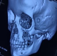
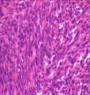
Abstract- Fibrosarcomas are rare but highly fulminant and aggressively malignant neoplasm which occurs as a result of mutated spindle-shaped fibroblasts. According to WHO, they are soft tissue sarcoma commonly present seldom at cutis. The etiology of such a malignancy is obscured nevertheless it has several predisposing factors like pre-existing pathology but the commonest of them is radiation. Mesenchymal malignancies were initially over-diagnosed and were the epicenter of diagnostic dilemma to the pathologist, but with the advent of Vimentin staining and immunohistochemistry, the diagnostic front has fairly advanced for fibrosarcomas. Incidence of Fibrosarcoma in Maxilla accounts for less than 0.1%, when compared to other head and neck malignancies, and is predominantly found between 2nd to 6th decades of life. Various approaches have been documented in the literature regarding its treatment, but aggressive surgical excision remains the mainstay of treating Fibrosarcomas. The role of adjuvant medicinal therapies have been well debated but do have a conclusive outcome. Through this article, we want to highlight and document a case, which occurred at an exceptionally young age, at a site that had no predisposing factors but has proven immunohistochemical diagnosis and was rapidly fulminating and aggressively malignant. The preoperative diagnosis of the patient was inconclusive reporting it to be a mesenchymal tumor, therefore, the patient was treated with the intraoral approach for wide surgical resection. The surgical specimen was subjected to immunohistochemistry with Vimentin staining which reported it to be a Fibrosarcomas, therefore the patient was advised chemotherapy, yet the prognosis is guarded.

Keywords: Fibrosarcoma, Maxilla

Corresponding Author How to Cite this Article To Browse
Bhavuk Vanza, Assistant Professor, Department of Oral & Maxillofacial Surgery, Government Dental College, Indore, Madhya Pradesh, India.
Email:
Vanza B, Rawat A, Yadav R, Patel U. Case Report: Fibrosarcoma in Maxilla in Youngest. Int J Med Res Rev. 2020;8(6):457-460.
Available From
https://ijmrr.medresearch.in/index.php/ijmrr/article/view/1180

Manuscript Received Review Round 1 Review Round 2 Review Round 3 Accepted
2020-04-08 2020-05-18 2020-06-08 2020-09-18
Conflict of Interest Funding Ethical Approval Plagiarism X-checker Note
No Nil Yes 7%

© 2020 by Bhavuk Vanza, Amit Rawat, Rashi Yadav, Umesh Patel and Published by Siddharth Health Research and Social Welfare Society. This is an Open Access article licensed under a Creative Commons Attribution 4.0 International License https://creativecommons.org/licenses/by/4.0/ unported [CC BY 4.0].

Introduction

Mesenchymal tumors have been extended over-diagnosed in past literature [1]. The paradigm shift is credited to the advent of vimentin sensitive immunohistochemistry, which has now become a gold standard for diagnosis of fibrosarcoma. Incidences of fibrosarcoma in the head and neck region have been 0.05% of all the malignancies [2].

As per the anamnestic, fibrosarcomas, occur in the 2nd to 6th decade of life, with a peak at around 30 years of age, with no distinct sex predilection and no distinct etiology [3]. However, fibrosarcomas are associated with mutated bone morphology due to pre-existing pathologies like fibrous dysplasia, Paget’s disease, chronic osteomyelitis, or irradiated jaw [4].

Clinically, lesions are pronounced in the long bone than in jaws. Fibrosarcomas, histologically, are classified as primary or Grade 1 where an abundance of spindle cells are evident with a herringbone pattern, scarce pleomorphism, and mitotic activity with well-differentiated cells.

Moderately differentiated or Grade 2 lesions, reveals a greater degree of cellular pleomorphism and a lesser degree of a herringbone pattern. Poorly differentiated or Grade 3 fibrosarcoma mimics features of other metastatizing connective tissue neoplasms like Malignant fibrous histiocytoma, Malignant Liposarcoma, Rhabdomyosarcomas [5,6], etc.

Clinically fibrosarcomas occur either primarily as tumors within the medullary cavity or secondarily at the periphery of bone, however, there are reported incidences of soft tissue tumors diagnosed as fibrosarcoma also. Treatment of such tumors, is radical surgery, along with radiation.

However, a high rate of recurrence makes it prudent to adopt the multimodal strategy. This article interests, as we report a case of Fibrosarcoma in the maxilla, without any predisposing etiopathology, at 6 years of age. To the best of our knowledge, it is the youngest case reported.

Case report

Patients of 6 years reported to our OPD, with the chief complaint of swelling on the left side of the check and mild pain for 20 days. On elaborating the history of presenting illness, parents revealed, that she had a small swelling for a month without any

pain. The swelling started growing rapidly to attain the current size of about 2X3 centimeters, with mild dull continuous aching pain in check for the past 20 days (Figure 1). The patient had no significant previous medical, dental history, or any reported allergies to known drugs.

None of the family members were diagnosed with malignancy nor did the patient’s mother was subjected to any kind of radiation during her pregnancy. Thorough physical examination revealed no significant abnormalities.

Loco-regionally, an oval-shaped swelling was present at the left buccal region measuring 2X3cms covered with normal-appearing skin extending superior inferiorly from malar prominence to corner of the mouth, anterior-posteriorly from nasolabial fold to 1 cm ahead of the preauricular region.

Mild tenderness was elicited on palpation with firm swelling detached from the overlying skin, however swelling appeared non-mobile, non-reducing, non-compressible, nodular, and single. The auscultatory finding was observantly negative.

The patient had adequate mouth opening, on availing which, an oval swelling present was at the pyriform region on the left side with well-defined borders, obliterating the vestibular depth and covered with inflamed gingiva.

Swelling extended superior-inferiorly from the depth of the buccal vestibule to the cervical margin of the teeth and antrio-posteriorly from ipsilateral deciduous maxillary canine to the permanent first molar.

The swelling was tender, firm, fixed to the underlying bone and overlying gingivomucosal tissue. Computed tomography revealed an expansile osteolytic lesion at the posteroinferior maxilla (Figure 2). Fine needle aspiration cytology did not yield fluid or cellular content.

With a provisional diagnosis of Spindel cell tumor and differential diagnosis of Giant cell granuloma, the patient was prepared for an excisional biopsy. Upon performing wide local excision of the lesion along with antral lining and floor of the orbit, the defect was reconstructed with Bachet’s flap.

Histopathological evaluation of the specimen confirmed Fibrosarcoma (Figure 3).


ijmrr_1180_01.jpg

Fig-1: Child View

ijmrr_1180_02.jpg

Fig-2: CT Image

ijmrr_1180_03.jpg

Fig-3: Histopathology

Discussion

According to SEERS a program of the National Cancer Institute, fibrosarcomas occurring in adults, accounts for 3.6% of all adult sarcomas, and depending upon the source, the patient’s sex may or may not play a role [7].

Sarcomas arising from fibroblasts are predominantly found in the areas adjoin or within the medullary bone. Documented anamnestic of Fibrosarcomas, reveals peak incidence occurs at 3rd decade of life, whereas, in our case, this has been diagnosed in 6 years old patient.

Wadhawan and colleagues proposed that, for the confirmation of fibrosarcoma, vimentin staining should be done and the Herringbone pattern is confirmatory, in the current case Herringbone pattern was observed on Histopathological evaluation and vimentin staining was positive as an Immunohistochemical marker [8,9].

A study conducted by Daniela Augsburger and colleagues in 2017 reports that the mainstay of treatment is generous excision with adequately wide margins, therefore even being a young patient she was undertaken for a wide local excision [4].

The role of chemotherapy is debatable, yet to avoid recurrence, is advocated to be supplemented with radical surgery, considering the age and incidence of recurrence, the patient was, therefore subjected to post-operative chemotherapy [10].

However current data is insufficient to establish a definite treatment plan at such young age, therefore the importance of this case report should not be challenged as it adds to the information regarding the age-related treatment of such fulminant, locally aggressive, and recurring malignancy.

Reference

  1. Weiss SW, Goldblum JR. Fibrosarcoma, In- Enzinger and Weiss’s Soft Tissue Tumors. 5th ed, New Delhi- Elsevier. 2008;303 30.
  2. Wadhwan V, Chaudhary MS, Gawande M. Fibrosarcoma of the oral cavity. Indian J Dent Res. 2010;21(2)295-298.

  1. Guillou L, Folpe AL. Fibroblastic and Fibrohistiocytic Tumors, In- Folpe AL, Inwards CY (eds), Bone and Soft Tissue Pathology. Elsevier, Philadelphia, PA. 2010.
  2. Augsburgerl D, Peter J, Nelson, Kalinski T, Udelnow A, Knosel T, Hofstette M, et al. Current diagnostics and treatment of fibrosarcoma– perspectives for future therapeutic targets and strategies. Oncotarget. 2017;8(61)104638-104653.
  3. Shrivastava S, Nayak SK, Nayak P, Sahu S. Fibrosarcoma of Maxilla- A case report. Journal of Oral & Maxillofacial Pathology. 2016;20(1)162.
  4. Thway K. Pathology of soft tissue sarcomas. Clinical Oncol. 2009;21(9)695-705.
  1. Toro JR, Travis LB, Wu HJ, Zhu K, Fletcher CD, Devesa SS. Incidence patterns of soft tissue sarcomas, regardless of primary site, in the surveillance, epidemiology and end results program, 1978–2001- An analysis of 26,758 cases. Int J Cancer. 2006;119(12)2922-30.
  2. Arora K. Soft tissue Tumors, Fibrosarcoma of soft tissue, adult.
  3. Wadhwan V, Chaudhary MS, Gawande M. Fibrosarcoma of the oral cavity. Indian J Dent Res. 2010;21(2)295 298.
  4. ESMO/European Sarcoma Network Working Group. Soft tissue and visceral sarcomas- ESMO Clinical Practice Guidelines for diagnosis, treatment and follow-up. Ann Oncol. 2014;29(4)iv51–iv67.