E-ISSN:2320-8686
P-ISSN:2321-127X

Research Article

Radiotherapy

International Journal of Medical Research and Review

2019 Volume 7 Number 6 November-December
Publisherwww.medresearch.in

Dosimetric comparison and clinical correlation between conventional four field radiotherapy versus three-dimensional conformal radiotherapy in cancer cervix

Agarwal S.1, Kumar Chauhan A.2*, Kumar P.3, Nigam J.4, Kumar P.5
DOI: https://doi.org/10.17511/ijmrr.2019.i06.13

1 Shubhi Agarwal, Senior Resident, Department of Radiation Oncology, Shri Ram Murti Smarak Institute of Medical Sciences, Bareilly, Uttar Pradesh, India.

2* Arvind Kumar Chauhan, Associate Professor, Department of Radiation Oncology, Shri Ram Murti Smarak Institute of Medical Sciences, Bareilly, Uttar Pradesh, India.

3 Piyush Kumar, Professor, Department of Radiation Oncology, Shri Ram Murti Smarak Institute of Medical Sciences, Bareilly, Uttar Pradesh, India.

4 Jitendra Nigam, Assistant Professor cum RSO, Department of Radiation Oncology, Shri Ram Murti Smarak Institute of Medical Sciences, Bareilly, Uttar Pradesh, India.

5 Pavan Kumar, , Department of Radiation Oncology, Shri Ram Murti Smarak Institute of Medical Sciences, Bareilly, Uttar Pradesh, India.

Introduction: With sectional imaging, wide variations are reported in pelvic anatomy of individual patients raising concerns over adequate coverage of target volume with conventional radiotherapy based on standard bony landmarks. Three-dimensional conformal radiotherapy (3DCRT) is reported to decrease normal tissue toxicity, along with decrease in chances of geographic miss. Materials and Methods: Fifty patients of cancer cervix underwent planning contrast enhanced CT scan. Target volumes & OAR were contoured. Patients were randomized into conventional & conformal arms. Conventional fields were planned using standard bony landmarks. Results: Field sizes used for the 3DCRT plans were significantly larger than those used for the conventional plans (p= 0.000). Optimal PTV coverage was significantly improved using 3DCRT as compared to conventional radiotherapy (p= 0.0001). Dose homogeneity in both arms were almost similar (p= 0.292), while conformity index was better in 3DCRT which was statistically significant between the groups (p= 0.000). Conclusion: The present study showed significantly better target volume coverage & dose homogeneity with 3DCRT which may translate into better local control & survival but longer follow up is required to validate it.

Keywords: Cancer cervix, Conventional radiotherapy, Three-dimensional conformal radiotherapy

Corresponding Author How to Cite this Article To Browse
Arvind Kumar Chauhan, Associate Professor, Department of Radiation Oncology, Shri Ram Murti Smarak Institute of Medical Sciences, Bareilly, Uttar Pradesh, India.
Email:
Agarwal S, Chauhan AK, Kumar P, Nigam J, Kumar P. Dosimetric comparison and clinical correlation between conventional four field radiotherapy versus three-dimensional conformal radiotherapy in cancer cervix. Int J Med Res Rev. 2019;7(6):526-535.
Available From https://ijmrr.medresearch.in/index.
php/ijmrr/article/view/1112

Manuscript Received Review Round 1 Review Round 2 Review Round 3 Accepted
2019-11-26 2019-12-04 2019-12-10 2019-12-14
Conflict of Interest Funding Ethical Approval Plagiarism X-checker Note
No Nil Yes 5%

© 2019 by Shubhi Agarwal, Arvind Kumar Chauhan, Piyush Kumar, Jitendra Nigam, Pavan Kumar and Published by Siddharth Health Research and Social Welfare Society. This is an Open Access article licensed under a Creative Commons Attribution 4.0 International License https://creativecommons.org/licenses/by/4.0/ unported [CC BY 4.0].

Introduction

Conventional radiotherapy or 2-dimensional radiotherapy uses bony landmarks to define the target volume for pelvic radiotherapy. Treatment is delivered either with anterior and posterior opposed fields or with a four-field box technique, which reduces the volume of small bowel in the treated volume [1,2].

However, these techniques, based on generic bony landmarks as surrogates for the clinical target volume (CTV), do not lend themselves to customized treatment planning using an individual patient’s CTV and results in inadequate coverage of lymph nodes and substantial irradiation of normal organs such as the small bowel, rectum and bone marrow [3,4,5].

Similarly, with MR imaging, the gross tumor definitions and positions during fractionated course of external radiation have questioned the conventional borders and margins, especially the antero‑posterior borders due to variable ante‑versions and ante‑flexions at uterus and bladder-rectum movements.

Three-dimensional conformal radiotherapy combines multiple radiation fields to deliver precise dose of radiation to the affected area. Tailoring each of the radiation fields to focus on the tumor delivers a high dose of radiation to the tumor and avoids nearby healthy tissue.

3DCRT has been shown to give better and more precise target coverage (20% reduction in the risk of a geographical miss) and has significantly reduced the volume of radiation-exposed bladder and bowel [3,4,5,6].

The aim of the present study was to evaluate dosimetric comparison and clinical correlation between Conventional Four Field Radiotherapy 1versus Three Dimensional Conformal Radiotherapy in cancer cervix; in terms of the doses received by the planning target volume and organs at risk (rectum, bladder, small intestine and femoral heads) in both groups, dosimetric comparison of Planning Target Volume (PTV) & Organs at risk (OARs) and Clinical correlation of dosimetry with tumour control and side effects.

Materials and Methods

Study setting: Department of Radiation Oncology, Shri Ram Murti Smarak Institute of Medical

Sciences, Bareilly

Duration: October 2015 to October 2017

Type of study: Prospective randomized controlled study

Study Population: Fifty patients of Carcinoma Cervix patients (Stage IB to IVA)

Study Tool: Dosimetric parameters and clinical correlation

Inclusion criteria

  1. Biopsy proven cancer cervix,
  2. Age > 18 years,
  3. Karnofsky performance scale above 70,
  4. Stage IB to IVA
  5. Normal hepatic, renal, and cardiopulmonary functions.

Exclusion Criteria

  1. Patients with Carcinoma cervix FIGO stage IVB,
  2. Metastatic disease and history of previously treated pelvic malignancy were excluded.

Pre-treatment evaluation was done by complete medical and physical examination including bimanual pelvic and rectal examination, cervical biopsy, baseline haematological tests (haemogram, renal function tests, liver function tests), chest radiography, Contrast enhanced whole abdomen, cystoscopy and proctosigmoidoscopy (only if clinically indicated) and Echocardiography.

Patients were randomized to either Conventional Radiotherapy Technique (Group A) or

3-Dimensional Conformal Radiotherapy Technique (Group B).

Radiotherapy Planning

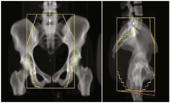
Conventional Radiotherapy (Fig-1): Planned by four field box technique (Antero-Posterior (AP), Postero-anterior (PA) and two opposing lateral fields) using standard bony landmarks.

  • AP/PA field: Superior: L4/L5 junction; Inferior: 3 cm distal to vaginal marker placed in vagina; Lateral: 1.5 to 2.0 cm beyond pelvic brim

Lateral field: Superior and inferior: As in AP field; Lateral: Anterior- Anterior border of pubic symphysis; Posterior- S2/S3 junction.


ijmrr_1112_01.jpg

Fig-1: Antero posterior and lateral fields based on bony landmarks for conventional four field box technique.

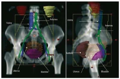
Three Dimensional Conformal Radiotherapy (Fig-2): Delineation of Gross Tumor Volume (GTV), Clinical target Volume1 (CTV1) (including GTV, uterus, vagina, bilateral parametrium), and CTV2 (Nodal CTV) (including pelvic lymph nodes-common iliac, external iliac, internal iliac, obturator and presacral) was done.

Planning Target Volume (PTV) was taken 1 cm beyond CTV (CTV1 + CTV2).

Radiotherapy dose delivered by Linear Accelerator was 50 Gy in 25 fractions at 200 cGy/day in 5 weeks. This was followed by 3 applications of intracavitary brachytherapy of 7 Gy/ fraction each to point A.

ijmrr_1112_02.jpg

Fig-2: Conformal RT volumes in cervical cancer.

Chemotherapy administration:Patients were administered Cisplatin (35mg/m2) on weekly basis during Radiotherapy. The patients were adequately hydrated with 2-2.5 liters of I.V. fluids and supplemented with KCL and MgSO4.Radiotherapy was delivered within 30 minutes of administration of Cisplatin.

Proper antiemetic therapy with 5-HT3 antagonist, dexamethasone, and ranitidine was given prior to chemotherapy administration.

Data Collection

  1. Dosimetric Assessment- For the conventional plans, the radiotherapy fields were generated based only on the bony digitally reconstructed radiograph (DRR) akin to X ray-simulator based planning.

Dose was prescribed at the isocenter, and beams were weighted equally as is done for conventional planning. Field sizes were recorded.

After treatment delivery all the conventional plans were analyzed retrospectively and PTV and organs at risk (OAR) were contoured.

Dose volume histograms (DVHs) were then analyzed for target volumes and organ at risk (urinary bladder, rectum, small bowel, and femoral heads).

Conformal plans were generated for optimal PTV coverage ensuring that 95% of the PTV received 95% of the prescribed dose. Dose was normalized at isocentre.

Subsequently, the field sizes were recorded, and the DVHs were analyzed for PTV and organs at risk (urinary bladder, rectum, small bowel, and femoral heads).

Dosimetric parameters evaluated were:

  • D95(Gy): Dose received by 95% of the planning target volume (PTV)
  • V95(%): Percentage of PTV receiving 95% of the prescribed dose, measure of conformity index of a plan.
  • PTV Average (Gy): Average dose received by PTV, measure of dose homogeneity of a plan.
  • Dmax (%): Maximum dose percentage received by PTV
  • Dmean (%): Mean dose percentage received by PTV
  • Organs at Risk (OAR) parameters that were assessed were:
  • Rectum Dmean (Gy): Mean dose received by the rectum.
  • Urinary Bladder Dmean (Gy): Mean dose received by the urinary bladder.

  • Small Bowel Dmean (Gy): Mean dose received by the small bowel.
  • Femoral heads Dmax (Gy): Maximun dose received by the femoral heads.
  1. Clinical response assessment- Clinical response was assessed during radiotherapy and every month after radiotherapy for up to 6 months. The patients were assessed for objective tumor response according to WHO criterion.

Complete response (CR)- Total tumor regression for at least 4 weeks; Partial response (PR)- 50% or more reduction in product of two major perpendiculars of the measurable tumor for at least 4 weeks; Stable disease (SD)- Less than 50% or more reduction to less than 25% increase in cross product; Progressive disease (PD)- Growth of measurable tumor by 25% or more or appearance of new lesion.

  1. Complication assessment- Treatment assessment was done on weekly basis during treatment and thereafter monthly basis on follow up. Complete blood counts and kidney function tests and liver function tests were repeated in all patients every week before each chemotherapy cycle.

Hematological toxicities were graded according to common toxicity criteria v4.03. Patients were assessed weekly during chemo radiation for acute radiation reactions. Radiation toxicity was assessed by RTOG acute and late morbidity scoring criteria. Follow up of all patients was done for atleast18 months, from the day of completion of treatment.

Data analysis- Collected data was analyzed using standard statistical methods and SPSS software version 20 to calculate level of significance using “p” value.

Descriptive statistical analysis has been carried out in the present study. Significance is assessed at 5% level of significance and statistical significance considered with p-value of <0.05. Chi-square test has been used to find the significance of haematological and radiation toxicities and clinical response on categorical scale between two groups. 95% Confidence Interval has been computed to find the significant features. Confidence Interval with lower limit more than 50% is associated with statistical significance. Student t test (two tailed, dependent) has been used to find the significance of dosimetric parameters on continuous scale between two groups.

Ethical consideration and permission: The study was approved by ethical committee. Prior to selection in the study, a written informed consent was taken by all the patients.

Patients were given the choice whether they want to participate in the study or not.

Results

Patient related, tumour related and treatment related characteristics have been shown in Table 1.

Table-1: Characteristics related to patient, tumour and treatment.

  Group A Group B
Patient related
Mean age (years) 50 (30-70) 51 (31-71)
Median age (years) 50 50
Pallor (%) 8 (32%) 11 (44%)
Co morbid Conditions
Hypertension 01 (4%) 00 (0%)
Diabetes mellitus 01 (4%) 00 (0%)
Tuberculosis 01 (4%) 01 (4%)
Tumor related
Stage
IB 02 (8%) 00 (0%)
IIA 05 (20%) 02 (8%)
IIB 13 (52%) 12 (48%)
IIIA 02 (8%) 03 (12%)
IIIB 03 (12%) 07 (28%)
IVA 00 (0%) 01 (4%)
Histopathology
Squamous Cell Ca 21 (84%) 23 (92%)
Adenocarcinoma 04 (16%) 02 (8%)
Histopathology Grading
Well Differentiated 02 (8%) 01 (4%)
Moderately Differentiated 21 (84%) 23 (92%)
Poorly Differentiated 02 (8%) 01 (4%)
Parametrial Invasion
Presence
Yes 17 (68) 21 (84)
No 08 (32) 04 (16)
Laterality
Unilateral 10 (54) 11 (52)
Bilateral 07 (46) 10 (48)
Extension
Less than pelvic wall 13 (68) 16 (72)
Upto pelvic wall 04 (32) 05(28)
Hydronephrosis
Present 02 (08) 04 (16)
Not Present 23 (92) 21 (84)

Lymph Node Involvement
Present 16 (64) 16 (64)
Not present 09 (36) 09 (36)
Treatment related
Chemotherapy Compliance
Less than 5 cycles 2 (8%) 4 (16%)
5 cycles 23 (92%) 21 (84%)
Overall Treatment Time
Within 8 weeks 19 19
More than 8 weeks 6 6
Median OTT (in days) 56 55

All patients underwent weekly assessment of radiation toxicities during treatment. No significant Grade III/IV haematological toxicity was seen in either group. Haemoglobin was maintained throughout the treatment by blood transfusion, oral hematinics and dietary advice. There were no significant acute and late reactions related to skin, vaginal mucosa, bladder, rectum and small intestine.

Field sizes used for the 3DCRT plans were significantly larger than those used for the conventional plans (p = 0.000) (Table 2). Optimal PTV coverage (95% of the PTV receiving 95% of the prescribed dose) was significantly improved using 3DCRT as compared to conventional radiotherapy (p value = 0.0001).

Table 2: Comparison of field sizes in both groups

Parameter Conventional (Mean) 3DCRT (Mean) p value
AP field area (cm2) 310.42 428.82 0.000
Lateral field area (cm2) 253.42 355.42 0.000

Dose homogeneity as assessed by average dose to PTV was not significantly better with 3DCRT (p = 0.292 for average dose to the PTV), while conformity index as assessed by volume receiving 95% of the prescribed dose, the difference was statistically significant between the two groups (p= 0.000). in addition, the mean dose to the planning target volume was increased significantly in the CT based plan when compared with the standard four field plan (p = 0.0001) (Table 3).

Table-3: Comparison of Dosimetric parameters of both groups

Parameter Conventional (Mean) 3DCRT (Mean) P-value
D95 19.15 49.46 0.0001
PTV Average 48.77 50.94 0.292
Dmax 107.84 107.15 0.266
Dmean 93.69 101.77 0.0001
V95 86.8 99.627 0.000

Differences in doses to the organs at risk (urinary bladder, and small bowel) were statistically significant across both groups (Table 4).

Table-4: Comparison of OAR dosimetric parameters of two groups

Parameter Conventional 3DCRT p value
Rectum Dmean (Gy) 49.39 48.26 0.082
Urinary Bladder Dmean (Gy) 50.53 49.04 0.024
Bowel Dmean (Gy) 18.86 27.25 0.000
Femoral head Dmax (Gy) 51.17 51.18 0.978

At the end of complete treatment, 80% of patients in group A and 64% of patients in group B had CR, but the difference was not statistically significant. At 18 months follow up, 96% of the patients in Group A and 100% of patients in group B had CR. 1 patient in Group A had progressive disease (Table 5).

Table-5: Response evaluation at end of treatment and 18 months follow up

Response evaluation Response Group A Group B p value
End of Treatment CR 20(80) 16(64) 0.207
PR 04(16) 09(36)
PD 01(04) 00(00)
18 Months follow up CR 24(96) 25(100) 0.312
PR 00(00) 00(00)
PD 01(04) 00(00)

CR- Complete Response, PR- Partial Response, PD- Progressive Disease.

Discussion

Three-dimensional conformal radiotherapy and intensity-modulated radiotherapy are now increasingly being used in the developed countries. These newer techniques have reported decrease in normal tissue toxicity, along with decrease in the chances of geographic miss. However, whether they are superior in terms of local control and survival also is yet to be demonstrated in larger trials.

By contrast, many centres in developing countries still prefer to use conventional X-ray-based planning using the standard bony landmarks. This is because in developing countries the number of state of art centres is very less and since patient load is high, X-ray-based planning is simple, less time consuming, and cost-effective as compared to three-dimensional CT-based planning.

However, randomized studies comparing volumetric planning versus conventional planning in carcinoma cervix are lacking [7].


Thus, before integrating volumetric planning in our routine practice, the present study was designed to compare conventional four field planning based on standard bony landmarks versus volumetric planning in patients of carcinoma cervix undergoing radiotherapy.

Field Sizes in conventional and 3D conformal planning

Greer et al [8] in their lymphangiographic study found that in 87% of the patients, the common iliac nodes were located proximal to the L5-S1 bifurcation, the conventional upper border of the pelvic portals and recommended that if coverage of the common iliac nodes is desired, the upper border should be moved to the L4- L5 junction. Pendlebury et al [9] also found that 62% of patients required alteration of the conventional pelvic portals based on lymphangiographic findings, with most requiring enlargement of one/more portals while in 20% patients, portals could actually be reduced.

They found that the lateral border of the AP/PA portals and the anterior border of the lateral portals were most often inadequate and recommended 2.5 cm margin from the pelvic brim for the former and 0.5 cm margin anterior to the symphysis pubis for the latter so as to cover 90% of the pelvic nodes. With the advent of CT simulation, it is possible to identify and contour the pelvic blood vessels, and these can then be used as surrogates for localizing the adjacent lymphatics and lymph nodes.

Other lymphangiographic studies by Zunino et al [1] and Bonin et al [10] on the other hand, found that lateral coverage of the external iliac nodes was insufficient on the AP/PA portals and recommended going 2.5 cm and 2.6 cm, respectively, beyond the pelvic brim. In an intra operative study using surgical clips, McAlpine et al [11] recommended that the superior border would need to be even higher at the L3- L4 junction to properly cover the common iliac nodes and also discovered that 26% of patients would have inadequate lateral coverage on the AP/PA portals.

Using noncontract CT images, a study by Finlay et al [6] found that had conventional portals alone been used for radiotherapy planning, the majority (95.4%) of subjects would have had at least one inadequate margin, the majority located superiorly though in around half the subjects, at least one margin would have been generous (>2 cm beyond the blood vessel), usually the lateral borders of the AP/PA portal.

Concurrently, large field sizes were called for, as was also observed in the present study. Field sizes used for the 3DCRT plans were significantly larger than those used for the conventional plans (p = 0.000 for both antero-posterior fields and lateral fields, significant).

This increase in field sizes in 3D conformal techniques could have led to increased doses to organs at risk and inferior sparing of the organs at risk. However, this was not the case because with the help of three dimensional imaging, just as PTV coverage is improved by nodal visualization, so also is the OAR sparing due to conformity of treatment fields, allowing much tighter blocking as compared to conventional planning where tighter blocking is deemed unsafe in terms of disease control.

Dosimetric analysis- Greer et al [8] reported the value of pelvic magnetic resonance imaging (MRI) in the design of pelvic fields of the box technique. In 25 patients with FIGO clinical Stages 1B - IVA, MRI was used primarily to define the treatment volume required to encompass the primary disease and its direct regional extensions, and only secondarily to assess the presence or absence of lymph node metastases.

The sagittal scans revealed that use of “conventional” or “standard” lateral radiation portals resulted in a failure to encompass all gross cancer extensions (marginal miss) in 24% patients. The use of conventional lateral portals resulted in an incomplete coverage (62.5%) of the uterine fundus, of whom three had gross cancer extension involving either the uterine cavity or the myometrium of the lower uterine segment.

The authors concluded that conventional lateral portal design, as in standard radiation oncology texts, may be suboptimal for a significant percentage of patients with locally advanced or bulky cervical cancer, and could be a contributing cause of failure to control pelvic disease.

Boss et al [12] performed MRI in 33 patients with gynecological cancer on 2 consecutive days to study the interfraction movement of the uterus and cervix. They observed that large movements of uterus occurred in the superior–inferior and anterior–posterior directions, although cervical displacement was less marked and recommended asymmetrical margin with CTV–PTV expansion of the uterus, cervix, and upper vagina of 15 mm AP, 15 mm SI, and 7 mm laterally.


Another study by Gulia A et al [7], observed that in 82% of patients, the standard four field based on bony landmarks failed to encompass the target volume defined by CT as compared with previous studies discussed earlier.

In conjunction with these studies, in the present study it was observed that in only three patients (12%) out of 25 patients, the whole of the planning target volume was encompassed by the standard four field box marked on bony landmarks. Target coverage was significantly improved using 3DCRT as compared to conventional RT (p = 0.0001 for dose to 95% of PTV). On the other hand, dose homogeneity within the PTV was better with 3DCRT as compared to conventional technique but it was not statistically significant (p = 0.292 for average dose to the PTV).

Also, conformity index as evaluated by V95 was also significantly better in 3DCRT group (p = 0.000). In addition, the mean dose to PTV was significantly higher in the CT‑based plan when compared with the standard four field plan. (p = 0.0001 for mean dose to PTV).

OAR Sparing- Silva et al reported that there was a significant increase in the maximum dose received by the OAR, the volume of bowel receiving 30 Gy, and a decrease in the bladder volume receiving 95% of the prescribed dose in the 3D plans as compared to conventional pelvic fields based on bony landmarks [13].

Włodarczyk H et al [14], also concluded that there were significant differences (p<0.05) in dose distributions in critical organs between the 3DCRT and 4 field conventional box techniques. The smallest volumes of critical organs were irradiated using the 4-portal conformal technique. The greatest volume of rectum and bladder was irradiated using the AP-PA conventional technique.

In conjunction to these findings, in the present study also doses to the organs at risk (rectum, urinary bladder, and small bowel) were significantly different across the 2 arms. Doses to the rectum were higher for the 3DCRT arm as compared to the conventional arm but it was statistically insignificant (p = 0.820 for maximum dose to rectum and p = 0.082 for mean rectal dose). But, doses to the urinary bladder were significantly higher for the conventional arm as compared to the 3DCRT arm (p=0.013 for maximum dose tourinary bladder and p=0.024 for mean dose received by urinary bladder, significant).

This increase in mean dose to urinary bladder in the conventional group also translated clinically into significantly higher acute and late bladder reactions on clinical assessment of the patients as discussed previously.

Also, doses to the small bowel were significantly higher for the 3DCRT arm as compared to the conventional arm (p = 0.000 for mean dose received by small bowel). This is attributable to the larger field sizes on the 3DCRT technique as compared to the bony landmark based conventional technique. But this increase in mean dose to the small bowel did not show any clinically significant difference between the small bowel toxicities in the two groups.

This is attributable to the fact that small bowel is a mobile organ and thus same loops of bowel will not be present in the radiation field in all the sessions of radiation therapy. Doses to the femoral heads were higher for the 3DCRT arm as compared to the conventional arm, but it was not statistically significant (p = 0.94 for mean dose received by femoral heads).

Response Evaluation: Although outcome of carcinoma cervix treated by radiotherapy are quite satisfactory in early stage disease and have also been greatly improved beyond the historical 30‑40% survival rate for advanced stage disease also, by addition of concurrent chemo radiation, it is still likely to be further improved by superior delineation and coverage of the pelvic lymph nodes. Geographic miss of the pelvic lymph nodes has serious consequences, especially in advanced stage disease [2].

In the present study at the end of complete treatment, 64% of patients in conformal and 80% of patients in conventional arm had CR, but the difference was not statistically significant. At 6th month follow up, 100% of the patients in conformal and 96% of patients in conventional arm had CR. 1 patient in conventional arm had progressive disease. In a study by Beadle et al [3] it was found that the majority (66%) of pelvic nodal failures were marginal; 71 out of 119 patients recurred above the treatment field, 2 had inguinal nodal failures while 2 other patients had recurrences both above the treatment field and in the inguinal lymph nodes.

This was one of the first studies to correlate the site of regional recurrence with respect to the treatment portals.


In the present study, use of CT simulation allowed superior visualization of the pelvic lymph nodes and improved the PTV coverage, mainly by reducing the chances of geographical miss to a minimum. This may translate into superior loco-regional control and even superior survival but longer follow up is needed for such results.

Limitations of present study: Small sample size and short follow up duration.

Conclusion

On dosimetric analysis, three- dimensional conformal radiotherapy gives significantly better PTV coverage when compared to conventional four field box technique. 3DCRT also requires significantly larger field sizes though doses to the OARs are not significantly higher compared to the conventional plans except for small bowel.

Thus, the improved delineation of the target, especially pelvic nodes, and the improved target coverage make 3DCRT a better technique as compared to four field box technique in cases of carcinoma cervix.

However, it remains to be seen whether 3DCRT will have a clinical benefit over conventional four field radiotherapy in terms of loco regional control and overall survival but the answer to the question requires longer follow-up and a larger group ofpatients.

What this study adds to existing knowledge?

Out of various definitive external beam radiotherapy techniques available for treatment carcinoma cervix like- 2D-CRT, 3D-CRT and IMRT; 2D and 3D-CRT are financially feasible for LMIC (Lower Middle-Income Countries) like India. 3DCRT not only reduces the chances of PTV miss as it is based on cross sectional imaging unlike 2DCRT which is based on bony landmarks but also it provides optimal PTV coverage which is comparable to advanced treatment planning techniques like IMRT at much lower financial expenses.

Author’s Contribution

Dr. Shubhi Agarwal: Data collection and assessment, drafting of manuscript

Dr. Arvind Kumar Chauhan: Verification of data, Manuscript correction and verification

Dr. Piyush Kumar: Designing study methodology, manuscript drafting and verification

Mr. Jitendra Nigam: Treatment planning

Dr. Pavan Kumar: Manuscript verification

Reference

  1. Zunino S, Rosato O, Lucino S, Jaurequi E, Rossi L, Valencia D. Anatomic study of the pelvis in carcinoma of the uterine cervix as related to the box technique. Int J Radiat Oncol Biol Phys. 1999;44(1)53-59.
  2. Kim RY, McGinnis S, Spencer SA, Meredith RF, Jennelle R, Salter MM. Conventional four-field pelvic radiotherapy technique without computer tomography-treatment planning in cancer of the cervix- Potential geographic miss and its impact on pelvic control. Int J Radiat Oncol Biol Phys. 1995;31(1)109-112.
  3. Beadle BM, Jhingran A, Yom SY, Ramirez PT, Eifel PJ. Patterns of regional recurrence after definitive chemoradiotherapy for cervical cancer. Int J Radiat Oncol Biol Phys. 2010;76(5)1396-1403.
  4. Gerstner N, Wachter S, Knocke TH, Fellner C, Wambersie A, P¨otter R. The benefit of Beam’s eye view-based 3D treatment planning for cervical cancer. Radiother Oncol. 1999;51(1)71-78.
  5. Olofsen-van Acht M JJ, Quint S, Seven M, van Santvoort JP, van den Berg HA, Logmans A, et al. Three-dimensional treatment planning for postoperative radiotherapy in patients with node-positive cervical cancer- Comparison between a conventional and a conformal technique. Strahlentherapie und Onkologie. 1999;175(9)462-469.

  1. Finlay MH, Ackerman I, Tirona RG, Hamilton P, Barbera L, Thomas G. Use of CT simulation for treatment of cervical cancer to assess the adequacy of lymph node coverage of conventional pelvic fields based on bony landmarks. Int J Radiat Oncol Biol Phys. 2006;64(1)205-209.
  2. Gulia A, Patel F, Rai B, Bansal A, Sharma S C. Conventional four field radiotherapy versus computed tomography-based treatment planning in cancer cervix- A dosimetric study. South Asian J Cancer. 2013;2(3)132-135.
  3. Greer BE, Koh WJ, Figge DC, Russell AH, Cain JM, Tamimi HK. Gynaecologic radiotherapy fields defined by intra-operative measurements. Gynecol Oncol. 1990;38(3)421-424.
  4. Pendlebury SC, Cahill S, Crandon AJ, Bull CA. Role of bipedal lymphangiogram in radiation treatment planning for cervix cancer. Int J Radiat Oncol Biol Phys. 1993;27(4)959-962.
  5. Bonin SR, Lanciano RM, Corn BW, Hogan WM, Hartz WH, Hanks GE. Bony landmarks are not an adequate substitute for lymphangiography in defining pelvic lymph node location for the treatment of cervical cancer with radiotherapy. Int J Radiat Oncol Biol Phys. 1996;34(1)167-172.
  1. Mc Alpine A, Schlaereth JB, Lim P, Chen D, Eisenkop SM, Spiritos NM. Radiation fields in gynecologic oncology- Correlation of soft tissue (surgical) to radiologic landmarks. Gynecol Oncol. 2004;92(1)25-30.
  2. Boss EA, Barentsz JO, Massuger LF, Boonstra H. The role of MR imaging in invasive cervical carcinoma. Eur Radiol. 2000;10(2)256-270.
  3. Silva ML, Fogaroli R, Maia MA, Pellizzon AC, Novaes PE, Chojniak R, et al. A Comparative Analysis of the Conventional Pelvic Fields based on Bony Landmarks and Contrast-Enhanced CT Simulation for the Treatment of Cervical Cancer. Int J Rad Oncol Biology Physics. 2007;69(3)S407.
  4. Włodarczyk H, Roszak A, Cikowska-Woźniak E, Bratos K, Wojciechowska-Łącka A. Does conformal therapy improve dose distribution in comparison to old techniques in tel eradiotherapy of cervical cancer patients?. Rep Pract Oncol Radiother. 2008;13(4)195-200.