E-ISSN:2320-8686
P-ISSN:2321-127X

Research Article

3D CRT

International Journal of Medical Research and Review

2019 Volume 7 Number 6 November-December
Publisherwww.medresearch.in

Dosimetric parameters of heart and left ventricle – comparison of 3D CRT and IMRT in left cancer breast

Mehta A.1, Kumar P.2*, Silambarasan N.3, Navitha S.4, Nigam J.5, Kumar P.6, Chauhan A.7
DOI: https://doi.org/10.17511/ijmrr.2019.i06.05

1 Ankita Mehta, Junior Resident, 2* Piyush Kumar, Professor and Head, 3 N.S. Silambarasan, Assistant Professor cum Medical Physicist, 4 S. Navitha, Assistant Professor cum Medical Physicist, 5 Jitendra Nigam, Assistant Professor cum Medical Physicist, 6 Pavan Kumar, Assistant Professor, 7 Arvind Chauhan, Associate Professor; 1,2,3,4,5,6,7author are affiliated with Department of Radiation Oncology, Shri Ram Murti Institute of Medical Sciences, Bareilly, Uttar Pradesh, India.

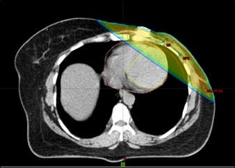
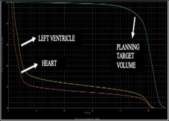
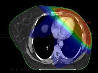
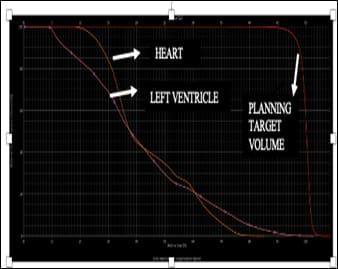
Background: Conformal Radiotherapy techniques adapting to the ballistics of delineated volumes allowed significant reduction in excess radiation induced mortality however the increasing number of long-term survivors and expanding use of cardiotoxic drug highlight the persistent need for maximal cardiac possible sparing. The low dose volume of left ventricle are better predictor of acute coronary events than mean heart dose. Materials and Methods: 38 post-MRM patients were randomized to treatment by 3Dimensional Conformal Radiotherapy (3D CRT) and Intensity Modulated Radiotherapy (IMRT) technique. Two tangential beams were used in 3D CRT technique while five to seven (mostly tangential beams) were used in inversely planned IMRT technique. The dose volume parameters of planning target volume, heart and left ventricle were compared. Results: The dosimetry of Planning target volume showed significantly better coverage in IMRT technique (D90, D95) however the D50 was comparable in both the techniques. In dosimetry of heart, the high dose volumes (V30, V40) were nearly comparable in both the techniques. The other dose volume parameters (V5, V10, V20, V25, D33, D67, D100) and the mean dose were significantly lesser in 3D CRT technique along with significantly better sparing of left ventricle (Dmean and V5). Conclusion: The dosimetry of target volume was better with IMRT technique, but this was accompanied by a huge increase in dose to whole heart and specifically the left ventricle which has strong potential to translate into an increased cardiotoxicity. A better distribution of the target region may be obtained by multiple segmentation of the two tangential fields in 3D CRT plans with further reduction in dose to heart and left ventricle.

Keywords: 3-Dimensional Conformal Radiotherapy (3D CRT), Intensity Modulated Radiotherapy (IMRT), Cardiotoxicity, Planning target volume, Heart, Left ventricle

Corresponding Author How to Cite this Article To Browse
Piyush Kumar, Professor and Head, Department of Radiation Oncology, Shri Ram Murti Institute of Medical Sciences, Bareilly, Uttar Pradesh, India.
Email:
Mehta A, Kumar P, Silambarasan NS, Navitha S, Nigam J, Kumar P, Chauhan A. Dosimetric parameters of heart and left ventricle – comparison of 3D CRT and IMRT in left cancer breast. Int J Med Res Rev. 2019;7(6):468-474.
Available From
https://ijmrr.medresearch.in/index.php/ijmrr/article/view/1096

Manuscript Received Review Round 1 Review Round 2 Review Round 3 Accepted
2019-11-04 2019-11-14 2019-11-20 2019-11-26
Conflict of Interest Funding Ethical Approval Plagiarism X-checker Note
No Nil Yes 9%

© 2019 by Ankita Mehta, Piyush Kumar, N.S. Silambarasan, S. Navitha, Jitendra Nigam, Pavan Kumar, Arvind Chauhan and Published by Siddharth Health Research and Social Welfare Society. This is an Open Access article licensed under a Creative Commons Attribution 4.0 International License https://creativecommons.org/licenses/by/4.0/ unported [CC BY 4.0].

Background

Breast cancer has the highest incidence and the leading cause of death of all malignancies in females [1]. Due to improvements in diagnostic modalities, wide implementation of screening programmes [2], advances in multimodality treatment, long-term cause-specific survival for all stages has shown significant improvement over the past few decades [3,4]. This has prompted an increased focus on long term treatment related toxicities. Long term serious sequelae like lethal cardiac events [5,6] increased risk of second malignancies are now the area of concern [7,8]. Large scale studies using older radiation techniques have shown the mortality of radiation induced heart disease to be severe enough to offset the survival advantage offered by radiotherapy [9]. Conformal radiotherapy techniques adapting to the ballistics and the energy to the delineated volumes with careful evaluation of the dose-volume distribution of critical organs allowed a drastic reduction in cardiac mortality.

As per QUANTEC (Quantitative Analyses of Normal tissue effects in the Clinic), the irradiated volume of the left ventricle was shown to be the most important predictor of a perfusion defect of heart [10]. The clinical correlation of dose being delivered to left ventricle was established in a large-scale study by Bogaard et al. with a significant dose-effect relationship observed for acute coronary events after irradiation over a follow up period of 9 years. The V5 of left ventricle was shown to be a better predictor than other dose volume parameters evaluated (ranging from V10 to V60). The model performance was significantly improved by replacing the conventional parameter of mean heart dose with the V5 of left ventricle [11]. The literature on optimal post-mastectomy external beam irradiation techniques in terms of cardiac sparing is controversial. This randomized study was designed to compare the dosimetric parameters of two conformal techniques in post mastectomy patients in terms of target volume, heart and left ventricle.

Materials and Methods

Study Setting: Department of Radiation Oncology, Shri Ram Murti Institute of Medical Sciences

Study design: Prospective, randomized controlled study.

Study population: Left sided Post MRM patients from September 2017 to October 2019.

Study tool: Dosimetric parameters were evaluated.

Duration and type of study:A total of 38 left sided patients who underwent MRM (Modified Radical Mastectomy) patients were recruited from September 2017 to October 2019. An equal number of patients were allocated to two arms (3D CRT and IMRT technique) by simple randomization.

Inclusion criteria

  1. Left sided Post MRM patients
  2. Conventional fractionation schedule – 50 Gy/25#
  3. Normal baseline 2D Echocardiography

Exclusion criteria

  1. No previous malignancy
  2. No previous history of thoracic radiotherapy
  3. Patients treated by Hypofractionation

Delineation:Simulation was done in supine position with arms overhead flexed at the elbow joint, abducted and externally rotated. Non contrast CT scan of 3 mm slice thickness were obtained. The delineation of clinical target volume and heart was done as per RTOG Breast Cancer atlas. The left ventricle was delineated as per cardiac contouring atlas [12]. Radio-opaque wires were used to mark the mastectomy scar, the inferior and lateral limit of the chest wall. A PTV margin of 5 mm was given to account for the setup errors.

Methodology of 3-Dimensional Conformal Radio-therapy (3D CRT):Two tangential beams were used for chest wall irradiation with gantry angles ranging from 2250 to 3150 on the lateral side and 450 to 1350 on the medial side. A separate field was used from 00 gantry angle for supraclavicular nodal irradiation. The isocentre was placed at the junction of tangential fields and supraclavicular field and half beam block technique with asymmetric jaws was used. Using the Beams eye view (BEV), the beam angles, were so chosen to optimize coverage of the PTV with maximum exclusion of the organs at risk. The fields extended a minimum of 1cm anteriorly from the chest wall to provide coverage of the “flash” region. Enhanced dynamic wedges and field in field technique were used when required for a homogenous dose distribution.

Methodology of Intensity modulated radiotherapy (IMRT): A total of 5 to 7 semi opposing (mostly tangential beams) were used in IMRT plans.


Inverse planning was done and beam optimisation was used to the achieve the desired objectives and constraints. Tissue inhomogeneities were considered in the beam optimisation process. The optimal planning objectives for both the techniques were specified as dose to planning target volume (PTV) between 95% and 107% levels relative to 100% prescription point.

The prescribed dose was 50 Gy in 25 fractions at 2 Gy per fraction using photons with a beam energy of 6 MV.

Data collection: Dose volume histograms were computed for the PTV, heart and left ventricle, analysed and compared between the two planning techniques. The following parameters for PTV were assessed- D90% and D95%, Dmean, V107%, D50%, D98% (Dnear-min) and D2% (Dnear-max), Homogeneity index (HI) (ICRU 83)[13], Conformity Index (CI) (The treated volume was taken as the volume encompassed by 95% isodose lines) (ICRU 62)[14].

The following dosimetric parameters for heart (D33%, D67%, D100%, Dmean, V5, V10, V20, V25, V30, V40) and left ventricle (Dmean, V5) were assessed.

Data analysis: Statistical significance was calculated using paired t test of unequal variances with a value of <0.05 considered significant.

Ethical consideration: Prior to selection in the study, an informed consent of all the patients was obtained. The patients were given the choice of whether they want to participate in the study or not.

Results

The dosimetry of PTV was better in IMRT technique with a significantly higher observed mean value of D90 and D95 however D50 was nearly comparable with no significant difference observed in both the techniques. The homogeneity index was also significantly better in IMRT technique. Although the conformity index was seen to be better in IMRT technique, but the difference was non-significant (Table 1) (Figure 1a & 1b).

Table-1: Dosimetric parameters of the planning target volume in two techniques (Mean±1 SD).

Parameters Variable 3D CRT vs IMRT p-value
PTV [Dose in Gy] D95% 40.55±4.88 vs. 46.17±5.53 0.002
D90% 44.76±1.62 vs 47.94±2.10 <0.0001
  Dmean 48.65±1.31 vs 49.64±1.23 0.02
D50% 49.54±1.04 vs. 49.99±0.78 0.14
D2% (Dnear-max) 53.65±1.38 vs 51.84±0.84 < 0.001
D98% (Dnear-min) 32.80±12.16 vs 43.34±9.10 0.01
V107% 4.33±4.06 vs. 0.23±0.79 < 0.0003
Conformity Index 1.26±0.28 vs. 1.19±0.13 0.36
Homogeneity index 0.42±0.24 vs. 0.17±0.19 0.001

Table-2: Table showing dosimetric parameters of the heart and left ventricle in two techniques (reported as Mean±1 SD).

Heart [Dose in Gy, Volume percentage] D33% 5.03±6.07 vs. 17.79±7.09 <0.001
D67% 1.63±0.50 vs. 8.60±5.22 <0.001
D100% 0.56±0.27 vs. 2.54±2.08 <0.001
V5 24.34±9.92 vs. 81.62±18.47 <0.001
V10 18.32±8.46 vs. 55.39±22.88 <0.001
V20 15.06±7.46 vs. 30.23±17.76 0.002
V25 13.76±7.12 vs. 23.13±13.58 0.01
V30 12.57±6.77 vs. 16.88±9.76 0.13
V40 9.77±5.61 vs. 8.54±6.58 0.54
Dmean 8.48±3.37 vs. 16.12±5.26 <0.001
Left ventricle [Dose in Gy, Volume percentage] V5 41.34±4.97 vs. 94.56±14.64 <0.001
Dmean 13.69±5.73 vs. 21.77±10.06 <0.001

ijmrr_1096_01.jpg

Fig-1(a): Dose colour wash of 3D CRT plan (Volume receiving 5 Gy).


ijmrr_1096_02.jpg

Fig-1b: Dose Volume Histogram of 3D CRT plan.

ijmrr_1096_03.jpg

Fig-2(a): Dose colour wash of IMRT plan (Volume receiving 5 Gy).

ijmrr_1096_04.jpg

Fig-2 (b): Dose Volume Histogram of IMRT plan.

In terms of dosimetry of heart, there was no significant difference observed in terms of high dose volumes (V25, V30, V40) in both the techniques.

However, the other dose volume parameters (V5, V10, V15, V20) and (D33, D67, D100) showed a clear advantage in 3D CRT technique with significantly lesser (p<0.01) observed mean values. A similar benefit was also observed in dosimetry of left ventricle with significant decrease observed in the mean dose and V5 (p<0.0001) (Table 2) (Figure 2a & Figure 2b).

Discussion

Intensity Modulated radiation therapy (IMRT) has proved to be superior to three-dimensional conformal radiation therapy (3DCRT) in head and neck, central nervous system, lung, prostate and various other sites. The majority of literature supports IMRT in terms of better dosimetry of the target region of interest over 3D-CRT for post mastectomy radiotherapy but its role in terms of sparing of surrounding critical structures has huge controversies. Our study also proved the superiority of IMRT technique in terms of a homogenous distribution owing to a significantly higher value of minimum dose and a lesser value of maximum dose.

The multibeam arrangement in IMRT technique led to adequate build up thickness in all the directions before the target volume ensuring a better coverage of superficial regions of chest wall in which were comparatively largely underdosed in 3D-CRT plans.

The dose volume parameters, D95% and D90% showed significantly higher mean observed values in IMRT technique however the D50% which is considered to be largely representative of dose distribution inside the entire PTV and also a better estimate of optimal coverage than other dose volume parameters showed no significant difference in two techniques [13].

The difficulties encountered in 3D-CRT technique were a suboptimal coverage of nodal region and the junction of two fields along with overdosage at the central region of chest wall. There was large dose heterogeneity and interindividual variation observed.

The delineated region of supraclavicular region also varied enormously with patient’s anatomy and the routine use of prescription at maximum depth did not optimally cover the target nodal volume for all the patient while it resulted in dose spillage into superficial lung in a proportion of patients.


In terms of dosimetry of heart, the dose to partial and whole volumes of heart (D33%, D67%, D100%) and the mean dose were significantly lesser in 3D-CRT technique. The other dose volume parameters (V5, V10, V20, V25) also showed an advantage in terms of better cardiac sparing in 3D-CRT technique. This was because of an indispensable increase in the entry dose owing to multibeam arrangement in IMRT plans. To our knowledge so far, this is the first small scale study comparing the dosimetry of left ventricle amongst the two techniques.

Although the mean dose and V5 of left ventricle were significantly reduced in 3D CRT technique but the observed values were much higher than the recommended dose constraints. The DEGRO breast cancer expert panel recommends the Dmean of LV (mean dose left ventricle) < 3 Gy; V5 (volume of LV receiving ≥5 Gy) < 17%. The literature on cardiac sparing radiotherapy techniques is controversial. Some studies have demonstrated a better cardiac sparing in terms of high dose volumes with IMRT technique [14,15,16] while a few others and our study have shown either equivalent or better results with 3D-CRT technique [17,18,19].

However, most of the studies have documented a clear disadvantage of IMRT technique in terms of low dose volumes which are now known to have a clear association with lethal cardiac events. The wide variation across the literature in terms of high dose volumes in IMRT technique is attributed to huge differences in planning techniques with large variation in the number and angle of beams (tangential and non- tangential) along with the priority defined in optimization process relative to the target region of interest and other critical organs at risk.

Rudat et al. in a study of post-MRM patients showed a significant reduction of the V35 of the heart by an average of 43% in IMRT plans (p < 0.01) along with reduction in the mean heart dose by an average of 20% (p=0.03) by using the same beam arrangement as of 3D-CRT plans [16]. A recent study done by Finazzi et al. validated our findings with a significantly higher value of mean heart dose (median 4.5 vs. 3.3 Gy; p < .01) seen in patients receiving adjuvant nodal irradiation by IMRT or VMAT technique over 3D-CRT technique. Despite these findings, their study showed a huge increase institutional time trends of IMRT/VMAT with an overall rate of 46.0% in 2015–2018 compared to only 5.6% in 2013-2014 [19].

In breast radiotherapy, simple maneuverers like treatment in prone position in whole breast irradiation [20,21,22] and treatment in DIBH (deep inspiration-breath holding) have shown promising results for sparing of lung and heart tissue.

Sripathi et al. in a small study showed 3DCRT in deep inspiration breath holding technique provided maximal cardiac sparing in patients with left-sided breast cancer obviating the need of IMRT technique [23]. A small-scale study by Helal A et al. comparing radiation techniques in patients who underwent breast conserving surgery showed a significant sparing of the surrounding critical structures with a more homogenous distribution by segmentation of the two tangential fields. The step and shoot optimized plans were generated by segmentation of two simple fields.

Five segments were generated for each field; segment one covering the PTV, segment two including PTV & excluding heart, segment three including PTV & excluding lung, segment four including PTV and excluding both heart and lung and segment five including PTV & excluding build up region. These plans showed a significant reduction of 12% in heart V5, 15% in V20, 21% in V30 and 53% in V40 over the conventional two beam plans [24].

These findings form the basis of future studies where the segmentation of multiple sub-fields within two tangential fields may allow a better dose coverage and homogeneity of the target regions along with better sparing of the heart and left ventricle which may help to achieve the recommended dose constraints.

Although a clear advantage of one technique over another cannot be established in the present study but it raises the concern about the shortcomings of IMRT technique which may led to a possible increase in mortality attributable to cardiac events. In this era of rapidly evolving radiation technology, there is a concern that IMRT in breast cancer is being widely implemented without evidence-based knowledge of its effects on long-term efficacy and morbidity [25].

In routine practise, the dose being delivered to left ventricle has not found widespread clinical adoption but given the emerging strong evidence on its clinical implications, a standardized contouring of cardiac substructures with the least possible dose to all cardiac segments needs to be implemented.


The risk of locoregional recurrence and cause specific breast cancer mortality needs to be individually weighed up against the baseline cardiac risk factors and the risk of radiation-induced cardiotoxicity. A routine follow-up with a cardiologist, including screening for valvular disease with echocardiography and coronary artery disease with computed tomography angiography or coronary artery calcium scoring is recommended in patients with a history of thoracic radiotherapy but wider adoption needs to be reinforced. A major limitation of the present study was less number of patients and short follow-up period. Ideally, both the plans should have been generated for all the patients to account for inter-individual anatomical variations, but this was not possible because of busy setup.

Conclusion

In this study, aiming to compare the two techniques, the target volumes were homogenized, and dose distribution was achieved to the desired limits in IMRT technique but with accompanying disadvantage of increase in the low dose volumes of heart and the left ventricle with no advantage of in terms of high dose volumes. There is no strong evidence to support growing practice of IMRT and the benefit observed could likely be achieved with simple segmentation creating multiple subfields within two tangential fields in 3D CRT technique.

What the study adds to the existing knowledge?

In routine practise of post-mastectomy radiotherapy, the dose being delivered to left ventricle is not given due weightage however it has huge clinical implications. The low dose volume of left ventricle is more important predictor of coronary events compared to dose to the whole heart. Before accepting the widespread use of IMRT, the indispensable increase in low dose volumes owing to the multibeam arrangement warrants serious consideration.

Author’s Contribution

Dr. Ankita Mehta: Collected data, Drafting the manuscript

Dr. Piyush Kumar: Verification of data, Correcting, verifying the manuscript

Mr. Silambarasan NS, Mrs. Navitha S., Mr. Jitendra Nigam J: Treatment planning

Dr. Pavan Kumar: Manuscript drafting

Dr. Arvind Kumar: Manuscript drafting

All authors contributed to final manuscript.

Acknowledgment

Authors would like to express their gratitude to patients and thanks to the team of radiation technologists and other staff who were involved in their treatment.

Reference

  1. Bray F, Ferlay J, Soerjomataram I, Siegel RL, Torre LA, Jemal A. Global cancer statistics 2018- GLOBOCAN estimates of incidence and mortality worldwide for 36 cancers in 185 countries. CA Cancer J Clin. 2018;68(6)394-424.
  2. Marmot MG, Altman DG, Cameron DA, Dewar JA, Thompson SG, Wilcox M. The benefits and harms of breast cancer screening- an independent review. Br J Cancer. 2013;108(11)2205-2240.
  3. Kanapuru B, Ershler WB, Hesdorffer C, Jemal A, Yates JW. Long-term survival of older breast cancer patients- population-based estimates over three decades. Breast Cancer Res Treat. 2012;134(2)853-857.
  4. Guarneri V, Conte PF. The curability of breast cancer and the treatment of advanced disease. Eur J Nucl Med Mol Imaging. 2004;31(1)S149-
  5. Shah C, Badiyan S, Berry S, Khan AJ, Goyal S, Schulte K, et al. Cardiac dose sparing and avoidance techniques in breast cancer radiotherapy. Radiotherap Oncol. 2014;112 (1)9-16.
  6. Lee MS, Finch W, Mahmud E. Cardiovascular complications of radiotherapy. Am J Cardiol. 2013;112(10)1688-1696.

  1. Boice Jr JD, Harvey EB, Blettner M, Stovall M, Flannery JT. Cancer in the contralateral breast after radiotherapy for breast cancer. N Engl J Med. 1992;326(12)781-785.
  2. Bazire L, De Rycke Y, Asselain B, Fourquet A, Kirova YM. Risks of second malignancies after breast cancer treatment- Long-term results. Cancer Radiother. 2017;21(1)10-15.
  3. Clarke M, Collins R, Darby S Davies C, Elphinstone P, Evans V, et al. Effects of radiotherapy and of differences in the extent of surgery for early breast cancer on local recurrence and 15-year survival- an overview of the randomised trials Early breast cancer trialists' collaborative group (EBCTCG). The Lancet. 2006;366(9503)2087-2106.
  4. Gagliardi G, Constine LS, Moiseenko V, Correa C, Pierce LJ, Allen AM, et al. Radiation dose–volume effects in the heart. Int J Radiation Oncology Biol Phys. 2010;76(3)77-85.
  5. van den Bogaard VA, Ta BD, van der Schaaf A, Bouma AB, Middag AM, Bantema-Joppe EJ, et al. Validation and Modification of a Prediction Model for Acute Cardiac Events in Patients with Breast Cancer Treated with Radio-therapy Based on Three-Dimensional Dose Distributions to Cardiac Substructures. J Clin Oncol. 2017;35(11)1171-1178.
  6. Duane F, Aznar MC, Bartlett F, Cutter DJ, Darby SC, Jagsi R, et al. Cardiac Contouring A cardiac contouring atlas for radiotherapy. Radiother Oncol. 2017;122(3)416-422.
  7. Vol I. Prescribing, recording, and reporting photon-beam intensity-modulated radiation therapy (IMRT)- contents. J ICRU. 2010;10.
  1. Menzel HG. International commission on radiation units and measurements. Journal of the ICRU. 2014;14 (2)1-2.
  2. Rastogi K, Sharma S, Gupta S, Agarwal N, Bhaskar S, Jain S. Dosimetric comparison of IMRT versus 3DCRT for post-mastectomy chest wall irradiation. J Radiat Oncol. 2018;36(1)71-78.
  3. Rudat V, Alaradi AA, Mohamed A, Khaled AY, Altuwaijri S. Tangential beam IMRT versus tangential beam 3D-CRT of the chest wall in postmastectomy breast cancer patients- A dosimetric comparison. Radiat Oncol J. 2011;6(1)26.
  4. Kausar M, Gurjar OP, Bagdare P, Gupta KL, Bhandari V, Naik A, et al. Dosimetric analysis of intensity-modulated radiotherapy and three-dimensional conformal radiotherapy for chest wall irradiation in breast cancer patients. J Radiotherap Pract. 2016;15(1)30-37.
  5. Aras S, İkizceli T, Aktan M. Dosimetric Comparison of Three-Dimensional Conformal Radiotherapy (3D-CRT) and Intensity Modulated Radiotherapy Techniques (IMRT) with Radiotherapy Dose Simulations for Left-Sided Mastectomy Patients. Eur J Breast Health. 2019;15(2)85-89.
  6. Finazzi T, Nguyen VT, Zimmermann F, Papachri-stofilou A. Impact of patient and treatment characteristics on heart and lung dose in adjuvant radiotherapy for left-sided breast cancer. J Rad Oncol. 2019;14(1)153.
  7. Buijsen J, Jager JJ, Bovendeerd J, Voncken R, Borger JH, Boersma LJ, et al. Prone breast irradiation for pendulous breasts. Radiother Oncol. 2007;82(3)337-340.

  1. Griem KL, Fetherston P, Kuznetsova M, Foster GS, Shott S, Chu J. Three-dimensional photon dosimetry- a comparison of treatment of the intact breast in the supine and prone position. Int J Radiat Oncol Biol Phys. 2003;57(3)891-899.
  2. Varga Z, Hideghéty K, Mező T, Nikolényi A, Thurzó L, Kahán Z. Individual positioning: a comparative study of adjuvant breast radiotherapy in the prone versus supine position. Int J Radiat Oncol Biol Phys. 2009;75(1)94-100.
  1. Sripathi LK, Ahlawat P, Simson DK, Khadanga CR, Kamarsu L, Surana SK, et al. Cardiac dose reduction with deep-inspiratory breath technique of radiotherapy for left-sided breast cancer. J Med Phys. 2017;42(3)123-127.
  2. Helal A, Elbatikhy R. Significant reductions in heart and lung doses using IMRT for left sided breast cancer patients. Pan Arab J Oncol. 2015;8(3)14-18.
  3. Kachnic LA, Powell SN. IMRT for Breast Cancer-Balancing Outcomes, Patient Selection, and Resource Utilization. J Natl Cancer Inst. 2011;103(10)777-779.